 日本語
日本語 繁體中文
繁體中文 简体中文
简体中文 English
Publications
Selected Publications

High-resolution CMOS MEA platform to study neurons at subcellular, cellular, and network levels

Presenting measurements of neuronal preparations with a novel CMOS-based microelectrode array at high-spatiotemporal-resolution on subcellular, cellular, and network level.
J. Müller, M. Ballini, P. Livi, Y. Chen, M. Radivojevic, A. Shadmani, V. Viswam, I. L. Jones, M. Fiscella, R. Diggelmann, A. Stettler, U. Frey, D. J. Bakkum, and A. Hierlemann, “High-resolution CMOS MEA platform to study neurons at subcellular, cellular, and network levels,” Lab Chip, vol. 15, no. 13, pp. 2767–2780, May 2015.
Revealing Neuronal Function through Microelectrode Array Recordings
Reviewing the current understanding of microelectrode signals and the techniques for analyzing them, with focus on the ongoing advancements in microelectrode technology (in vivo and in vitro) and recent advanced microelectrode array measurement methods that facilitate the understanding of single neurons and network function.
M. E. J. Obien, K. Deligkaris, T. Bullmann, D. J. Bakkum, and U. Frey, “Revealing Neuronal Function through Microelectrode Array Recordings,” Front. Neurosci., 8:423, Jan 2015.

A 1024-Channel CMOS Microelectrode Array With 26,400 Electrodes for Recording and Stimulation of Electrogenic Cells In Vitro
A high-resolution CMOS-based microelectrode array featuring 1,024 low-noise readout channels, 26,400 electrodes at a density of 3,265 electrodes per mm2, including on-chip 10bit ADCs and consuming only 75 mW.
M. Ballini, J. Muller, P. Livi, Y. Chen, U. Frey, A. Stettler, A. Shadmani, V. Viswam, I. L. Jones, D. Jackel, M. Radivojevic, M. K. Lewandowska, W. Gong, M. Fiscella, D. J. Bakkum, F. Heer, and A. Hierlemann, “A 1024-Channel CMOS Microelectrode Array With 26,400 Electrodes for Recording and Stimulation of Electrogenic Cells In Vitro,” IEEE Journal of Solid-State Circuits, vol. 49, no. 11, pp. 2705-2719, 2014.

Tracking axonal action potential propagation on a high-density microelectrode array across hundreds of sites
Demonstrating a method to electrically visualize action potential propagation on axons and revealing
large variations in velocity.
D. J. Bakkum, U. Frey, M. Radivojevic, T. L. Russell, J. Muller, M. Fiscella, H. Takahashi, and A. Hierlemann, “Tracking axonal action potential propagation on a high-density microelectrode array across hundreds of sites,” Nature Communications, 4:2181, Jul 2013.

Microelectronic System for High-Resolution Mapping of Extracellular Electric Fields Applied to Brain Slices
Recording and modeling extracellular action potentials of Purkinje cells at subcellular resolution.
U. Frey, U. Egert, F. Heer, S. Hafizovic, and A. Hierlemann, “Microelectronic System for High-Resolution Mapping of Extracellular Electric Fields Applied to Brain Slices,” Biosensors and Bioelectronics, vol. 24, no. 7, pp. 2191-2198, 2009.

Modulation of Cardiomyocyte Electrical Properties Using Regulated Bone Morphogenetic Protein-2 Expression
Controlling BMP-2 expression to modulate the electrophysiological properties of cardiomyocytes using an HD-MEA for detailed monitoring.
C. D. Sanchez-Bustamante, U. Frey, J. M. Kelm, A. Hierlemann, and M. Fussenegger,
“Modulation of Cardiomyocyte Electrical Properties Using Regulated Bone Morphogenetic Protein-2 Expression,” Tissue Engineering Part A, vol. 14, no. 12, pp. 1969-1988, 2008.
All Publications
| 41. | McSweeney, Danny; Gabriel, Rafael; Jin, Kang; Pang, Zhiping P; Aronow, Bruce; and Pak, ChangHui: CASK loss of function differentially regulates neuronal maturation and synaptic function in human induced cortical excitatory neurons. In: iScience, 2022. (Type: Journal Article | Abstract | Links | BibTeX) @article{McSweeney2022b, title = {CASK loss of function differentially regulates neuronal maturation and synaptic function in human induced cortical excitatory neurons}, author = {Danny McSweeney and Rafael Gabriel and Kang Jin and Zhiping P. Pang and Bruce Aronow and and ChangHui Pak}, url = {https://www.cell.com/iscience/fulltext/S2589-0042(22)01459-6?_returnURL=https%3A%2F%2Flinkinghub.elsevier.com%2Fretrieve%2Fpii%2FS2589004222014596%3Fshowall%3Dtrue}, doi = {https://doi.org/10.1016/j.isci.2022.105187}, year = {2022}, date = {2022-10-21}, journal = {iScience}, abstract = {Loss-of-function (LOF) mutations in CASK cause severe developmental pheno- types, including microcephaly with pontine and cerebellar hypoplasia, X-linked in- tellectual disability, and autism. Unraveling the pathological mechanisms of CASK-related disorders has been challenging owing to limited human cellular models to study the dynamic roles of this molecule during neuronal maturation and synapse development. Here, we investigate cell-autonomous functions of CASK in cortical excitatory induced neurons (iNs) generated from CASK knockout (KO) isogenic human embryonic stem cells (hESCs) using gene expression, mor- phometrics, and electrophysiology. While immature CASK KO iNs show robust neuronal outgrowth, mature CASK KO iNs display severe defects in syn- aptic transmission and synchronized network activity without compromising neuronal morphology and synapse numbers. In the developing human cortical excitatory neurons, CASK functions to promote both structural integrity and establishment of cortical excitatory neuronal networks. These results lay the foundation for future studies identifying suppressors of such phenotypes rele- vant to human patients.}, keywords = {}, pubstate = {published}, tppubtype = {article} } Loss-of-function (LOF) mutations in CASK cause severe developmental pheno- types, including microcephaly with pontine and cerebellar hypoplasia, X-linked in- tellectual disability, and autism. Unraveling the pathological mechanisms of CASK-related disorders has been challenging owing to limited human cellular models to study the dynamic roles of this molecule during neuronal maturation and synapse development. Here, we investigate cell-autonomous functions of CASK in cortical excitatory induced neurons (iNs) generated from CASK knockout (KO) isogenic human embryonic stem cells (hESCs) using gene expression, mor- phometrics, and electrophysiology. While immature CASK KO iNs show robust neuronal outgrowth, mature CASK KO iNs display severe defects in syn- aptic transmission and synchronized network activity without compromising neuronal morphology and synapse numbers. In the developing human cortical excitatory neurons, CASK functions to promote both structural integrity and establishment of cortical excitatory neuronal networks. These results lay the foundation for future studies identifying suppressors of such phenotypes rele- vant to human patients. |
| 42. | Kagan, Brett J; Kitchen, Andy C; Tran, Nhi T; Habibollahi, Forough; Khajehnejad, Moein; Parker, Bradyn J; Bhat, Anjali; Rollo, Ben; Razi, Adeel; Friston, Karl J: In vitro neurons learn and exhibit sentience when embodied in a simulated game-world. In: Neuron, 2022. (Type: Journal Article | Abstract | Links | BibTeX) @article{Kagan2022, title = {In vitro neurons learn and exhibit sentience when embodied in a simulated game-world}, author = {Brett J. Kagan and Andy C. Kitchen and Nhi T. Tran and Forough Habibollahi and Moein Khajehnejad and Bradyn J. Parker and Anjali Bhat and Ben Rollo and Adeel Razi and Karl J. Friston}, url = {https://www.cell.com/neuron/fulltext/S0896-6273(22)00806-6?_returnURL=https%3A%2F%2Flinkinghub.elsevier.com%2Fretrieve%2Fpii%2FS0896627322008066%3Fshowall%3Dtrue#articleInformation}, doi = {https://doi.org/10.1016/j.neuron.2022.09.001}, year = {2022}, date = {2022-10-12}, journal = {Neuron}, abstract = {Integrating neurons into digital systems may enable performance infeasible with silicon alone. Here, we develop DishBrain, a system that harnesses the inherent adaptive computation of neurons in a structured environment. In vitro neural networks from human or rodent origins are integrated with in silico computing via a high-density multielectrode array. Through electrophysiological stimulation and recording, cultures are embedded in a simulated game-world, mimicking the arcade game ‘‘Pong.’’ Applying implications from the theory of active inference via the free energy principle, we find apparent learning within five minutes of real-time gameplay not observed in control conditions. Further experiments demonstrate the importance of closed-loop structured feedback in eliciting learning over time. Cultures display the ability to self-organize activity in a goal-directed manner in response to sparse sensory information about the consequences of their actions, which we term synthetic biological intelligence. Future applications may provide further insights into the cellular correlates of intelligence.}, keywords = {}, pubstate = {published}, tppubtype = {article} } Integrating neurons into digital systems may enable performance infeasible with silicon alone. Here, we develop DishBrain, a system that harnesses the inherent adaptive computation of neurons in a structured environment. In vitro neural networks from human or rodent origins are integrated with in silico computing via a high-density multielectrode array. Through electrophysiological stimulation and recording, cultures are embedded in a simulated game-world, mimicking the arcade game ‘‘Pong.’’ Applying implications from the theory of active inference via the free energy principle, we find apparent learning within five minutes of real-time gameplay not observed in control conditions. Further experiments demonstrate the importance of closed-loop structured feedback in eliciting learning over time. Cultures display the ability to self-organize activity in a goal-directed manner in response to sparse sensory information about the consequences of their actions, which we term synthetic biological intelligence. Future applications may provide further insights into the cellular correlates of intelligence. |
| 43. | Habibey, Rouhollah; Striebel, Johannes; Schmieder, Felix; Czarske, Jürgen; Busskamp, Volker: Long-term morphological and functional dynamics of human stem cell-derived neuronal networks on high-density micro-electrode arrays. In: Frontiers in Neuroscience, 2022. (Type: Journal Article | Abstract | Links | BibTeX) @article{Habibey2022, title = {Long-term morphological and functional dynamics of human stem cell-derived neuronal networks on high-density micro-electrode arrays}, author = {Rouhollah Habibey and Johannes Striebel and Felix Schmieder and Jürgen Czarske and Volker Busskamp}, url = {https://www.frontiersin.org/articles/10.3389/fnins.2022.951964/full}, doi = {10.3389/fnins.2022.951964}, year = {2022}, date = {2022-10-04}, journal = {Frontiers in Neuroscience}, abstract = {Comprehensive electrophysiological characterizations of human induced pluripotent stem cell (hiPSC)-derived neuronal networks are essential to determine to what extent these in vitro models recapitulate the functional features of in vivo neuronal circuits. High-density micro-electrode arrays (HD-MEAs) offer non-invasive recording with the best spatial and temporal resolution possible to date. For 3 months, we tracked the morphology and activity features of developing networks derived from a transgenic hiPSC line in which neurogenesis is inducible by neurogenic transcription factor overexpression. Our morphological data revealed large-scale structural changes from homogeneously distributed neurons in the first month to the formation of neuronal clusters over time. This led to a constant shift in position of neuronal cells and clusters on HD-MEAs and corresponding changes in spatial distribution of the network activity maps. Network activity appeared as scarce action potentials (APs), evolved as local bursts with longer duration and changed to network-wide synchronized bursts with higher frequencies but shorter duration over time, resembling the emerging burst features found in the developing human brain. Instantaneous firing rate data indicated that the fraction of fast spiking neurons (150–600 Hz) increases sharply after 63 days post induction (dpi). Inhibition of glutamatergic synapses erased burst features from network activity profiles and confirmed the presence of mature excitatory neurotransmission. The application of GABAergic receptor antagonists profoundly changed the bursting profile of the network at 120 dpi. This indicated a GABAergic switch from excitatory to inhibitory neurotransmission during circuit development and maturation. Our results suggested that an emerging GABAergic system at older culture ages is involved in regulating spontaneous network bursts. In conclusion, our data showed that long-term and continuous microscopy and electrophysiology readouts are crucial for a meaningful characterization of morphological and functional maturation in stem cell-derived human networks. Most importantly, assessing the level and duration of functional maturation is key to subject these human neuronal circuits on HD-MEAs for basic and biomedical applications.}, keywords = {}, pubstate = {published}, tppubtype = {article} } Comprehensive electrophysiological characterizations of human induced pluripotent stem cell (hiPSC)-derived neuronal networks are essential to determine to what extent these in vitro models recapitulate the functional features of in vivo neuronal circuits. High-density micro-electrode arrays (HD-MEAs) offer non-invasive recording with the best spatial and temporal resolution possible to date. For 3 months, we tracked the morphology and activity features of developing networks derived from a transgenic hiPSC line in which neurogenesis is inducible by neurogenic transcription factor overexpression. Our morphological data revealed large-scale structural changes from homogeneously distributed neurons in the first month to the formation of neuronal clusters over time. This led to a constant shift in position of neuronal cells and clusters on HD-MEAs and corresponding changes in spatial distribution of the network activity maps. Network activity appeared as scarce action potentials (APs), evolved as local bursts with longer duration and changed to network-wide synchronized bursts with higher frequencies but shorter duration over time, resembling the emerging burst features found in the developing human brain. Instantaneous firing rate data indicated that the fraction of fast spiking neurons (150–600 Hz) increases sharply after 63 days post induction (dpi). Inhibition of glutamatergic synapses erased burst features from network activity profiles and confirmed the presence of mature excitatory neurotransmission. The application of GABAergic receptor antagonists profoundly changed the bursting profile of the network at 120 dpi. This indicated a GABAergic switch from excitatory to inhibitory neurotransmission during circuit development and maturation. Our results suggested that an emerging GABAergic system at older culture ages is involved in regulating spontaneous network bursts. In conclusion, our data showed that long-term and continuous microscopy and electrophysiology readouts are crucial for a meaningful characterization of morphological and functional maturation in stem cell-derived human networks. Most importantly, assessing the level and duration of functional maturation is key to subject these human neuronal circuits on HD-MEAs for basic and biomedical applications. |
| 44. | Kumar, Sreedhar S; Gänswein, Tobias; Buccino, Alessio P; Xue, Xiaohan; Bartram, Julian; Emmenegger, Vishalini; Hierlemann, Andreas: Tracking axon initial segment plasticity using high-density microelectrode arrays: A computational study. In: Frontiers in Neuroinformatics, 2022. (Type: Journal Article | Abstract | Links | BibTeX) @article{Kumar2022, title = {Tracking axon initial segment plasticity using high-density microelectrode arrays: A computational study}, author = {Sreedhar S. Kumar and Tobias Gänswein and Alessio P. Buccino and Xiaohan Xue and Julian Bartram and Vishalini Emmenegger and Andreas Hierlemann}, url = {https://www.frontiersin.org/articles/10.3389/fninf.2022.957255/full}, doi = {10.3389/fninf.2022.957255}, year = {2022}, date = {2022-10-03}, journal = {Frontiers in Neuroinformatics}, abstract = {Despite being composed of highly plastic neurons with extensive positive feedback, the nervous system maintains stable overall function. To keep activity within bounds, it relies on a set of negative feedback mechanisms that can induce stabilizing adjustments and that are collectively termed “homeostatic plasticity.” Recently, a highly excitable microdomain, located at the proximal end of the axon—the axon initial segment (AIS)—was found to exhibit structural modifications in response to activity perturbations. Though AIS plasticity appears to serve a homeostatic purpose, many aspects governing its expression and its functional role in regulating neuronal excitability remain elusive. A central challenge in studying the phenomenon is the rich heterogeneity of its expression (distal/proximal relocation, shortening, lengthening) and the variability of its functional role. A potential solution is to track AISs of a large number of neurons over time and attempt to induce structural plasticity in them. To this end, a promising approach is to use extracellular electrophysiological readouts to track a large number of neurons at high spatiotemporal resolution by means of high-density microelectrode arrays (HD-MEAs). However, an analysis framework that reliably identifies specific activity signatures that uniquely map on to underlying microstructural changes is missing. In this study, we assessed the feasibility of such a task and used the distal relocation of the AIS as an exemplary problem. We used sophisticated computational models to systematically explore the relationship between incremental changes in AIS positions and the specific consequences observed in simulated extracellular field potentials. An ensemble of feature changes in the extracellular fields that reliably characterize AIS plasticity was identified. We trained models that could detect these signatures with remarkable accuracy. Based on these findings, we propose a hybrid analysis framework that could potentially enable high-throughput experimental studies of activity-dependent AIS plasticity using HD-MEAs.}, keywords = {}, pubstate = {published}, tppubtype = {article} } Despite being composed of highly plastic neurons with extensive positive feedback, the nervous system maintains stable overall function. To keep activity within bounds, it relies on a set of negative feedback mechanisms that can induce stabilizing adjustments and that are collectively termed “homeostatic plasticity.” Recently, a highly excitable microdomain, located at the proximal end of the axon—the axon initial segment (AIS)—was found to exhibit structural modifications in response to activity perturbations. Though AIS plasticity appears to serve a homeostatic purpose, many aspects governing its expression and its functional role in regulating neuronal excitability remain elusive. A central challenge in studying the phenomenon is the rich heterogeneity of its expression (distal/proximal relocation, shortening, lengthening) and the variability of its functional role. A potential solution is to track AISs of a large number of neurons over time and attempt to induce structural plasticity in them. To this end, a promising approach is to use extracellular electrophysiological readouts to track a large number of neurons at high spatiotemporal resolution by means of high-density microelectrode arrays (HD-MEAs). However, an analysis framework that reliably identifies specific activity signatures that uniquely map on to underlying microstructural changes is missing. In this study, we assessed the feasibility of such a task and used the distal relocation of the AIS as an exemplary problem. We used sophisticated computational models to systematically explore the relationship between incremental changes in AIS positions and the specific consequences observed in simulated extracellular field potentials. An ensemble of feature changes in the extracellular fields that reliably characterize AIS plasticity was identified. We trained models that could detect these signatures with remarkable accuracy. Based on these findings, we propose a hybrid analysis framework that could potentially enable high-throughput experimental studies of activity-dependent AIS plasticity using HD-MEAs. |
| 45. | Lee, Jihyun; Gänswein, Tobias; Ulusan, Hasan; Emmenegger, Vishalini; Saguner, Ardan M; Duru, Firat; and Hierlemann, Andreas: Repeated and On-Demand Intracellular Recordings of Cardiomyocytes Derived from Human Induced Pluripotent Stem Cells. In: ACS Sensors, 2022. (Type: Journal Article | Abstract | Links | BibTeX) @article{Lee2022, title = {Repeated and On-Demand Intracellular Recordings of Cardiomyocytes Derived from Human Induced Pluripotent Stem Cells}, author = {Jihyun Lee and Tobias Gänswein and Hasan Ulusan and Vishalini Emmenegger and Ardan M. Saguner and Firat Duru and and Andreas Hierlemann}, url = {https://pubs.acs.org/doi/10.1021/acssensors.2c01678}, doi = {https://doi.org/10.1021/acssensors.2c01678}, year = {2022}, date = {2022-09-27}, journal = {ACS Sensors}, abstract = {Pharmaceutical compounds may have cardiotoxic properties, triggering potentially life-threatening arrhythmi- as. To investigate proarrhythmic effects of drugs, the patch clamp technique has been used as the gold standard for charac- terizing the electrophysiology of cardiomyocytes in vitro. However, the applicability of this technology for drug screening is limited, as it is complex to use and features low throughput. Recent studies have demonstrated that 3D-nanostructured electrodes enable to obtain intracellular signals from many cardiomyocytes in parallel; however, the tedious electrode fab- rication and limited measurement duration still remain major issues for cardiotoxicity testing. Here, we demonstrate how porous Pt-black electrodes, arranged in high-density microelectrode arrays, can be used to record intracellular-like signals of cardiomyocytes at large-scale repeatedly over an extended period of time. The developed technique, which yields highly parallelized electroporations by using stimulation voltages around 1 Volt peak-to-peak amplitude, enabled intracellular-like recordings at high success rates without causing significant alteration in key electrophysiological features. In a proof of concept study, we investigated electrophysiological modulations induced by two clinically applied drugs, nifedipine and quinidine. As the obtained results were in good agreement with previously published data, we are confident that the devel- oped technique has the potential to be routinely used in in vitro platforms for cardiotoxicity screening.}, keywords = {}, pubstate = {published}, tppubtype = {article} } Pharmaceutical compounds may have cardiotoxic properties, triggering potentially life-threatening arrhythmi- as. To investigate proarrhythmic effects of drugs, the patch clamp technique has been used as the gold standard for charac- terizing the electrophysiology of cardiomyocytes in vitro. However, the applicability of this technology for drug screening is limited, as it is complex to use and features low throughput. Recent studies have demonstrated that 3D-nanostructured electrodes enable to obtain intracellular signals from many cardiomyocytes in parallel; however, the tedious electrode fab- rication and limited measurement duration still remain major issues for cardiotoxicity testing. Here, we demonstrate how porous Pt-black electrodes, arranged in high-density microelectrode arrays, can be used to record intracellular-like signals of cardiomyocytes at large-scale repeatedly over an extended period of time. The developed technique, which yields highly parallelized electroporations by using stimulation voltages around 1 Volt peak-to-peak amplitude, enabled intracellular-like recordings at high success rates without causing significant alteration in key electrophysiological features. In a proof of concept study, we investigated electrophysiological modulations induced by two clinically applied drugs, nifedipine and quinidine. As the obtained results were in good agreement with previously published data, we are confident that the devel- oped technique has the potential to be routinely used in in vitro platforms for cardiotoxicity screening. |
| 46. | Al-Absi, Abdel-Rahman; Thambiappaa, Sakeerthi Kethees; Khanc, Ahmad Raza; Glerup, Simon; Sanchez, Connie; Landau, Anne M; Nyengaard, Jens R: Df(h22q11)/+ mouse model exhibits reduced binding levels of GABAA receptors and structural and functional dysregulation in the inhibitory and excitatory networks of hippocampus. In: Molecular and Cellular Neuroscience, 2022. (Type: Journal Article | Abstract | Links | BibTeX) @article{Al-Absi2022, title = {Df(h22q11)/+ mouse model exhibits reduced binding levels of GABAA receptors and structural and functional dysregulation in the inhibitory and excitatory networks of hippocampus}, author = {Abdel-Rahman Al-Absi and Sakeerthi Kethees Thambiappaa and Ahmad Raza Khanc and Simon Glerup and Connie Sanchez and Anne M. Landau and Jens R. Nyengaard}, url = {https://www.sciencedirect.com/science/article/pii/S1044743122000756?via%3Dihub}, doi = {https://doi.org/10.1016/j.mcn.2022.103769}, year = {2022}, date = {2022-08-18}, journal = {Molecular and Cellular Neuroscience}, abstract = {The 22q11.2 hemizygous deletion confers high risk for multiple neurodevelopmental disorders. Inhibitory signaling, largely regulated through GABAA receptors, is suggested to serve a multitude of brain functions that are disrupted in the 22q11.2 deletion syndrome. We investigated the putative deficit of GABAA receptors and the potential substrates contributing to the inhibitory and excitatory dysregulations in hippocampal networks of the Df(h22q11)/+ mouse model of the 22q11.2 hemizygous deletion. The Df(h22q11)/+ mice exhibited impairments in several hippocampus-related functional domains, represented by impaired spatial memory and sensory gating functions. Autoradiography using the [3H]muscimol tracer revealed a significant reduction in GABAA receptor binding in the CA1 and CA3 subregions, together with a loss of GAD67+ interneurons in CA1 of Df(h22q11)/+ mice. Furthermore, electro- physiology recordings exhibited significantly higher neuronal activity in CA3, in response to the GABAA receptor antagonist, bicuculline, as compared with wild type mice. Density and volume of dendritic spines in pyramidal neurons were reduced and Sholl analysis also showed a reduction in the complexity of basal dendritic tree in CA1 and CA3 subregions of Df(h22q11)/+ mice. Overall, our findings demonstrate that hemizygous deletion in the 22q11.2 locus leads to dysregulations in the inhibitory circuits, involving reduced binding levels of GABAA receptors, in addition to functional and structural modulations of the excitatory networks of hippocampus.}, keywords = {}, pubstate = {published}, tppubtype = {article} } The 22q11.2 hemizygous deletion confers high risk for multiple neurodevelopmental disorders. Inhibitory signaling, largely regulated through GABAA receptors, is suggested to serve a multitude of brain functions that are disrupted in the 22q11.2 deletion syndrome. We investigated the putative deficit of GABAA receptors and the potential substrates contributing to the inhibitory and excitatory dysregulations in hippocampal networks of the Df(h22q11)/+ mouse model of the 22q11.2 hemizygous deletion. The Df(h22q11)/+ mice exhibited impairments in several hippocampus-related functional domains, represented by impaired spatial memory and sensory gating functions. Autoradiography using the [3H]muscimol tracer revealed a significant reduction in GABAA receptor binding in the CA1 and CA3 subregions, together with a loss of GAD67+ interneurons in CA1 of Df(h22q11)/+ mice. Furthermore, electro- physiology recordings exhibited significantly higher neuronal activity in CA3, in response to the GABAA receptor antagonist, bicuculline, as compared with wild type mice. Density and volume of dendritic spines in pyramidal neurons were reduced and Sholl analysis also showed a reduction in the complexity of basal dendritic tree in CA1 and CA3 subregions of Df(h22q11)/+ mice. Overall, our findings demonstrate that hemizygous deletion in the 22q11.2 locus leads to dysregulations in the inhibitory circuits, involving reduced binding levels of GABAA receptors, in addition to functional and structural modulations of the excitatory networks of hippocampus. |
| 47. | Buccino Alessio Paolo; Damart, Tanguy; Bartram Julian; Mandge Darshan; Xue Xiaohan; Zbili Mickael; Gänswein Tobias; Jaquier Aurélien; Emmenegger Vishalini; Markram Henry; Hierlemann Andreas; Van Geit Werner. : A multi-modal fitting approach to construct single-neuron models with patch clamp and high-density microelectrode arrays. In: bioRxiv, 2022. (Type: Journal Article | Abstract | Links | BibTeX) @article{Buccino2022, title = {A multi-modal fitting approach to construct single-neuron models with patch clamp and high-density microelectrode arrays}, author = {Buccino, Alessio Paolo; Damart, Tanguy; Bartram, Julian; Mandge, Darshan; Xue, Xiaohan; Zbili, Mickael; Gänswein, Tobias; Jaquier, Aurélien; Emmenegger, Vishalini; Markram, Henry; Hierlemann, Andreas; Van Geit, Werner.}, doi = {https://doi.org/10.1101/2022.08.03.502468}, year = {2022}, date = {2022-08-11}, journal = {bioRxiv}, abstract = {In computational neuroscience, multicompartment models are among the most biophysically realistic representations of single neurons. Constructing such models usually involves the use of the patch-clamp technique to record somatic voltage signals under different experimental conditions. The experimental data are then used to fit the many parameters of the model. While patching of the soma is currently the gold-standard approach to build multicompartment models, several studies have also evidenced a richness of dynamics in dendritic and axonal sections. Recording from the soma alone makes it hard to observe and correctly parameterize the activity of non-somatic compartments. In order to provide a richer set of data as input to multicompartment models, we here investigate the combination of somatic patch-clamp recordings with recordings of high-density micro-electrode arrays (HD-MEAs). HD-MEAs enable the observation of extracellular potentials and neural activity of neuronal compartments at sub-cellular resolution. In this work, we introduce a novel framework to combine patch-clamp and HD-MEA data to construct multicompartment models. We first validate our method on a ground-truth model with known parameters and show that the use of features extracted from extracellular signals, in addition to intracellular ones, yields models enabling better fits than using intracellular features alone. We also demonstrate our procedure using experimental data by constructing cell models from in vitro cell cultures. The proposed multi-modal fitting procedure has the potential to augment the modeling efforts of the computational neuroscience community and to provide the field with neuronal models that are more realistic and can be better validated. Author Summary Multicompartment models are one of the most biophysically detailed representations of single neurons. The vast majority of these models are built using experimental data from somatic recordings. However, neurons are much more than just their soma and one needs recordings from distal neurites to build an accurate model. In this article, we combine the patch-clamp technique with extracellular high-density microelectrode arrays (HD-MEAs) to compensate this shortcoming. In fact, HD-MEAs readouts allow one to record the neuronal signal in the entire axonal arbor. We show that the proposed multi-modal strategy is superior to the use of patch clamp alone using an existing model as ground-truth. Finally, we show an application of this strategy on experimental data from cultured neurons.}, keywords = {}, pubstate = {published}, tppubtype = {article} } In computational neuroscience, multicompartment models are among the most biophysically realistic representations of single neurons. Constructing such models usually involves the use of the patch-clamp technique to record somatic voltage signals under different experimental conditions. The experimental data are then used to fit the many parameters of the model. While patching of the soma is currently the gold-standard approach to build multicompartment models, several studies have also evidenced a richness of dynamics in dendritic and axonal sections. Recording from the soma alone makes it hard to observe and correctly parameterize the activity of non-somatic compartments. In order to provide a richer set of data as input to multicompartment models, we here investigate the combination of somatic patch-clamp recordings with recordings of high-density micro-electrode arrays (HD-MEAs). HD-MEAs enable the observation of extracellular potentials and neural activity of neuronal compartments at sub-cellular resolution. In this work, we introduce a novel framework to combine patch-clamp and HD-MEA data to construct multicompartment models. We first validate our method on a ground-truth model with known parameters and show that the use of features extracted from extracellular signals, in addition to intracellular ones, yields models enabling better fits than using intracellular features alone. We also demonstrate our procedure using experimental data by constructing cell models from in vitro cell cultures. The proposed multi-modal fitting procedure has the potential to augment the modeling efforts of the computational neuroscience community and to provide the field with neuronal models that are more realistic and can be better validated. Author Summary Multicompartment models are one of the most biophysically detailed representations of single neurons. The vast majority of these models are built using experimental data from somatic recordings. However, neurons are much more than just their soma and one needs recordings from distal neurites to build an accurate model. In this article, we combine the patch-clamp technique with extracellular high-density microelectrode arrays (HD-MEAs) to compensate this shortcoming. In fact, HD-MEAs readouts allow one to record the neuronal signal in the entire axonal arbor. We show that the proposed multi-modal strategy is superior to the use of patch clamp alone using an existing model as ground-truth. Finally, we show an application of this strategy on experimental data from cultured neurons. |
| 48. | Xue, Xiaohan; Buccino, Alessio Paolo; Kumar, Sreedhar Saseendran; Hierlemann, Andreas; Bartram, Julian: Inferring monosynaptic connections from paired dendritic spine Ca2+ imaging and large-scale recording of extracellular spiking. In: Journal of Neural Engineering, 2022. (Type: Journal Article | Abstract | Links | BibTeX) @article{Xue2022b, title = {Inferring monosynaptic connections from paired dendritic spine Ca2+ imaging and large-scale recording of extracellular spiking}, author = {Xiaohan Xue and Alessio Paolo Buccino and Sreedhar Saseendran Kumar and Andreas Hierlemann and Julian Bartram}, doi = {https://doi.org/10.1088/1741-2552/ac8765}, year = {2022}, date = {2022-08-11}, journal = {Journal of Neural Engineering}, abstract = {Techniques to identify monosynaptic connections between neurons have been vital for neuroscience research, facilitating important advancements concerning network topology, synaptic plasticity, and synaptic integration, among others. Here, we introduce a novel approach to identify and monitor monosynaptic connections using high-resolution dendritic spine Ca2+ imaging combined with simultaneous large-scale recording of extracellular electrical activity by means of high-density microelectrode arrays (HD-MEAs). We introduce an easily adoptable analysis pipeline that associates the imaged spine with its presynaptic unit and test it on in vitro recordings. The method is further validated and optimized by simulating synaptically-evoked spine Ca2+ transients based on measured spike trains in order to obtain simulated ground-truth connections. The proposed approach offers unique advantages as i) it can be used to identify monosynaptic connections with an accurate localization of the synapse within the dendritic tree, ii) it provides precise information of presynaptic spiking, and iii) postsynaptic spine Ca2+ signals and, finally, iv) the non-invasive nature of the proposed method allows for long-term measurements. The analysis toolkit together with the rich data sets that were acquired are made publicly available for further exploration by the research community.}, keywords = {}, pubstate = {published}, tppubtype = {article} } Techniques to identify monosynaptic connections between neurons have been vital for neuroscience research, facilitating important advancements concerning network topology, synaptic plasticity, and synaptic integration, among others. Here, we introduce a novel approach to identify and monitor monosynaptic connections using high-resolution dendritic spine Ca2+ imaging combined with simultaneous large-scale recording of extracellular electrical activity by means of high-density microelectrode arrays (HD-MEAs). We introduce an easily adoptable analysis pipeline that associates the imaged spine with its presynaptic unit and test it on in vitro recordings. The method is further validated and optimized by simulating synaptically-evoked spine Ca2+ transients based on measured spike trains in order to obtain simulated ground-truth connections. The proposed approach offers unique advantages as i) it can be used to identify monosynaptic connections with an accurate localization of the synapse within the dendritic tree, ii) it provides precise information of presynaptic spiking, and iii) postsynaptic spine Ca2+ signals and, finally, iv) the non-invasive nature of the proposed method allows for long-term measurements. The analysis toolkit together with the rich data sets that were acquired are made publicly available for further exploration by the research community. |
| 49. | Sharf Tal; Molen, Tjitse; Glasauer Stella; Guzman Elmer; Buccino Alessio; Luna Gabriel; Cheng Zhuowei; Audouard Morgane; Ranasinghe Kamalini; Kudo Kiwamu; Nagarajan Srikantan; Tovar Kenneth; Petzold Linda; Hierlemann Andreas; Hansma Paul; ; Kosik, Kenneth; : Functional neuronal circuitry and oscillatory dynamics in human brain organoids. In: Nature Communications, 2022. (Type: Journal Article | Abstract | Links | BibTeX) @article{Sharf2022, title = {Functional neuronal circuitry and oscillatory dynamics in human brain organoids}, author = {Sharf, Tal; Molen, Tjitse; Glasauer, Stella; Guzman, Elmer; Buccino, Alessio; Luna, Gabriel; Cheng, Zhuowei; Audouard, Morgane; Ranasinghe, Kamalini; Kudo, Kiwamu; Nagarajan, Srikantan; Tovar, Kenneth; Petzold, Linda; Hierlemann, Andreas; Hansma, Paul; and Kosik, Kenneth; }, doi = {https://doi.org/10.1038/s41467-022-32115-4}, year = {2022}, date = {2022-07-29}, journal = {Nature Communications}, abstract = {Human brain organoids replicate much of the cellular diversity and developmental anatomy of the human brain. However, the physiology of neuronal circuits within organoids remains under-explored. With high-density CMOS microelectrode arrays and shank electrodes, we captured spontaneous extracellular activity from brain organoids derived from human induced pluripotent stem cells. We inferred functional connectivity from spike timing, revealing a large number of weak connections within a skeleton of significantly fewer strong connections. A benzodiazepine increased the uniformity of firing patterns and decreased the relative fraction of weakly connected edges. Our analysis of the local field potential demonstrate that brain organoids contain neuronal assemblies of sufficient size and functional connectivity to co-activate and generate field potentials from their collective transmembrane currents that phase-lock to spiking activity. These results point to the potential of brain organoids for the study of neuropsychiatric diseases, drug action, and the effects of external stimuli upon neuronal networks.}, keywords = {}, pubstate = {published}, tppubtype = {article} } Human brain organoids replicate much of the cellular diversity and developmental anatomy of the human brain. However, the physiology of neuronal circuits within organoids remains under-explored. With high-density CMOS microelectrode arrays and shank electrodes, we captured spontaneous extracellular activity from brain organoids derived from human induced pluripotent stem cells. We inferred functional connectivity from spike timing, revealing a large number of weak connections within a skeleton of significantly fewer strong connections. A benzodiazepine increased the uniformity of firing patterns and decreased the relative fraction of weakly connected edges. Our analysis of the local field potential demonstrate that brain organoids contain neuronal assemblies of sufficient size and functional connectivity to co-activate and generate field potentials from their collective transmembrane currents that phase-lock to spiking activity. These results point to the potential of brain organoids for the study of neuropsychiatric diseases, drug action, and the effects of external stimuli upon neuronal networks. |
| 50. | Idrees, Saad; Baumann, Matthias-Philipp; Korympidou, Maria M; Schubert, Timm; Kling, Alexandra; Franke, Katrin; Hafed, Ziad M; Franke, Felix; Münch, Thomas A: Suppression without inhibition: how retinal computation contributes to saccadic suppression. In: Communications Biology, 2022. (Type: Journal Article | Abstract | Links | BibTeX) @article{Idrees2022, title = {Suppression without inhibition: how retinal computation contributes to saccadic suppression}, author = {Saad Idrees and Matthias-Philipp Baumann and Maria M. Korympidou and Timm Schubert and Alexandra Kling and Katrin Franke and Ziad M. Hafed and Felix Franke and Thomas A. Münch }, url = {https://www.nature.com/articles/s42003-022-03526-2}, year = {2022}, date = {2022-07-12}, journal = {Communications Biology}, abstract = {Visual perception remains stable across saccadic eye movements, despite the concurrent strongly disruptive visual flow. This stability is partially associated with a reduction in visual sensitivity, known as saccadic suppression, which already starts in the retina with reduced ganglion cell sensitivity. However, the retinal circuit mechanisms giving rise to such sup- pression remain unknown. Here, we describe these mechanisms using electrophysiology in mouse, pig, and macaque retina, 2-photon calcium imaging, computational modeling, and human psychophysics. We find that sequential stimuli, like those that naturally occur during saccades, trigger three independent suppressive mechanisms in the retina. The main mechanism is triggered by contrast-reversing sequential stimuli and originates within the receptive field center of ganglion cells. It does not involve inhibition or other known sup- pressive mechanisms like saturation or adaptation. Instead, it relies on temporal filtering of the inherently slow response of cone photoreceptors coupled with downstream non- linearities. Two further mechanisms of suppression are present predominantly in ON ganglion cells and originate in the receptive field surround, highlighting another disparity between ON and OFF ganglion cells. The mechanisms uncovered here likely play a role in shaping the retinal output following eye movements and other natural viewing conditions where sequential stimulation is ubiquitous.}, keywords = {}, pubstate = {published}, tppubtype = {article} } Visual perception remains stable across saccadic eye movements, despite the concurrent strongly disruptive visual flow. This stability is partially associated with a reduction in visual sensitivity, known as saccadic suppression, which already starts in the retina with reduced ganglion cell sensitivity. However, the retinal circuit mechanisms giving rise to such sup- pression remain unknown. Here, we describe these mechanisms using electrophysiology in mouse, pig, and macaque retina, 2-photon calcium imaging, computational modeling, and human psychophysics. We find that sequential stimuli, like those that naturally occur during saccades, trigger three independent suppressive mechanisms in the retina. The main mechanism is triggered by contrast-reversing sequential stimuli and originates within the receptive field center of ganglion cells. It does not involve inhibition or other known sup- pressive mechanisms like saturation or adaptation. Instead, it relies on temporal filtering of the inherently slow response of cone photoreceptors coupled with downstream non- linearities. Two further mechanisms of suppression are present predominantly in ON ganglion cells and originate in the receptive field surround, highlighting another disparity between ON and OFF ganglion cells. The mechanisms uncovered here likely play a role in shaping the retinal output following eye movements and other natural viewing conditions where sequential stimulation is ubiquitous. |
| 51. | Wang, F; Li, S; Wang, T Y; Lopez, G A; Antoshechkin, I; Chou, T F: P97/VCP ATPase inhibitors can rescue p97 mutation-linked motor neuron degeneration. In: Brain Communications, 2022. (Type: Journal Article | Abstract | Links | BibTeX) @article{Wang2022, title = {P97/VCP ATPase inhibitors can rescue p97 mutation-linked motor neuron degeneration}, author = {F. Wang and S. Li and T. Y. Wang and G. A. Lopez and I. Antoshechkin and T.F. Chou}, url = {https://academic.oup.com/braincomms/article/4/4/fcac176/6632805}, doi = {https://doi.org/10.1093/braincomms/fcac176}, year = {2022}, date = {2022-07-06}, journal = {Brain Communications}, abstract = {Mutations in p97/VCP cause two motor neuron diseases: inclusion body myopathy associated with Paget disease of bone and frontotemporal dementia and familial amyotrophic lateral sclerosis. How p97 mutations lead to motor neuron degeneration is, however, unknown. Here we used patient-derived induced pluripotent stem cells to generate p97 mutant motor neurons. We reduced the genetic background variation by comparing mutant motor neurons to its isogenic wild type lines. Proteomic analysis reveals that p97R155H/+ motor neurons upregulate several cell cycle proteins at Day 14, but this effect diminishes by Day 20. Molecular changes linked to delayed cell cycle exit are observed in p97 mutant motor neurons. We also find that two p97 inhibitors, CB-5083 and NMS-873, restore some dysregulated protein levels. In addition, two p97 inhibitors and a food and drug administration-approved cyclin-dependent kinase 4/6 inhibitor, Abemaciclib, can rescue motor neuron death. Overall, we successfully used iPSC-derived motor neurons, identified dysregulated proteome and transcriptome and showed that p97 inhibitors rescue phenotypes in this disease model.}, keywords = {}, pubstate = {published}, tppubtype = {article} } Mutations in p97/VCP cause two motor neuron diseases: inclusion body myopathy associated with Paget disease of bone and frontotemporal dementia and familial amyotrophic lateral sclerosis. How p97 mutations lead to motor neuron degeneration is, however, unknown. Here we used patient-derived induced pluripotent stem cells to generate p97 mutant motor neurons. We reduced the genetic background variation by comparing mutant motor neurons to its isogenic wild type lines. Proteomic analysis reveals that p97R155H/+ motor neurons upregulate several cell cycle proteins at Day 14, but this effect diminishes by Day 20. Molecular changes linked to delayed cell cycle exit are observed in p97 mutant motor neurons. We also find that two p97 inhibitors, CB-5083 and NMS-873, restore some dysregulated protein levels. In addition, two p97 inhibitors and a food and drug administration-approved cyclin-dependent kinase 4/6 inhibitor, Abemaciclib, can rescue motor neuron death. Overall, we successfully used iPSC-derived motor neurons, identified dysregulated proteome and transcriptome and showed that p97 inhibitors rescue phenotypes in this disease model. |
| 52. | Schröter Manuel; Wang, Congwei; Terrigno Marco; Hornauer Philipp; Huang Ziqiang; Jagasia Ravi; Hierlemann Andreas : Functional imaging of brain organoids using high-density microelectrode arrays. In: MRS Bulletin, 2022. (Type: Journal Article | Abstract | Links | BibTeX) @article{Schroter2022, title = {Functional imaging of brain organoids using high-density microelectrode arrays}, author = {Schröter, Manuel; Wang, Congwei; Terrigno, Marco; Hornauer, Philipp; Huang, Ziqiang; Jagasia, Ravi; Hierlemann, Andreas}, url = {https://link.springer.com/article/10.1557/s43577-022-00282-w}, year = {2022}, date = {2022-06-30}, journal = {MRS Bulletin}, abstract = {Studies have provided evidence that human cerebral organoids (hCOs) recapitulate fundamental milestones of early brain development, but many important questions regarding their functionality and electrophysiological properties persist. High-density microelectrode arrays (HD-MEAs) represent an attractive analysis platform to perform functional studies of neuronal networks at the cellular and network scale. Here, we use HD-MEAs to derive large-scale electrophysiological recordings from sliced hCOs. We record the activity of hCO slices over several weeks and probe observed neuronal dynamics pharmacologically. Moreover, we present results on how the obtained recordings can be spike-sorted and subsequently studied across scales. For example, we show how to track single neurons across several days on the HD-MEA and how to infer axonal action potential velocities. We also infer putative functional connectivity from hCO recordings. The introduced methodology will contribute to a better understanding of developing neuronal networks in brain organoids and provide new means for their functional characterization.}, keywords = {}, pubstate = {published}, tppubtype = {article} } Studies have provided evidence that human cerebral organoids (hCOs) recapitulate fundamental milestones of early brain development, but many important questions regarding their functionality and electrophysiological properties persist. High-density microelectrode arrays (HD-MEAs) represent an attractive analysis platform to perform functional studies of neuronal networks at the cellular and network scale. Here, we use HD-MEAs to derive large-scale electrophysiological recordings from sliced hCOs. We record the activity of hCO slices over several weeks and probe observed neuronal dynamics pharmacologically. Moreover, we present results on how the obtained recordings can be spike-sorted and subsequently studied across scales. For example, we show how to track single neurons across several days on the HD-MEA and how to infer axonal action potential velocities. We also infer putative functional connectivity from hCO recordings. The introduced methodology will contribute to a better understanding of developing neuronal networks in brain organoids and provide new means for their functional characterization. |
| 53. | Xu, Jax H; Yao, Yao; Yao, Fenyong; Chen, Jiehui; Li, Meishi; Xianfa, ; Yang, ; Li, Sheng; Lu, Fangru; Hu, Ping; He, Shuijin; Peng, Guangdun; Jing, Naihe: Generation of functional posterior spinal motor neurons from hPSCs-derived human spinal cord neural progenitor cells. In: BioRxiv, 2022. (Type: Journal Article | Abstract | Links | BibTeX) @article{Xu2022, title = {Generation of functional posterior spinal motor neurons from hPSCs-derived human spinal cord neural progenitor cells}, author = {Jax H. Xu and Yao Yao and Fenyong Yao and Jiehui Chen and Meishi Li and Xianfa and Yang and Sheng Li and Fangru Lu and Ping Hu and Shuijin He and Guangdun Peng and Naihe Jing}, url = {https://www.biorxiv.org/content/10.1101/2022.06.26.495599v1}, doi = {https://doi.org/10.1101/2022.06.26.495599}, year = {2022}, date = {2022-06-27}, journal = {BioRxiv}, abstract = {Spinal motor neurons deficiency results in a series of devastating disorders such as amyotrophic lateral sclerosis (ALS), spinal muscular atrophy (SMA) and spinal cord injury (SCI). These disorders are currently incurable, while human pluripotent stem cells (hPSCs)-derived spinal motor neurons are promising but suffered from low-efficiency, functional immaturity and lacks of posterior cellular identity. In this study, we have established human spinal cord neural progenitor cells (hSCNPCs) via hPSCs differentiated neuromesodermal progenitors (NMPs) and demonstrated the hSCNPCs can be continuously expanded up to 40 passages. hSCNPCs can be rapidly differentiated into posterior spinal motor neurons with high efficiency. The functional maturity has been examined in detail. Moreover, a co-culture scheme which is compatible for both neural and muscular differentiation is developed to mimic the neuromuscular junction (NMJ) formation in vitro. Together, these studies highlight the potential avenues for generating clinically relevant spinal motor neurons and modeling neuromuscular diseases through our defined hSCNPCs.}, keywords = {}, pubstate = {published}, tppubtype = {article} } Spinal motor neurons deficiency results in a series of devastating disorders such as amyotrophic lateral sclerosis (ALS), spinal muscular atrophy (SMA) and spinal cord injury (SCI). These disorders are currently incurable, while human pluripotent stem cells (hPSCs)-derived spinal motor neurons are promising but suffered from low-efficiency, functional immaturity and lacks of posterior cellular identity. In this study, we have established human spinal cord neural progenitor cells (hSCNPCs) via hPSCs differentiated neuromesodermal progenitors (NMPs) and demonstrated the hSCNPCs can be continuously expanded up to 40 passages. hSCNPCs can be rapidly differentiated into posterior spinal motor neurons with high efficiency. The functional maturity has been examined in detail. Moreover, a co-culture scheme which is compatible for both neural and muscular differentiation is developed to mimic the neuromuscular junction (NMJ) formation in vitro. Together, these studies highlight the potential avenues for generating clinically relevant spinal motor neurons and modeling neuromuscular diseases through our defined hSCNPCs. |
| 54. | Sommer Daniel ; Rajkumar, Sandeep; Seidel Mira; Aly Amr; Ludolph Albert; Ho Ritchie; Boeckers Tobias; Catanese Alberto. : Aging-Dependent Altered Transcriptional Programs Underlie Activity Impairments in Human C9orf72-Mutant Motor Neurons. In: Frontiers in Molecular Neuroscience, 2022. (Type: Journal Article | Abstract | Links | BibTeX) @article{Sommer2022, title = {Aging-Dependent Altered Transcriptional Programs Underlie Activity Impairments in Human C9orf72-Mutant Motor Neurons}, author = {Sommer, Daniel ; Rajkumar, Sandeep; Seidel, Mira; Aly, Amr; Ludolph, Albert; Ho, Ritchie; Boeckers, Tobias; Catanese, Alberto.}, url = {https://www.frontiersin.org/articles/10.3389/fnmol.2022.894230/full}, year = {2022}, date = {2022-06-14}, journal = {Frontiers in Molecular Neuroscience}, abstract = {Amyotrophic Lateral Sclerosis (ALS) is an incurable neurodegenerative disease characterized by dysfunction and loss of upper and lower motor neurons (MN). Despite several studies identifying drastic alterations affecting synaptic composition and functionality in different experimental models, the specific contribution of impaired activity to the neurodegenerative processes observed in ALS-related MN remains controversial. In particular, contrasting lines of evidence have shown both hyper- as well as hypoexcitability as driving pathomechanisms characterizing this specific neuronal population. In this study, we combined high definition multielectrode array (HD-MEA) techniques with transcriptomic analysis to longitudinally monitor and untangle the activity-dependent alterations arising in human C9orf72-mutant MN. We found a time-dependent reduction of neuronal activity in ALSC9orf72 cultures occurring as synaptic contacts undergo maturation and matched by a significant loss of mutant MN upon aging. Notably, ALS-related neurons displayed reduced network synchronicity most pronounced at later stages of culture, suggesting synaptic imbalance. In concordance with the HD-MEA data, transcriptomic analysis revealed an early up-regulation of synaptic terms in ALSC9orf72 MN, whose expression was decreased in aged cultures. In addition, treatment of older mutant cells with Apamin, a K+ channel blocker previously shown to be neuroprotective in ALS, rescued the time-dependent loss of firing properties observed in ALSC9orf72 MN as well as the expression of maturity-related synaptic genes. All in all, this study broadens the understanding of how impaired synaptic activity contributes to MN degeneration in ALS by correlating electrophysiological alterations to aging-dependent transcriptional programs. }, keywords = {}, pubstate = {published}, tppubtype = {article} } Amyotrophic Lateral Sclerosis (ALS) is an incurable neurodegenerative disease characterized by dysfunction and loss of upper and lower motor neurons (MN). Despite several studies identifying drastic alterations affecting synaptic composition and functionality in different experimental models, the specific contribution of impaired activity to the neurodegenerative processes observed in ALS-related MN remains controversial. In particular, contrasting lines of evidence have shown both hyper- as well as hypoexcitability as driving pathomechanisms characterizing this specific neuronal population. In this study, we combined high definition multielectrode array (HD-MEA) techniques with transcriptomic analysis to longitudinally monitor and untangle the activity-dependent alterations arising in human C9orf72-mutant MN. We found a time-dependent reduction of neuronal activity in ALSC9orf72 cultures occurring as synaptic contacts undergo maturation and matched by a significant loss of mutant MN upon aging. Notably, ALS-related neurons displayed reduced network synchronicity most pronounced at later stages of culture, suggesting synaptic imbalance. In concordance with the HD-MEA data, transcriptomic analysis revealed an early up-regulation of synaptic terms in ALSC9orf72 MN, whose expression was decreased in aged cultures. In addition, treatment of older mutant cells with Apamin, a K+ channel blocker previously shown to be neuroprotective in ALS, rescued the time-dependent loss of firing properties observed in ALSC9orf72 MN as well as the expression of maturity-related synaptic genes. All in all, this study broadens the understanding of how impaired synaptic activity contributes to MN degeneration in ALS by correlating electrophysiological alterations to aging-dependent transcriptional programs. |
| 55. | Cheng, Zhuowei; Guzman, Elmer; van der Molen, Tjitse; Sharf, Tal; Hansma, Paul K; Kosik, Kenneth S; Petzold, Linda; Tovar, Kenneth R: Automated detection of extracellular action potentials from single neurons. In: bioRxiv, 2022. (Type: Journal Article | Abstract | Links | BibTeX) @article{Cheng2023, title = {Automated detection of extracellular action potentials from single neurons}, author = {Zhuowei Cheng and Elmer Guzman and Tjitse van der Molen and Tal Sharf and Paul K. Hansma and Kenneth S Kosik and Linda Petzold and Kenneth R Tovar}, url = {https://www.biorxiv.org/content/10.1101/2022.06.06.494896v1.abstract}, doi = {https://doi.org/10.1101/2022.06.06.494896}, year = {2022}, date = {2022-06-06}, journal = {bioRxiv}, abstract = {Multi-electrode arrays (MEAs) non-invasively record extracellular action potentials (eAPs, also known as spikes) from hundreds of neurons simultaneously. However, because extracellular electrodes sample from the local electrical field, each electrode can detect eAPs from multiple nearby neurons. Interpreting spike trains at individual electrodes of high-density arrays requires spike sorting, a computational process which groups eAPs from single ’units’ based on assumptions of how spike waveforms correlate with different neuronal sources. Additionally, when experimental conditions result in changes to eAP waveforms, spike sorting routines may have difficulty correlating eAPs from multiple neurons at single electrodes before and after such waveform changes. We present here a novel, empirical method for unambiguously isolating eAPs from individual, uniquely identifiable neurons, based on automated multi- point detection of action potential propagation. This method is insensitive to changes in eAP waveform morphology because it makes no assumptions about the relationship between spike waveform and neuronal source. Our algorithm for automated detection of action potential propagation produces a ’fingerprint’ that uniquely identifies those spikes from each neuron. By unambiguously isolating eAPs from multiple neurons in each recording, on a range of platforms and experimental preparations, our method now enables high-content screening with contemporary MEAs. We outline the limitations and strengths of propagation-based isolation of eAPs from single neurons and propose how our automated method complements spike sorting and could be adapted to in vivo use.}, keywords = {}, pubstate = {published}, tppubtype = {article} } Multi-electrode arrays (MEAs) non-invasively record extracellular action potentials (eAPs, also known as spikes) from hundreds of neurons simultaneously. However, because extracellular electrodes sample from the local electrical field, each electrode can detect eAPs from multiple nearby neurons. Interpreting spike trains at individual electrodes of high-density arrays requires spike sorting, a computational process which groups eAPs from single ’units’ based on assumptions of how spike waveforms correlate with different neuronal sources. Additionally, when experimental conditions result in changes to eAP waveforms, spike sorting routines may have difficulty correlating eAPs from multiple neurons at single electrodes before and after such waveform changes. We present here a novel, empirical method for unambiguously isolating eAPs from individual, uniquely identifiable neurons, based on automated multi- point detection of action potential propagation. This method is insensitive to changes in eAP waveform morphology because it makes no assumptions about the relationship between spike waveform and neuronal source. Our algorithm for automated detection of action potential propagation produces a ’fingerprint’ that uniquely identifies those spikes from each neuron. By unambiguously isolating eAPs from multiple neurons in each recording, on a range of platforms and experimental preparations, our method now enables high-content screening with contemporary MEAs. We outline the limitations and strengths of propagation-based isolation of eAPs from single neurons and propose how our automated method complements spike sorting and could be adapted to in vivo use. |
| 56. | Noh Seungmin ; Jeon, Sungwoong; Kim Eunhee; Oh Untaek; Park Danbi; Park Sun Hwa; Kim Sung Won; Pané Salvador; Nelson Bradley; Kim Jin-young; Choi Hongsoo; : A Biodegradable Magnetic Microrobot Based on Gelatin Methacrylate for Precise Delivery of Stem Cells with Mass Production Capability. In: Small, 2022. (Type: Journal Article | Abstract | Links | BibTeX) @article{Noh2022, title = {A Biodegradable Magnetic Microrobot Based on Gelatin Methacrylate for Precise Delivery of Stem Cells with Mass Production Capability}, author = {Noh, Seungmin ; Jeon, Sungwoong; Kim, Eunhee; Oh, Untaek; Park, Danbi; Park, Sun Hwa; Kim, Sung Won; Pané, Salvador; Nelson, Bradley; Kim, Jin-young; Choi, Hongsoo;}, url = {https://onlinelibrary.wiley.com/doi/10.1002/smll.202107888}, doi = {10.1002/smll.202107888}, year = {2022}, date = {2022-05-23}, journal = {Small}, abstract = {A great deal of research has focused on small-scale robots for biomedical applications and minimally invasive delivery of therapeutics (e.g., cells, drugs, and genes) to a target area. Conventional fabrication methods, such as two-photon polymerization, can be used to build sophisticated micro- and nanorobots, but the long fabrication cycle for a single microrobot has limited its practical use. This study proposes a biodegradable spherical gelatin methacrylate (GelMA) microrobot for mass production in a microfluidic channel. The proposed microrobot is fabricated in a flow-focusing droplet generator by shearing a mixture of GelMA, photoinitiator, and superparamagnetic iron oxide nanoparticles (SPIONs) with a mixture of oil and surfactant. Human nasal turbinate stem cells (hNTSCs) are loaded on the GelMA microrobot, and the hNTSC-loaded microrobot shows precise rolling motion in response to an external rotating magnetic field. The microrobot is enzymatically degraded by collagenase, and released hNTSCs are proliferated and differentiated into neuronal cells. In addition, the feasibility of the GelMA microrobot as a cell therapeutic delivery system is investigated by measuring electrophysiological activity on a multielectrode array. Such a versatile and fully biodegradable microrobot has the potential for targeted stem cell delivery, proliferation, and differentiation for stem cell-based therapy.}, keywords = {}, pubstate = {published}, tppubtype = {article} } A great deal of research has focused on small-scale robots for biomedical applications and minimally invasive delivery of therapeutics (e.g., cells, drugs, and genes) to a target area. Conventional fabrication methods, such as two-photon polymerization, can be used to build sophisticated micro- and nanorobots, but the long fabrication cycle for a single microrobot has limited its practical use. This study proposes a biodegradable spherical gelatin methacrylate (GelMA) microrobot for mass production in a microfluidic channel. The proposed microrobot is fabricated in a flow-focusing droplet generator by shearing a mixture of GelMA, photoinitiator, and superparamagnetic iron oxide nanoparticles (SPIONs) with a mixture of oil and surfactant. Human nasal turbinate stem cells (hNTSCs) are loaded on the GelMA microrobot, and the hNTSC-loaded microrobot shows precise rolling motion in response to an external rotating magnetic field. The microrobot is enzymatically degraded by collagenase, and released hNTSCs are proliferated and differentiated into neuronal cells. In addition, the feasibility of the GelMA microrobot as a cell therapeutic delivery system is investigated by measuring electrophysiological activity on a multielectrode array. Such a versatile and fully biodegradable microrobot has the potential for targeted stem cell delivery, proliferation, and differentiation for stem cell-based therapy. |
| 57. | Sato Yuya; Yamamoto, Hideaki; Kato Hideyuki; Tanii Takashi; Sato Shigeo; Hirano-Iwata Ayumi : Microfluidic cell engineering on high-density microelectrode arrays for assessing structure-function relationships in living neuronal networks. In: 2022. (Type: Journal Article | Abstract | Links | BibTeX) @article{Sato2022, title = {Microfluidic cell engineering on high-density microelectrode arrays for assessing structure-function relationships in living neuronal networks}, author = {Sato, Yuya; Yamamoto, Hideaki; Kato, Hideyuki; Tanii, Takashi; Sato, Shigeo; Hirano-Iwata, Ayumi}, url = {https://arxiv.org/abs/2205.04342}, year = {2022}, date = {2022-05-11}, abstract = {Neuronal networks in dissociated culture combined with cell engineering technology offer a pivotal platform to constructively explore the relationship between structure and function in living neuronal networks. Here, we fabricated defined neuronal networks possessing a modular architecture on high-density microelectrode arrays (HD-MEAs), a state-of-the-art electrophysiological tool for recording neural activity with high spatial and temporal resolutions. We first established a surface coating protocol using a cell-permissive hydrogel to stably attach polydimethylsiloxane microfluidic film on the HD-MEA. We then recorded the spontaneous neural activity of the engineered neuronal network, which revealed an important portrait of the engineered neuronal network--modular architecture enhances functional complexity by reducing the excessive neural correlation between spatially segregated modules. The results of this study highlight the impact of HD-MEA recordings combined with cell engineering technologies as a novel tool in neuroscience to constructively assess the structure-function relationships in neuronal networks.}, keywords = {}, pubstate = {published}, tppubtype = {article} } Neuronal networks in dissociated culture combined with cell engineering technology offer a pivotal platform to constructively explore the relationship between structure and function in living neuronal networks. Here, we fabricated defined neuronal networks possessing a modular architecture on high-density microelectrode arrays (HD-MEAs), a state-of-the-art electrophysiological tool for recording neural activity with high spatial and temporal resolutions. We first established a surface coating protocol using a cell-permissive hydrogel to stably attach polydimethylsiloxane microfluidic film on the HD-MEA. We then recorded the spontaneous neural activity of the engineered neuronal network, which revealed an important portrait of the engineered neuronal network--modular architecture enhances functional complexity by reducing the excessive neural correlation between spatially segregated modules. The results of this study highlight the impact of HD-MEA recordings combined with cell engineering technologies as a novel tool in neuroscience to constructively assess the structure-function relationships in neuronal networks. |
| 58. | Shimba Kenta; Asahina, Takahiro; Sakai Koji; Kotani Kiyoshi; Jimbo Yasuhiko : Recording Saltatory Conduction Along Sensory Axons Using a High-Density Microelectrode Array. In: Frontiers in Neuroscience, 2022. (Type: Journal Article | Abstract | Links | BibTeX) @article{Shimba2022, title = {Recording Saltatory Conduction Along Sensory Axons Using a High-Density Microelectrode Array}, author = {Shimba, Kenta; Asahina, Takahiro; Sakai, Koji; Kotani, Kiyoshi; Jimbo, Yasuhiko}, url = {https://www.frontiersin.org/articles/10.3389/fnins.2022.854637/full}, year = {2022}, date = {2022-04-18}, journal = {Frontiers in Neuroscience}, abstract = {In bottom-up neuroscience, questions on neural information processing are addressed by engineering small but reproducible biological neural networks of defined network topology in vitro. The network topology can be controlled by culturing neurons within polydimethylsiloxane (PDMS) microstructures that are combined with microelectrode arrays (MEAs) for electric access to the network. However, currently used glass MEAs are limited to 256 electrodes and pose a limitation to the spatial resolution as well as the design of more complex microstructures. The use of high density complementary metal-oxide-semiconductor (CMOS) MEAs greatly increases the spatial resolution, enabling sub-cellular readout and stimulation of neurons in defined neural networks. Unfortunately, the non-planar surface of CMOS MEAs complicates the attachment of PDMS microstructures. To overcome the problem of axons escaping the microstructures through the ridges of the CMOS MEA, we stamp-transferred a thin film of hexane-diluted PDMS onto the array such that the PDMS filled the ridges at the contact surface of the microstructures without clogging the axon guidance channels. This method resulted in 23 % of structurally fully connected but sealed networks on the CMOS MEA of which about 45 % showed spiking activity in all channels. Moreover, we provide an impedance-based method to visualize the exact location of the microstructures on the MEA and show that our method can confine axonal growth within the PDMS microstructures. Finally, the high spatial resolution of the CMOS MEA enabled us to show that action potentials follow the unidirectional topology of our circular multi-node microstructure.}, keywords = {}, pubstate = {published}, tppubtype = {article} } In bottom-up neuroscience, questions on neural information processing are addressed by engineering small but reproducible biological neural networks of defined network topology in vitro. The network topology can be controlled by culturing neurons within polydimethylsiloxane (PDMS) microstructures that are combined with microelectrode arrays (MEAs) for electric access to the network. However, currently used glass MEAs are limited to 256 electrodes and pose a limitation to the spatial resolution as well as the design of more complex microstructures. The use of high density complementary metal-oxide-semiconductor (CMOS) MEAs greatly increases the spatial resolution, enabling sub-cellular readout and stimulation of neurons in defined neural networks. Unfortunately, the non-planar surface of CMOS MEAs complicates the attachment of PDMS microstructures. To overcome the problem of axons escaping the microstructures through the ridges of the CMOS MEA, we stamp-transferred a thin film of hexane-diluted PDMS onto the array such that the PDMS filled the ridges at the contact surface of the microstructures without clogging the axon guidance channels. This method resulted in 23 % of structurally fully connected but sealed networks on the CMOS MEA of which about 45 % showed spiking activity in all channels. Moreover, we provide an impedance-based method to visualize the exact location of the microstructures on the MEA and show that our method can confine axonal growth within the PDMS microstructures. Finally, the high spatial resolution of the CMOS MEA enabled us to show that action potentials follow the unidirectional topology of our circular multi-node microstructure. |
| 59. | Hornauer Philipp; Prack, Gustavo; Anastasi Nadia; Ronchi Silvia; Kim Taehoon; Donner Christian; Fiscella Michele; Borgwardt Karsten; Taylor Verdon; Jagasia Ravi; Roqueiro Damian; Hierlemann Andreas; : Downregulating α-synuclein in iPSC-derived dopaminergic neurons mimics 2 electrophysiological phenotype of the A53T mutation. In: bioRxiv, 2022. (Type: Journal Article | Abstract | Links | BibTeX) @article{Hornauer2022, title = {Downregulating α-synuclein in iPSC-derived dopaminergic neurons mimics 2 electrophysiological phenotype of the A53T mutation}, author = {Hornauer, Philipp; Prack, Gustavo; Anastasi, Nadia; Ronchi, Silvia; Kim, Taehoon; Donner, Christian; Fiscella, Michele; Borgwardt, Karsten; Taylor, Verdon; Jagasia, Ravi; Roqueiro, Damian; Hierlemann, Andreas;}, url = {https://www.biorxiv.org/content/10.1101/2022.03.31.486582v1.full}, year = {2022}, date = {2022-04-01}, journal = {bioRxiv}, abstract = {Parkinson’s disease (PD) is a common debilitating neurodegenerative disorder, characterized by a progressive loss of dopaminergic (DA) neurons. Mutations, gene dosage increase, and single nucleotide polymorphisms in the α-synuclein-encoding gene SNCA either cause or increase the risk for PD. However, neither the function of α-synuclein in health and disease, nor its role throughout development is fully understood. Here, we introduce DeePhys, a new tool that allows for data-driven functional phenotyping of neuronal cell lines by combining electrophysiological features inferred from high-density microelectrode array (HD-MEA) recordings with a robust machine learning workflow. We apply DeePhys to human induced pluripotent stem cell (iPSC)-derived DA neuron-astrocyte co-cultures harboring the prominent SNCA mutation A53T and an isogenic control line. Moreover, we demonstrate how DeePhys can facilitate the assessment of cellular and network-level electrophysiological features to build functional phenotypes and to evaluate potential treatment interventions. We find that electrophysiological features across all scales proved to be highly specific for the A53T phenotype, enabled to predict the genotype and age of individual cultures with high accuracy, and revealed a mutant-like phenotype after downregulation of α-synuclein.}, keywords = {}, pubstate = {published}, tppubtype = {article} } Parkinson’s disease (PD) is a common debilitating neurodegenerative disorder, characterized by a progressive loss of dopaminergic (DA) neurons. Mutations, gene dosage increase, and single nucleotide polymorphisms in the α-synuclein-encoding gene SNCA either cause or increase the risk for PD. However, neither the function of α-synuclein in health and disease, nor its role throughout development is fully understood. Here, we introduce DeePhys, a new tool that allows for data-driven functional phenotyping of neuronal cell lines by combining electrophysiological features inferred from high-density microelectrode array (HD-MEA) recordings with a robust machine learning workflow. We apply DeePhys to human induced pluripotent stem cell (iPSC)-derived DA neuron-astrocyte co-cultures harboring the prominent SNCA mutation A53T and an isogenic control line. Moreover, we demonstrate how DeePhys can facilitate the assessment of cellular and network-level electrophysiological features to build functional phenotypes and to evaluate potential treatment interventions. We find that electrophysiological features across all scales proved to be highly specific for the A53T phenotype, enabled to predict the genotype and age of individual cultures with high accuracy, and revealed a mutant-like phenotype after downregulation of α-synuclein. |
| 60. | Sawada, Haruto; Wake, Naoki; Sasabuchi, Kazuhiro; Takamatsu, Jun; Takahashi, Hirokazu; Ikeuchi, Katsushi: Design strategies for controlling neuron-connected robots using reinforcement learning. In: arXiv, 2022. (Type: Journal Article | Abstract | Links | BibTeX) @article{Sawada2022, title = {Design strategies for controlling neuron-connected robots using reinforcement learning}, author = {Haruto Sawada and Naoki Wake and Kazuhiro Sasabuchi and Jun Takamatsu and Hirokazu Takahashi and Katsushi Ikeuchi}, url = {https://arxiv.org/abs/2203.15290}, doi = {https://doi.org/10.48550/arXiv.2203.15290}, year = {2022}, date = {2022-03-29}, journal = {arXiv}, abstract = {Despite the growing interest in robot control utilizing the computation of biological neurons, context-dependent behavior by neuron-connected robots remains a challenge. Context-dependent behavior here is defined as behavior that is not the result of a simple sensory-motor coupling, but rather based on an understanding of the task goal. This paper proposes design principles for training neuron-connected robots based on task goals to achieve context-dependent behavior. First, we employ deep reinforcement learning (RL) to enable training that accounts for goal achievements. Second, we propose a neuron simulator as a probability distribution based on recorded neural data, aiming to represent physiologically valid neural dynamics while avoiding complex modeling with high computational costs. Furthermore, we propose to update the simulators during the training to bridge the gap between the simulation and the real settings. The experiments showed that the robot gradually learned context-dependent behaviors in pole balancing and robot navigation tasks. Moreover, the learned policies were valid for neural simulators based on novel neural data, and the task performance increased by updating the simulators during training. These results suggest the effectiveness of the proposed design principle for the context-dependent behavior of neuron-connected robots.}, keywords = {}, pubstate = {published}, tppubtype = {article} } Despite the growing interest in robot control utilizing the computation of biological neurons, context-dependent behavior by neuron-connected robots remains a challenge. Context-dependent behavior here is defined as behavior that is not the result of a simple sensory-motor coupling, but rather based on an understanding of the task goal. This paper proposes design principles for training neuron-connected robots based on task goals to achieve context-dependent behavior. First, we employ deep reinforcement learning (RL) to enable training that accounts for goal achievements. Second, we propose a neuron simulator as a probability distribution based on recorded neural data, aiming to represent physiologically valid neural dynamics while avoiding complex modeling with high computational costs. Furthermore, we propose to update the simulators during the training to bridge the gap between the simulation and the real settings. The experiments showed that the robot gradually learned context-dependent behaviors in pole balancing and robot navigation tasks. Moreover, the learned policies were valid for neural simulators based on novel neural data, and the task performance increased by updating the simulators during training. These results suggest the effectiveness of the proposed design principle for the context-dependent behavior of neuron-connected robots. |
Lewandowska, Marta K; Bogatikov, Evgenii; Hierlemann, Andreas; Punga, Anna Rostedt Contribution 700.13 , Society for Neuroscience (SfN) Meeting San Diego, CA, USA, 2018. Abstract | Links | BibTeX | タグ: ETH-CMOS-MEA, Physiology Fiscella, Michele; Leary, Noelle; Ronchi, Silvia; Hierlemann, Andreas Contribution 700.13 , Society for Neuroscience (SfN) Meeting San Diego, CA, USA, 2018. Abstract | Links | BibTeX | タグ: HD-MEA, IPSC Emmenegger, Vishalini; Bartram, Julian; Sitnikov, Sergey; Hierlemann, Andreas Contribution 203.05 , Society for Neuroscience (SfN) Meeting, San Diego, CA, USA, 2018. Abstract | Links | BibTeX | タグ: ETH-CMOS-MEA Ronchi, Silvia; Fiscella, Michele; Marchetti, Camilla; Viswam, Vijay; Müller, Jan; Frey, Urs; Hierlemann, Andreas Single-neuron sub-cellular-resolution electrical stimulation with high-density microelectrode arrays Conference Contribution 174.05 , Society for Neuroscience (SfN) Meeting San Diego, CA, USA, 2018. Abstract | Links | BibTeX | タグ: ETH-CMOS-MEA, Stimulation Bartram, Julian; Schroter, Manuel; Ronchi, Silvia; Emmenegger, Vishalini; Muller, Jan; Hierlemann, Andreas Mechanisms of homeostatic synaptic plasticity Conference Contribution 037.11 , Society for Neuroscience (SfN) Meeting San Diego, CA, USA, 2018. Abstract | Links | BibTeX | タグ: HD-MEA Yuan, Xinyue; Emmenegger, Vishalini; Obien, Marie Engelene J; Hierlemann, Andreas; Frey, Urs paper 6065 , IEEE Biomedical Circuits and Systems Conference (BioCAS) Cleveland, Ohio, USA, 2018. Abstract | Links | BibTeX | タグ: ETH-CMOS-MEA Lewandowska, Marta K; Bogatikov, Evgenii; Hierlemann, Andreas; Punga, Anna Rostedt Long-Term High-Density Extracellular Recordings Enable Studies of Muscle Cell Physiology Journal Article Frontiers in Physiology, 9 , 2018. Abstract | Links | BibTeX | タグ: ETH-CMOS-MEA, Muscle Diggelmann, Roland; Fiscella, Michele; Hierlemann, Andreas; Franke, Felix Automatic Spike Sorting Algorithm for High-Density Microelectrode Arrays Journal Article Journal of Neurophysiology, 120 (4), 2018. Abstract | Links | BibTeX | タグ: MEA Technology Ronchi, Silvia; Fiscella, Michele; Muller, Jan; Viswam, Vijay; Frey, Urs; Hierlemann, Andreas Single-cell electrical stimulation with CMOS-based high-density microelectrode arrays Conference 11th International Meeting on Substrate Integrated Microelectrode Arrays (MEA Meeting) Reutlingen, Germany, 2018. Abstract | Links | BibTeX | タグ: ETH-CMOS-MEA, Stimulation Obien, Marie Engelene J; Zorzi, Giulio; Fiscella, Michele; Leary, Noelle; Hierlemann, Andreas Comparison of axonal-conduction velocity in developing primary cells and human iPSC-derived neurons Conference 11th International Meeting on Substrate Integrated Microelectrode Arrays (MEA Meeting) Reutlingen, Germany, 2018. Abstract | Links | BibTeX | タグ: ETH-CMOS-MEA, MaxOne Zorzi, Giulio; Obien, Marie Engelene J; Fiscella, Michele; Leary, Noelle; Hierlemann, Andreas Automatic extraction of axonal arbor morphology applied to h-iPSC-derived neurons Conference 11th International Meeting on Substrate Integrated Microelectrode Arrays (MEA Meeting) Reutlingen, Germany, 2018. Abstract | Links | BibTeX | タグ: ETH-CMOS-MEA, MaxOne Bounik, Raziyeh; Gusmaroli, Massimiliano; Viswam, Vijay; Modena, Mario M; Hierlemann, Andreas COMSOL modeling of an integrated impedance sensor in a hanging-drop platform Conference 11th International Meeting on Substrate Integrated Microelectrode Arrays (MEA Meeting) Reutlingen, Germany, 2018. Abstract | Links | BibTeX | タグ: ETH-CMOS-MEA, Microtissue Yuan, Xinyue; Hierlemann, Andreas; Frey, Urs 11th International Meeting on Substrate Integrated Microelectrode Arrays (MEA Meeting) Reutlingen, Germany, 2018. Abstract | Links | BibTeX | タグ: ETH-CMOS-MEA, Stimulation Fiscella, Michele; Leary, Noelle; Ronchi, Silvia; Hierlemann, Andreas 11th International Meeting on Substrate Integrated Microelectrode Arrays (MEA Meeting) Reutlingen, Germany, 2018. Abstract | Links | BibTeX | タグ: HD-MEA, IPSC Urwyler, Cedar; Bounik, Raziyeh; Viswam, Vijay; Hierlemann, Andreas Electrical impedance tomography on high-density microelectrode arrays Conference 11th International Meeting on Substrate Integrated Microelectrode Arrays (MEA Meeting) Reutlingen, Germany, 2018. Abstract | Links | BibTeX | タグ: HD-MEA Schroter, Manuel; Girr, Monika; Boos, Julia Alicia; Renner, Magdalena; Gazorpak, Mahshid; Gong, Wei; Bartram, Julian; Muller, Jan; Hierlemann, Andreas Mapping neuronal network dynamics in developing cerebral organoids Conference 11th International Meeting on Substrate Integrated Microelectrode Arrays (MEA Meeting) Reutlingen, Germany, 2018. Abstract | Links | BibTeX | タグ: Neuronal Networks, Organoids Bartram, Julian; Schroter, Manuel; Ronchi, Silvia; Emmenegger, Vishalini; Muller, Jan; Hierlemann, Andreas 11th International Meeting on Substrate Integrated Microelectrode Arrays (MEA Meeting) Reutlingen, Germany, 2018. Abstract | Links | BibTeX | タグ: HD-MEA da Drinnenberg Antonia; Franke, Felix; Morikawa Rei Jüttner; Hillier Daniel; Hantz Peter; Hierlemann Andreas; Azeredo Silveira Rava; Roska Botond K; How diverse retinal functions arise from feedback at the first visual synapse Journal Article Neuron, 99 (1), pp. 117-134, 2018. Abstract | Links | BibTeX | タグ: Retina Fiscella, Michele; Leary, Noelle; Ronchi, Silvia; Hierlemann, Andreas W-2151 , International Society for Stem Cell Research (ISSCR) Annual Meeting Melbourne, Australia, 2018. Abstract | Links | BibTeX | タグ: HD-MEA, IPSC Bakkum Douglas J; Radivojevic, Milos; Obien Marie Engelene; Jaeckel David; Frey Urs; Takahashi Hirokazu; Hierlemann Andreas The axon initial segment drives the neuron's extracellular action potential Journal Article bioRxiv, pp. 1-30, 2018. Abstract | Links | BibTeX | タグ: ETH-CMOS-MEA Viswam, Vijay; Obien, Marie Engelene J; Frey, Urs; Franke, Felix; Hierlemann, Andreas Acquisition of Bioelectrical Signals with Small Electrodes Conference 2017 IEEE Biomedical Circuits and Systems Conference (BioCAS) Turin, Italy, 2017. Abstract | Links | BibTeX | タグ: Electrodes Radivojevic, Milos; Franke, Felix; Altermatt, Michael; Müller, Jan; Hierlemann, Andreas; Bakkum, Douglas J Tracking individual action potentials throughout mammalian axonal arbors Journal Article eLife, 6 , pp. 1-23, 2017, ISSN: 2050-084X. Abstract | Links | BibTeX | タグ: Data Analysis, ETH-CMOS-MEA, Neuronal Networks, Stimulation Tajima Satohiro; Mita, Takeshi; Bakkum Douglas Takahashi Hirokazu; Toyoizumi Taro J; Locally embedded presages of global network bursts Journal Article National Academy of Sciences, pp. 1-6, 2017, ISSN: 0027-8424. Abstract | Links | BibTeX | タグ: Neuronal Networks Shein-Idelson, Mark; Pammer, Lorenz; Hemberger, Mike; Laurent, Gilles Large-scale mapping of cortical synaptic projections with extracellular electrode arrays Journal Article Nature Methods, 14 (9), pp. 882–889, 2017, ISSN: 1548-7091. Abstract | Links | BibTeX | タグ: Brain Slice, MaxOne, Neuronal Networks Obien, Marie Engelene J; Zorzi, Giulio; Hierlemann, Andreas Mapping neuron cluster development based on axonal action potential propagation Conference The 40th Annual Meeting of the Japan Neuroscience Society Chiba, Japan, 2017. BibTeX | タグ: Action Potential, Neuronal Networks Diggelmann, Roland; Fiscella, Michele; Hierlemann, Andreas; Franke, Felix 26th Annual Computational Neuroscience Meeting (CNS2017) Antwerp, Belgium , 2017. Abstract | Links | BibTeX | タグ: Spike Sorting Viswam, Vijay; Bounik, Raziyeh; Shadmani, Amir; Dragas, Jelena; Obien, Marie Engelene J; Muller, Jan; Chen, Yihui; Hierlemann, Andreas 19th International Conference on Solid-State Sensors, Actuators and Microsystems (TRANSDUCERS) Kaohsiung, Taiwan, 2017, ISSN: 2167-0021. Abstract | Links | BibTeX | タグ: Brain Slice, ETH-CMOS-MEA, HD-MEA Frey, Urs; Obien, Marie Engelene J; Muller, Jan; Hierlemann, Andreas IEEE International Symposium on Circuits and Systems (ISCAS Baltimore, MD, USA, 2017, ISSN: 2379-447X. Abstract | Links | BibTeX | タグ: HD-MEA, In-Vitro Hillier, Daniel; Fiscella, Michele; Drinnenberg, Antonia; Trenholm, Stuart; Rompani, Santiago B; Raics, Zoltan; Katona, Gergely; Jüttner, Josephine; Hierlemann, Andreas; Rozsa, Balazs; Roska, Botond Causal evidence for retina-dependent and -independent visual motion computations in mouse cortex Journal Article Nature Neuroscience, 20 (7), pp. 960–968, 2017, ISSN: 1097-6256. Abstract | Links | BibTeX | タグ: MaxOne, Retina Bullmann Torsten; Radivojevic, Milos; Huber Stefan Deligkaris Kosmas; Hierlemann Andreas; Frey Urs T: Network Analysis Of High-Density Microelectrode Recordings Journal Article bioRxiv , (139436), pp. 1-23, 2017. Abstract | Links | BibTeX | タグ: ETH-CMOS-MEA Dragas, Jelena; Viswam, Vijay; Shadmani, Amir; Chen, Yihui; Bounik, Raziyeh; Stettler, Alexander; Radivojevic, Milos; Geissler, Sydney; Obien, Marie Engelene J; Müller, Jan; Hierlemann, Andreas IEEE journal of solid-state circuits, 52 (6), pp. 1576-1590, 2017, ISSN: 0018-9200. Abstract | Links | BibTeX | タグ: ETH-CMOS-MEA, MEA Technology Jäckel, David; Bakkum, Douglas J; Russell, Thomas L; Müller, Jan; Radivojevic, Milos; Frey, Urs; Franke, Felix; Hierlemann, Andreas Combination of High-density Microelectrode Array and Patch Clamp Recordings to Enable Studies of Multisynaptic Integration Journal Article Scientific Reports, 7 (1), pp. 978, 2017, ISSN: 2045-2322. Abstract | Links | BibTeX | タグ: ETH-CMOS-MEA, Neuronal Networks Seichepine, Florent; Rothe, Jorg; Dudina, Alexandra; Hierlemann, Andreas; Frey, Urs Dielectrophoresis‐Assisted Integration of 1024 Carbon Nanotube Sensors into a CMOS Microsystem Journal Article Advanced Materials, 29 (17), 2017. Abstract | Links | BibTeX | タグ: ETH-CMOS-MEA Yada, Yuichiro; Mita, Takeshi; Sanada, Akihiro; Yano, Ryuichi; Kanzaki, Ryohei; Bakkum, Douglas J; Hierlemann, Andreas; Takahashi, Hirokazu Development of neural population activity toward self-organized criticality Journal Article Neuroscience, 343 , pp. 55-65, 2017, ISSN: 0306-4522. Abstract | Links | BibTeX | タグ: ETH-CMOS-MEA, Neuronal Networks Gong, Wei; Sencar, Jure; Bakkum, Douglas J; Jäckel, David; Obien, Marie Engelene J; Radivojevic, Milos; Hierlemann, Andreas Multiple single-unit long-term tracking on organotypic hippocampal slices using high-density microelectrode arrays Journal Article Frontiers in Neuroscience, 10 , pp. 1-16, 2016, ISSN: 1662453X. Abstract | Links | BibTeX | タグ: Brain Slice, ETH-CMOS-MEA Deligkaris, Kosmas; Bullmann, Torsten; Frey, Urs Frontiers in Neuroscience, 10 , pp. 421, 2016, ISSN: 1662-453X. Abstract | Links | BibTeX | タグ: ETH-CMOS-MEA, Neuronal Networks Radivojevic, Milos; Jäckel, David; Altermatt, Michael; Müller, Jan; Viswam, Vijay; Hierlemann, Andreas; Bakkum, Douglas J Electrical Identification and Selective Microstimulation of Neuronal Compartments Based on Features of Extracellular Action Potentials Journal Article Scientific Reports, 6 (1), pp. 1-20, 2016, ISSN: 2045-2322. Abstract | Links | BibTeX | タグ: ETH-CMOS-MEA, Neuronal Networks, Stimulation Lewandowska, Marta K; Radivojevic, Milos; Jäckel, David; Müller, Jan; Hierlemann, Andreas Cortical axons, isolated in channels, display activity-dependent signal modulation as a result of targeted stimulation Journal Article Frontiers in Neuroscience, 10 , pp. 83, 2016, ISSN: 1662453X. Abstract | Links | BibTeX | タグ: MaxOne, Neuronal Networks, u-Tunnels Franke, Felix; Fiscella, Michele; Sevelev, Maksim; Roska, Botond; Hierlemann, Andreas; Azeredo da Silveira, Rava Structures of Neural Correlation and How They Favor Coding Journal Article Neuron, 89 (2), pp. 409-422, 2016, ISSN: 10974199. Abstract | Links | BibTeX | タグ: Data Analysis, ETH-CMOS-MEA, Neuronal Networks, Retina Yonehara, Keisuke; Fiscella, Michele; Drinnenberg, Antonia; Esposti, Federico; Trenholm, Stuart; Krol, Jacek; Franke, Felix; Scherf, Brigitte Gross; Kusnyerik, Akos; Müller, Jan; Szabo, Arnold; Jüttner, Josephine; Cordoba, Francisco; Reddy, Ashrithpal Police; Németh, János; Nagy, Zoltán Zsolt; Munier, Francis; Hierlemann, Andreas; Roska, Botond Congenital Nystagmus Gene FRMD7 Is Necessary for Establishing a Neuronal Circuit Asymmetry for Direction Selectivity Journal Article Neuron, 89 (1), pp. 177-193, 2016, ISSN: 10974199. Abstract | Links | BibTeX | タグ: ETH-CMOS-MEA, Retina Jones, Ian L; Russell, Thomas L; Farrow, Karl; Fiscella, Michele; Franke, Felix; Müller, Jan; Jäckel, David; Hierlemann, Andreas A method for electrophysiological characterization of hamster retinal ganglion cells using a high-density CMOS microelectrode array Journal Article Frontiers in Neuroscience, 9 , pp. 360, 2015, ISSN: 1662453X. Abstract | Links | BibTeX | タグ: ETH-CMOS-MEA, Retina Fiscella, Michele; Franke, Felix; Farrow, Karl; Müller, Jan; Roska, Botond; Azeredo da Silveira, Rava ; Hierlemann, Andreas Visual coding with a population of direction-selective neurons Journal Article Journal of Neurophysiology, 114 (4), pp. 2485-2499, 2015, ISSN: 0022-3077. Abstract | Links | BibTeX | タグ: Data Analysis, ETH-CMOS-MEA, Retina Müller, Jan; Ballini, Marco; Livi, Paolo; Chen, Yihui; Radivojevic, Milos; Shadmani, Amir; Viswam, Vijay; Jones, Ian L; Fiscella, Michele; Diggelmann, Roland; Stettler, Alexander; Frey, Urs; Bakkum, Douglas J; Hierlemann, Andreas High-resolution CMOS MEA platform to study neurons at subcellular, cellular, and network levels Journal Article Lab Chip, 15 (13), pp. 2767-2780, 2015, ISSN: 1473-0197. Abstract | Links | BibTeX | タグ: MaxOne, Neuronal Networks Krol, Jacek; Krol, Ilona; Alvarez, Claudia Patricia Patino; Fiscella, Michele; Hierlemann, Andreas; Roska, Botond; Filipowicz, Witold A network comprising short and long noncoding RNAs and RNA helicase controls mouse retina architecture. Journal Article Nature Communications, 6 , pp. 7305, 2015, ISSN: 2041-1723. Abstract | Links | BibTeX | タグ: MaxOne, Retina Okawa, Satoru; Mita, Takeshi; Bakkum, Douglas J; Frey, Urs; Hierlemann, Andreas; Kanzaki, Ryohei; Takahashi, Hirokazu Chronic Co-Variation of Neural Network Configuration and Activity in Mature Dissociated Cultures Journal Article Electronics and Communications in Japan, 98 (5), pp. 34-42, 2015, ISSN: 1942-9541. Abstract | Links | BibTeX | タグ: ETH-CMOS-MEA, Neuronal Networks Lewandowska, Marta K; Bakkum, Douglas J; Rompani, Santiago B; Hierlemann, Andreas Recording large extracellular spikes in microchannels along many axonal sites from individual neurons Journal Article PLoS ONE, 10 (3), pp. 1-24, 2015, ISSN: 19326203. Abstract | Links | BibTeX | タグ: ETH-CMOS-MEA, Neuronal Networks Dragas, Jelena; Jäckel, David; Hierlemann, Andreas; Franke, Felix Complexity Optimization and High-Throughput Low-Latency Hardware Implementation of a Multi-Electrode Spike-Sorting Algorithm Journal Article IEEE Transactions on Neural Systems and Rehabilitation Engineering, 23 (2), pp. 149–158, 2015, ISSN: 15344320. Abstract | Links | BibTeX | タグ: Spike Sorting Obien, Marie Engelene J; Deligkaris, Kosmas; Bullmann, Torsten; Bakkum, Douglas J; Frey, Urs Revealing neuronal function through microelectrode array recordings Journal Article Frontiers in Neuroscience, 9 , pp. 423, 2015, ISSN: 1662453X. Abstract | Links | BibTeX | タグ: MEA Technology, Review Ballini, Marco; Müller, Jan; Livi, Paolo; Chen, Yihui; Frey, Urs; Stettler, Alexander; Shadmani, Amir; Viswam, Vijay; Jones, Ian L; Jäckel, David; Radivojevic, Milos; Lewandowska, Marta K; Gong, Wei; Fiscella, Michele; Bakkum, Douglas J; Heer, Flavio; Hierlemann, Andreas A 1024-channel CMOS microelectrode array with 26,400 electrodes for recording and stimulation of electrogenic cells in vitro Journal Article IEEE Journal of Solid-State Circuits, 49 (11), pp. 2705-2719, 2014, ISSN: 00189200. Abstract | Links | BibTeX | タグ: MaxOne, MEA Technology Bakkum, Douglas J; Radivojevic, Milos; Frey, Urs; Franke, Felix; Hierlemann, Andreas; Takahashi, Hirokazu Parameters for burst detection Journal Article Frontiers in Computational Neuroscience, 7 , pp. 1-12, 2014, ISSN: 1662-5188. Abstract | Links | BibTeX | タグ: Data Analysis, ETH-CMOS-MEA
2018
![]()
title = {Long-term high-density extracellular recordings enable studies of muscle cell physiology and pathology},
author = {Marta K. Lewandowska and Evgenii Bogatikov and Andreas Hierlemann and Anna Rostedt Punga},
url = {https://abstractsonline.com/pp8/#!/4649/presentation/24936},
year = {2018},
date = {2018-11-07},
volume = {Contribution 700.13},
address = {San Diego, CA, USA},
organization = {Society for Neuroscience (SfN) Meeting},
abstract = {Skeletal (voluntary) muscle is the most abundant tissue in the body, thus making it an important biomedical research subject. Studies of neuromuscular transmission, including disorders of defective ion channels or receptors in autoimmune or genetic neuromuscular disorders, require high spatial resolution and an ability to acquire repeated recordings over time in order to track pharmacological interventions. Preclinical techniques for studying diseases of neuromuscular transmission can be enhanced by physiologic ex vivo models of tissue-tissue and cell-cell interactions. We present a method, which we used to follow the development of primary skeletal muscle cells from myoblasts into mature contracting myofibers over more than two months. In contrast to most previous studies, the muscles do not detach from the surface but instead form functional networks between the myofibers, whose electrical signals we observed over the entire culturing period. Primary cultures of mouse myoblasts differentiated into contracting myofibers on a chip that contains an array of 26,400 platinum electrodes at a density of 3,265 electrodes per mm2. Our ability to track extracellular action potentials at subcellular resolution enables discovery of the origin of possible failure mechanisms in muscle diseases. This system in turn enables creation of a novel electrophysiological platform for establishing ex vivo disease models.},
keywords = {ETH-CMOS-MEA, Physiology},
pubstate = {published},
tppubtype = {conference}
}
![]()
title = {Electrophysiological phenotype characterization of human iPSC-derived dopaminergic neuronal lines by means of high-resolution microelectrode array},
author = {Michele Fiscella and Noelle Leary and Silvia Ronchi and Andreas Hierlemann },
url = {https://www.abstractsonline.com/pp8/#!/4649/presentation/24924},
year = {2018},
date = {2018-11-07},
volume = {Contribution 700.13},
address = {San Diego, CA, USA},
organization = {Society for Neuroscience (SfN) Meeting},
abstract = {High-resolution-microelectrode-array (HD-MEA) technology enables to study neuronal dynamics at different scales, ranging from axonal physiology to network connectivity [1]. We have used this HD-MEA technology to characterize and compare the electrical phenotypes of commercially available human dopaminergic neurons (iCell DopaNeurons, MyCell DopaNeurons A53T α-synuclein, Cellular Dynamics International, Madison, WI, US). Furthermore, we have studied the effect of human astrocytes (iCell Astrocytes, Cellular Dynamics International, Madison, WI, US) on neural-culture development. Astrocyte/neuron co-cultures showed higher signal amplitudes and higher firing rates than neural cultures without astrocytes. Adding astrocytes to neural cultures changed the whole culture morphology by promoting cell clustering. Interestingly, astrocyte/neuron co-cultures showed a lower sample-to-sample variability across multiple HD-MEA recordings compared to neural cultures without astrocytes. We compared action potential propagation velocities along axons between dopaminergic A53T α-synuclein neurons and the wild-type isogenic control cell line. We found that in both, wild-type and disease-model neurons, axonal action potential propagation velocities were lower than in rat primary cortical neurons [2]. Furthermore, we found different axonal-action-potential-velocity-development profiles of A53T α-synuclein dopaminergic neurons and the wild-type counterpart. Finally, we were able to precisely evoke action potentials in individual single human neurons by subcellular-resolution electrical stimulation. HD-MEA systems enable to access novel electrophysiological parameters of iPSC-derived neurons, which can be potentially used as biomarkers for phenotype screening and drug testing.},
keywords = {HD-MEA, IPSC},
pubstate = {published},
tppubtype = {conference}
}
![]()
title = {Investigating the analog modulation of action-potential waveforms in axonal arbors of cortical neurons using whole-cell patch-clamp recordings and high-density microelectrode arrays},
author = {Vishalini Emmenegger and Julian Bartram and Sergey Sitnikov and Andreas Hierlemann},
url = {https://abstractsonline.com/pp8/#!/4649/presentation/9323},
year = {2018},
date = {2018-11-04},
volume = {Contribution 203.05},
publisher = {Society for Neuroscience (SfN) Meeting},
address = {San Diego, CA, USA},
abstract = {Analog-digital facilitation (ADF) is a type of short-term plasticity, where the subthreshold membrane potential in the presynaptic element enhances the spike-evoked synaptic response. Most cases of ADF have been induced by long (0.3-10 s) subthreshold depolarization of the soma, while in few cases, transient (15-200 ms) hyperpolarization has been evoked immediately before the action potential (AP). In both cases, somatic membrane fluctuations modulate the biophysical properties of voltage-gated ion channels causing changes in the AP waveform, which result in larger release of neurotransmitters. However, it is still unknown, whether the modulation of the AP waveform changes with increasing distance from the soma and differs in different axonal arbors, and whether such modulation affects the velocity of AP propagation.
Here, we used CMOS-based high-density microelectrode arrays (HD-MEA) with 26,400 microelectrodes, which enabled unprecedented high-resolution access to investigating axonal signaling at multiple sites simultaneously, thus providing in-depth information on the propagation of AP. The subthreshold depolarization and hyperpolarization of the presynaptic cell was performed using whole-cell patch-clamp recordings from cells in low-density cortical cultures, plated on a HD-MEA that allowed to trace the effects of such manipulations on AP propagation characteristics. Array-wide spike-triggered average signals were computed, and their spatiotemporal distribution was reconstructed.
In order to study the changes in extracellular AP waveforms, we first induced pharmacological modulations using dendrotoxin and carbamazepine, which increased AP width and amplitude. As a next step, we evoked AP broadening and amplitude changes by subthreshold depolarization and hyperpolarization. We detected the extracellular AP waveforms directly under the soma and traced them throughout the axon during various time spans and for different holding potentials. We found that changes in AP propagation velocity were correlated to the detected AP broadening. Our preliminary data evidenced changes in AP waveforms with increasing distance from the soma along the axon, but further experiments on synaptically coupled neurons using paired recordings will be performed to better understand the influence of AP modulation on ADF.
Information encoding in neuronal circuits is probably contingent on both, temporal spike patterns and spike waveforms. In the light of the latter being typically disregarded in computational models, the study of the analog modulation of AP waveforms will contribute to a better understanding of neuronal information processing.},
keywords = {ETH-CMOS-MEA},
pubstate = {published},
tppubtype = {conference}
}
Here, we used CMOS-based high-density microelectrode arrays (HD-MEA) with 26,400 microelectrodes, which enabled unprecedented high-resolution access to investigating axonal signaling at multiple sites simultaneously, thus providing in-depth information on the propagation of AP. The subthreshold depolarization and hyperpolarization of the presynaptic cell was performed using whole-cell patch-clamp recordings from cells in low-density cortical cultures, plated on a HD-MEA that allowed to trace the effects of such manipulations on AP propagation characteristics. Array-wide spike-triggered average signals were computed, and their spatiotemporal distribution was reconstructed.
In order to study the changes in extracellular AP waveforms, we first induced pharmacological modulations using dendrotoxin and carbamazepine, which increased AP width and amplitude. As a next step, we evoked AP broadening and amplitude changes by subthreshold depolarization and hyperpolarization. We detected the extracellular AP waveforms directly under the soma and traced them throughout the axon during various time spans and for different holding potentials. We found that changes in AP propagation velocity were correlated to the detected AP broadening. Our preliminary data evidenced changes in AP waveforms with increasing distance from the soma along the axon, but further experiments on synaptically coupled neurons using paired recordings will be performed to better understand the influence of AP modulation on ADF.
Information encoding in neuronal circuits is probably contingent on both, temporal spike patterns and spike waveforms. In the light of the latter being typically disregarded in computational models, the study of the analog modulation of AP waveforms will contribute to a better understanding of neuronal information processing.![]()
title = {Single-neuron sub-cellular-resolution electrical stimulation with high-density microelectrode arrays},
author = {Silvia Ronchi and Michele Fiscella and Camilla Marchetti and Vijay Viswam and Jan Müller and Urs Frey and Andreas Hierlemann},
url = {https://abstractsonline.com/pp8/#!/4649/presentation/24382},
year = {2018},
date = {2018-11-04},
volume = {Contribution 174.05},
address = {San Diego, CA, USA},
organization = {Society for Neuroscience (SfN) Meeting},
abstract = {Non-invasive electrical stimulation is a consolidated technique to study and control neural activity in the brain and peripheral nervous system. It is used, e.g., for controlling Parkinson's disease or to induce sensation in paralyzed patients (Armenta Salas et al., 2018), as well as for attempts to restore vision (Fan et al., 2018; Grosberg et al., 2017) and hearing (Wilson & Dorman, 2008). A common requirement in electrical stimulation is the precise and controlled stimulation of individual targeted neurons. For achieving this purpose, it is necessary that electrodes can stimulate and record extracellular signals at sub-cellular resolution. Furthermore, it is important to design efficient stimulation pulses to be delivered to the neurons. In the present work we used an CMOS-based high-density microelectrode array (HD-MEA) (Ballini et al., 2014), featuring 26’400 bidirectional electrodes with a pitch of 17.5 µm, which was designed for in-vitro applications. This high-resolution was used to test electrical stimulation parameters in vitro, which then could potentially be adapted to elicit single-cell action potentials in vivo. In this work we used different stimulation parameters, such as waveforms, amplitudes and durations (Grosberg et al., 2017; Wagenaar, Pine, & Potter, 2004), using 5x9 µm² electrodes to target sub-cellular structures in single neurons. E-18 Wistar rat cortical neurons were stimulated at days-in-vitro 10, 15, 20 and 25 using randomized voltage and current stimulation modalities. Axon initial segments of individual neurons (Radivojevic et al., 2016) were targeted for stimulation, enabled by the HD-MEA device. We found that voltage biphasic anodic-cathodic waveforms were less efficient than biphasic cathodic-anodic waveforms in eliciting action potentials in a single neuron. Moreover, it was possible to detect action potentials directly at the cell soma, which ensured a reliable confirmation of successful neuron stimulation. Finally, HD-MEA technology enabled to elicit action potentials in single-neurons embedded in high-density cell cultures. The obtained results can be used to optimize in vivo single-cell targeting for stimulation and read-out.},
keywords = {ETH-CMOS-MEA, Stimulation},
pubstate = {published},
tppubtype = {conference}
}
![]()
title = {Mechanisms of homeostatic synaptic plasticity},
author = {Julian Bartram and Manuel Schroter and Silvia Ronchi and Vishalini Emmenegger and Jan Muller and Andreas Hierlemann},
url = {https://www.abstractsonline.com/pp8/#!/4649/presentation/3968},
year = {2018},
date = {2018-11-03},
volume = {Contribution 037.11},
address = {San Diego, CA, USA},
organization = {Society for Neuroscience (SfN) Meeting},
abstract = {Homeostatic plasticity is a crucial set of mechanisms acting at typically slow temporal scales in order to stabilize neuronal spike rates. Despite the functional significance of such processes, revealing the precise induction mechanisms has proven to be difficult, as the roles of postsynaptic spiking and synaptic activity are still debated. For a clearer picture of the induction process to emerge, information about synaptic efficacies of multiple inputs needs to be combined with accurate information about spiking activities of the respective presynaptic cells and the postsynaptic cell during the induction of homeostatic plasticity. In this study, we were able to achieve such measurements by performing combined high-density microelectrode array (HD-MEA) and whole-cell patch-clamp recordings in cultures of primary cortical neurons. Homeostatic plasticity was induced by pharmacological alteration of global network spiking and synaptic transmission with TTX or CNQX. Monosynaptic connections between neurons - here with a focus on excitatory connections between pyramidal cells - were identified by correlating presynaptic spiking activity (HD-MEA recordings) with postsynaptic subthreshold responses (patch current-clamp recordings). Presynaptic spiking was spontaneously observed or could be induced via the stimulation capabilities of the HD-MEA system. This experimental approach enabled us to link changes in synaptic efficacy with the respective pre- and postsynaptic spike patterns, recorded during the induction phase, which sheds new light on the rules and mechanisms of homeostatic synaptic plasticity at excitatory synapses.
Financial support through the ERC Advanced Grant 694829 “neuroXscales” is gratefully acknowledged.},
keywords = {HD-MEA},
pubstate = {published},
tppubtype = {conference}
}
Financial support through the ERC Advanced Grant 694829 “neuroXscales” is gratefully acknowledged.
title = {Dual-Mode Microelectrode Array Featuring 20k Electrodes and High SNR for Extracellular Recording of Neural Networks},
author = {Xinyue Yuan and Vishalini Emmenegger and Marie Engelene J. Obien and Andreas Hierlemann and Urs Frey},
url = {https://www.epapers.org/biocas2018/ESR/paper_details.php?PHPSESSID=ok076vdjtkiu7kett65d8hk0g0&paper_id=6056},
year = {2018},
date = {2018-10-17},
volume = {paper 6065},
address = {Cleveland, Ohio, USA},
organization = {IEEE Biomedical Circuits and Systems Conference (BioCAS)},
abstract = {In recent electrophysiological studies, CMOS-based high-density microelectrode arrays (HD-MEA) have been widely used for studies of both in-vitro and in-vivo neuronal signals and network behavior. Yet, an open issue in MEA design concerns the tradeoff between signal-to-noise ratio (SNR) and number of readout channels. Here we present a new HD-MEA design in 0.18 μm CMOS technology, consisting of 19,584 electrodes at a pitch of 18.0 μm. By combing two readout structures,namely active-pixel-sensor (APS) and switch-matrix (SM) on a single chip, the dual-mode HD-MEA is capable of recording simultaneously from the entire array and achieving high signal-to-noise-ratio recordings on a subset of electrodes. The APS readout circuits feature a noise level of 10.9 μVrms for the action potential band (300 Hz - 5 kHz), while the noise level for the switch-matrix readout is 3.1 μVrms. },
keywords = {ETH-CMOS-MEA},
pubstate = {published},
tppubtype = {conference}
}

title = {Long-Term High-Density Extracellular Recordings Enable Studies of Muscle Cell Physiology },
author = {Marta K. Lewandowska and Evgenii Bogatikov and Andreas Hierlemann and Anna Rostedt Punga},
url = {https://www.frontiersin.org/article/10.3389/fphys.2018.01424 },
doi = {10.3389/fphys.2018.01424},
year = {2018},
date = {2018-10-09},
journal = {Frontiers in Physiology},
volume = {9},
abstract = {Skeletal (voluntary) muscle is the most abundant tissue in the body, thus making it an important biomedical research subject. Studies of neuromuscular transmission, including disorders of ion channels or receptors in autoimmune or genetic neuromuscular disorders, require high-spatial-resolution measurement techniques and an ability to acquire repeated recordings over time in order to track pharmacological interventions. Preclinical techniques for studying diseases of neuromuscular transmission can be enhanced by physiologic ex vivo models of tissue-tissue and cell-cell interactions. Here, we present a method, which allows tracking the development of primary skeletal muscle cells from myoblasts into mature contracting myotubes over more than 2 months. In contrast to most previous studies, the myotubes did not detach from the surface but instead formed functional networks between the myotubes, whose electrical signals were observed over the entire culturing period. Primary cultures of mouse myoblasts differentiated into contracting myotubes on a chip that contained an array of 26,400 platinum electrodes at a density of 3,265 electrodes per mm2. Our ability to track extracellular action potentials at subcellular resolution enabled study of skeletal muscle development and kinetics, modes of spiking and spatio-temporal relationships between muscles. The developed system in turn enables creation of a novel electrophysiological platform for establishing ex vivo disease models.
},
keywords = {ETH-CMOS-MEA, Muscle},
pubstate = {published},
tppubtype = {article}
}

title = {Automatic Spike Sorting Algorithm for High-Density Microelectrode Arrays},
author = {Roland Diggelmann and Michele Fiscella and Andreas Hierlemann and Felix Franke},
url = {https://www.physiology.org/doi/pdf/10.1152/jn.00803.2017},
doi = {10.1152/jn.00803.2017},
year = {2018},
date = {2018-09-12},
journal = {Journal of Neurophysiology},
volume = {120},
number = {4},
abstract = {High-density microelectrode arrays (HD-MEAs) can be used to record extracellular action potentials from hundreds to thousands of neurons simultaneously. Efficient spike-sorters have to be developed to cope with such large data volumes. Most existing spike sorting methods for single electrodes or small multi-electrodes, however, suffer from the "curse of dimensionality", and cannot be directly applied to recordings with hundreds of electrodes. This holds particularly true for the standard reference spike sorting algorithm, principal-component-analysis-based feature extraction, followed by k-means or expectation maximization clustering, against which most spike-sorters are evaluated. We present a spike sorting algorithm that circumvents the dimensionality problem by sorting local groups of electrodes independently using classical spike sorting approaches. It is scalable to any number of recording electrodes and well suited for parallel computing. The combination of data pre-whitening before the principal-component-analysis-based extraction and a parameter-free clustering algorithm obviated the need for parameter adjustments. We evaluated its performance using surrogate data in which we systematically varied spike amplitudes and spike rates and which were generated by inserting template spikes into the voltage traces of real recordings. In a direct comparison, our algorithm could compete with existing state-of-the-art spike sorters in terms of sensitivity and precision, while parameter adjustment or manual cluster curation were not required.},
keywords = {MEA Technology},
pubstate = {published},
tppubtype = {article}
}

title = {Single-cell electrical stimulation with CMOS-based high-density microelectrode arrays},
author = {Silvia Ronchi and Michele Fiscella and Jan Muller and Vijay Viswam and Urs Frey and Andreas Hierlemann},
url = {https://www.frontiersin.org/10.3389/conf.fncel.2018.38.00086/event_abstract},
doi = {10.3389/conf.fncel.2018.38.00086},
year = {2018},
date = {2018-07-04},
address = {Reutlingen, Germany},
organization = {11th International Meeting on Substrate Integrated Microelectrode Arrays (MEA Meeting)},
abstract = {The main goal of this work was to explore electrical stimulation parameters that reproducibly and precisely elicit action potentials in single neurons (Wagenaar et al. 2004). We compared voltage and current modalities’ and their efficacy in activating single neurons; we also studied the related stimulation artifacts. For our studies, we used a CMOS-based MEA featuring 26400 electrodes at 17.5 µm pitch (Ballini et al. 2014). },
keywords = {ETH-CMOS-MEA, Stimulation},
pubstate = {published},
tppubtype = {conference}
}
title = {Comparison of axonal-conduction velocity in developing primary cells and human iPSC-derived neurons},
author = {Marie Engelene J. Obien and Giulio Zorzi and Michele Fiscella and Noelle Leary and Andreas Hierlemann},
url = {https://www.frontiersin.org/10.3389/conf.fncel.2018.38.00095/event_abstract},
doi = {10.3389/conf.fncel.2018.38.00095},
year = {2018},
date = {2018-07-04},
address = {Reutlingen, Germany},
organization = {11th International Meeting on Substrate Integrated Microelectrode Arrays (MEA Meeting)},
abstract = {Neurons communicate through action potentials propagating along axons. In developing cell cultures, axonal arbor outgrowth indicates the formation of synaptic connections between neurons, which form networks. As axons regulate the transfer of information, we hypothesize that axonal conduction characteristics, e.g., axonal action potential amplitude and propagation velocity, may be indicative of the maturation state of cells and the strength of interneuronal connections.},
keywords = {ETH-CMOS-MEA, MaxOne},
pubstate = {published},
tppubtype = {conference}
}
title = {Automatic extraction of axonal arbor morphology applied to h-iPSC-derived neurons},
author = {Giulio Zorzi and Marie Engelene J. Obien and Michele Fiscella and Noelle Leary and Andreas Hierlemann},
url = {https://www.frontiersin.org/10.3389/conf.fncel.2018.38.00049/event_abstract},
doi = {10.3389/conf.fncel.2018.38.00049},
year = {2018},
date = {2018-07-04},
address = {Reutlingen, Germany},
organization = {11th International Meeting on Substrate Integrated Microelectrode Arrays (MEA Meeting)},
abstract = {Neurons derived from human induced pluripotent stem cells (h-iPSCs) offer tremendous opportunities to investigate the mechanisms involved in brain function and to model neurodegenerative diseases. Analyzing the behavior of h-iPSC-derived neurons that represent the phenotypes of human neurological disorders paves the way for the development of physiologically-relevant models and assays for drug discovery. In this framework, we utilize a CMOS-based high-density microelectrode array (HD-MEA, MaxWell Biosystems) to investigate h-iPSC neurons at sub-cellular resolution. Recording extracellular action potentials (EAPs or spikes) of cultured neurons through microelectrode arrays (MEAs) is a well-established technique for extracting valuable features of neuronal function and network connectivity (Obien et al., Frontiers in Neuroscience, 2015). },
keywords = {ETH-CMOS-MEA, MaxOne},
pubstate = {published},
tppubtype = {conference}
}
title = {COMSOL modeling of an integrated impedance sensor in a hanging-drop platform},
author = {Raziyeh Bounik and Massimiliano Gusmaroli and Vijay Viswam and Mario M. Modena and Andreas Hierlemann},
url = {https://www.frontiersin.org/10.3389/conf.fncel.2018.38.00083/event_abstract},
doi = {10.3389/conf.fncel.2018.38.00083},
year = {2018},
date = {2018-07-04},
address = {Reutlingen, Germany},
organization = {11th International Meeting on Substrate Integrated Microelectrode Arrays (MEA Meeting)},
abstract = {Traditional dish-based, two-dimensional cell cultures have limited prediction capability for drug testing, whereas three-dimensional spherical microtissues (spheroids) and organoids much more accurately replicate physiological conditions of cells in the respective tissue [1,2]. Such spheroids can be formed and cultured in microphysiological multi-tissue formats by using the hanging-drop technology as depicted in Fig. 1 [3]. Like most other microfluidic platforms, the hanging-drop platform still requires a microscope for visual inspection and considerable time for doing off-line measurements, as the spheroids/media have to be harvested from the microfluidic device for labeling and chemical analysis. It would be beneficial to have an integrated on-line multi-functional sensor as an additional readout, located directly at the tissue sites in the hanging-drop platform, so that measurements can be performed in situ and without harvesting medium or the tissue and without interrupting the overall culturing process. },
keywords = {ETH-CMOS-MEA, Microtissue},
pubstate = {published},
tppubtype = {conference}
}
title = {Dual-mode Microelectrode Array with 20k-electrodes and High SNR for High-Throughput Extracellular Recording and Stimulation},
author = {Xinyue Yuan and Andreas Hierlemann and Urs Frey},
url = {https://https://www.frontiersin.org/Community/AbstractDetails.aspx?ABS_DOI=10.3389/conf.fncel.2018.38.00088&eid=5473&sname=MEA_Meeting_2018_%7C_11th_International_Meeting_on_Substrate_Integrated_Microelectrode_Arrays},
doi = {10.3389/conf.fncel.2018.38.00088},
year = {2018},
date = {2018-07-04},
address = {Reutlingen, Germany},
organization = {11th International Meeting on Substrate Integrated Microelectrode Arrays (MEA Meeting)},
abstract = {Recording and analysis of neuronal signals can provide much insight into how neurons process information and communicate with each other. Recent advancements of microelectrode-array (MEA) technology provide unprecedented means to study neuronal signals and network behavior in in vitro and in vivo applications [1], [2]. The trade-off between noise performance, power consumption and electrode density, however, remains a major challenge in MEA design. To balance this tradeoff, we designed a Dual-mode (DM) MEA that combines two major types of readout schemes, i.e., the active-pixel-sensor (APS) and switch-matrix (SM) schemes, in order to achieve high electrode density and high signal-to-noise ratio (SNR) at the same time. Based on a previous prototype [3], the new DM-MEA has shown to be a useful tool for in-vitro neuroscience studies, especially for network studies},
keywords = {ETH-CMOS-MEA, Stimulation},
pubstate = {published},
tppubtype = {conference}
}
title = {Electrophysiological phenotype characterization of human iPSC-derived dopaminergic neuronal lines by means of high-resolution microelelectrode arrays},
author = {Michele Fiscella and Noelle Leary and Silvia Ronchi and Andreas Hierlemann},
url = {https://www.frontiersin.org/Community/AbstractDetails.aspx?ABS_DOI=10.3389/conf.fncel.2018.38.00014&eid=5473&sname=MEA_Meeting_2018_%7C_11th_International_Meeting_on_Substrate_Integrated_Microelectrode_Arrays},
doi = {10.3389/conf.fncel.2018.38.00014},
year = {2018},
date = {2018-07-04},
address = {Reutlingen, Germany},
organization = {11th International Meeting on Substrate Integrated Microelectrode Arrays (MEA Meeting)},
abstract = {High-resolution-microelectrode-array (MEA) technology enables to study neuronal dynamics at different scales, ranging from axonal physiology to network connectivity (Müller et. al, Lab on a Chip, 2015). We have used this MEA technology to characterize and compare the electrical phenotypes of commercially available human dopaminergic neurons (iCell DopaNeurons, MyCell DopaNeurons A53T α-synuclein, Cellular Dynamics International, Madison, WI, US). Furthermore, we have studied the effect of human astrocytes (iCell Astrocytes, Cellular Dynamics International, Madison, WI, US) on neural culture development.
Astrocyte/neuron co-cultures showed higher signal amplitudes and higher firing rates than neural cultures without astrocytes. Adding astrocytes to neural cultures changed the whole culture morphology by promoting cell clustering. Interestingly, astrocyte/neuron co-cultures showed a lower sample-to-sample variability across multiple MEA recording sessions compared to neural cultures without astrocytes.
We compared velocities of action potential propagation along axons between dopaminergic A53T α-synuclein neurons and the wild-type isogenic control cell line. We found that in both, wild-type and disease-model neurons, axonal action potential propagation velocities were lower than, for example, in rat primary cortical neurons (Bakkum et. al, Nature Communications, 2013). Furthermore, we found different axonal action-potential-velocity development profiles of A53T α-synuclein dopaminergic neurons and the wild-typecell line. Finally, we were able to precisely and reproducibly evoke action potentials in individual single human IPSC-derived neurons through subcellular-resolution electrical stimulation.
High-resolution MEA systems enable to access novel electrophysiological parameters of iPSC-derived neurons, which can be potentially used as biomarkers for phenotype screening and drug testing.},
keywords = {HD-MEA, IPSC},
pubstate = {published},
tppubtype = {conference}
}
Astrocyte/neuron co-cultures showed higher signal amplitudes and higher firing rates than neural cultures without astrocytes. Adding astrocytes to neural cultures changed the whole culture morphology by promoting cell clustering. Interestingly, astrocyte/neuron co-cultures showed a lower sample-to-sample variability across multiple MEA recording sessions compared to neural cultures without astrocytes.
We compared velocities of action potential propagation along axons between dopaminergic A53T α-synuclein neurons and the wild-type isogenic control cell line. We found that in both, wild-type and disease-model neurons, axonal action potential propagation velocities were lower than, for example, in rat primary cortical neurons (Bakkum et. al, Nature Communications, 2013). Furthermore, we found different axonal action-potential-velocity development profiles of A53T α-synuclein dopaminergic neurons and the wild-typecell line. Finally, we were able to precisely and reproducibly evoke action potentials in individual single human IPSC-derived neurons through subcellular-resolution electrical stimulation.
High-resolution MEA systems enable to access novel electrophysiological parameters of iPSC-derived neurons, which can be potentially used as biomarkers for phenotype screening and drug testing.
title = {Electrical impedance tomography on high-density microelectrode arrays},
author = {Cedar Urwyler and Raziyeh Bounik and Vijay Viswam and Andreas Hierlemann },
url = {https://www.frontiersin.org/10.3389/conf.fncel.2018.38.00084/event_abstract},
doi = {10.3389/conf.fncel.2018.38.00084},
year = {2018},
date = {2018-07-04},
address = {Reutlingen, Germany},
organization = {11th International Meeting on Substrate Integrated Microelectrode Arrays (MEA Meeting)},
abstract = {Electrical impedance tomography (EIT) is a non-invasive, label-free imaging technique that enables to reconstruct the conductivity distribution in a body from a series of impedance measurements. Impedance measurements can be used to determine the position, morphology, and growth of cells or tissues, as well as pathological signs, e.g., precancerous tissue conditions (Gersing 1999). The newest high-density microelectrode array (MEA) system developed in our group features 59,760 integrated electrodes (Dragas et al. 2017). The chip features a variety of electrophysiological functions: Action-potential recording (2048 channels), cyclic voltammetry (28 channels), local-field-potential recording (32 channels) and extracellular stimulation (16 channels) [Fig 1A]. The chip can also measure impedance through 32 channels, which enables EIT measurements. We were able to establish a proof of concept for EIT (Viswam et al. 2017). The current goal of this project is to develop an impedance measurement protocol and an appropriate reconstruction algorithm that allow for single-cell-resolution impedance imaging.},
keywords = {HD-MEA},
pubstate = {published},
tppubtype = {conference}
}
title = {Mapping neuronal network dynamics in developing cerebral organoids},
author = {Manuel Schroter and Monika Girr and Julia Alicia Boos and Magdalena Renner and Mahshid Gazorpak and Wei Gong and Julian Bartram and Jan Muller and Andreas Hierlemann},
url = {https://www.frontiersin.org/10.3389/conf.fncel.2018.38.00066/event_abstract},
doi = {10.3389/conf.fncel.2018.38.00066},
year = {2018},
date = {2018-07-04},
address = {Reutlingen, Germany},
organization = {11th International Meeting on Substrate Integrated Microelectrode Arrays (MEA Meeting)},
abstract = {Cerebral organoids represent an attractive, novel model system to study early brain development in vitro (Di Lullo and Kriegstein, 2017). Although recent evidence shows that cerebral organoids do recapitulate fundamental milestones of early brain morphogenesis (Lancaster and Knoblich, 2014), the emergence and functionality of brain-organoid neuronal connectivity has not been studied systematically yet. In this study, we apply high-density micro-electrode arrays (MEAs) to record from developing mouse cerebral organoids and characterize their spontaneous neuronal activity. Results provide first evidence on the potential of MEAs as a platform to study the role of spontaneous neuronal activity during brain organoid development and formation of functional microcircuits. },
keywords = {Neuronal Networks, Organoids},
pubstate = {published},
tppubtype = {conference}
}
title = {Probing synaptic connectivity and function using high-density microelectrode arrays and whole-cell patch-clamp recordings},
author = {Julian Bartram and Manuel Schroter and Silvia Ronchi and Vishalini Emmenegger and Jan Muller and Andreas Hierlemann},
url = {https://www.frontiersin.org/10.3389/conf.fncel.2018.38.00085/5473/MEA_Meeting_2018_%7C_11th_International_Meeting_on_Substrate_Integrated_Microelectrode_Arrays/all_events/event_abstract},
doi = {10.3389/conf.fncel.2018.38.00085},
year = {2018},
date = {2018-07-04},
address = {Reutlingen, Germany},
organization = {11th International Meeting on Substrate Integrated Microelectrode Arrays (MEA Meeting)},
abstract = {Synaptic efficacy and synapse number of monosynaptic connections between neurons are often regulated by the spiking activity of the respective pre- and postsynaptic cell. Progress towards a better understanding of the rules and mechanisms that underlie such modifications has been limited due to the difficulties associated with simultaneously studying plasticity at multiple synaptic inputs. Here, we provide a solution to this problem by combining cutting-edge high-density microelectrode array (HD-MEA) technology with the patch-clamp technique. While the latter allows for accurate measurement of postsynaptic currents or potentials, evoked by individual synaptic activation, the HD-MEA technology provides large-scale information about unit activity and allows for selective stimulation of neurons, including multiple presynaptic cells. The proposed approach has been applied to comprehensively examine forms of homeostatic plasticity – a collection of crucial processes acting at different temporal scales in order to stabilize neuronal firing rates. We report on a characterization of classic synaptic scaling operating in mature cortical networks and propose a novel model for the study of homeostatic plasticity during natural network states.
},
keywords = {HD-MEA},
pubstate = {published},
tppubtype = {conference}
}
title = {How diverse retinal functions arise from feedback at the first visual synapse},
author = {Drinnenberg, Antonia; Franke, Felix; Morikawa, Rei K; Jüttner; Hillier, Daniel; Hantz, Peter; Hierlemann, Andreas; Azeredo da Silveira, Rava; Roska, Botond},
url = {https://www.cell.com/neuron/fulltext/S0896-6273(18)30469-0},
doi = {10.1016/j.neuron.2018.06.001},
year = {2018},
date = {2018-06-21},
journal = {Neuron},
volume = {99},
number = {1},
pages = {117-134},
abstract = {Many brain regions contain local interneurons of distinct types. How does an interneuron type contribute to the input-output transformations of a given brain region? We addressed this question in the mouse retina by chemogenetically perturbing horizontal cells, an interneuron type providing feedback at the first visual synapse, while monitoring the light-driven spiking activity in thousands of ganglion cells, the retinal output neurons. We uncovered six reversible perturbation-induced effects in the response dynamics and response range of ganglion cells. The effects were enhancing or suppressive, occurred in different response epochs, and depended on the ganglion cell type. A computational model of the retinal circuitry reproduced all perturbation-induced effects and led us to assign specific functions to horizontal cells with respect to different ganglion cell types. Our combined experimental and theoretical work reveals how a single interneuron type can differentially shape the dynamical properties of distinct output channels of a brain region.},
keywords = {Retina},
pubstate = {published},
tppubtype = {article}
}

title = {Electrophysiological phenotype characterization of human iPSC-derived dopaminergic neuronal lines by means of high-resolution microelelectrode arrays},
author = {Michele Fiscella and Noelle Leary and Silvia Ronchi and Andreas Hierlemann },
url = {http://www.isscr.org/docs/default-source/2018-melbourne-ann-mtng/66670-isscr-abstracts_with-links.pdf?sfvrsn=4&utm_source=ISSCR-Informz&utm_medium=email&utm_campaign=default},
year = {2018},
date = {2018-06-20},
volume = {W-2151},
address = {Melbourne, Australia},
organization = {International Society for Stem Cell Research (ISSCR) Annual Meeting},
abstract = {High-resolution-microelectrode-array (MEA) technology enables to study neuronal dynamics at different scales, ranging from axonal physiology to network connectivity (Müller et. al, Lab on a Chip, 2015). We have used this MEA technology to characterize and compare the electrical phenotypes of commercially available human dopaminergic neurons (iCell DopaNeurons, MyCell DopaNeurons A53T α-synuclein, Cellular Dynamics International, Madison, WI, US). Furthermore, we have studied the effect of human astrocytes (iCell Astrocytes, Cellular Dynamics International, Madison, WI, US) on neural culture development. Astrocyte/neuron co- cultures showed higher signal amplitudes and higher firing rates than neural cultures without astrocytes. Adding astrocytes to neural cultures changed the whole culture morphology by promoting cell clustering. Interestingly, astrocyte/neuron co-cultures showed a lower sample-to-sample variability across multiple MEA recordings compared to neural cultures without astrocytes. We compared action potential propagation velocities along axons between dopaminergic A53T α-synuclein neurons and the wild-type isogenic control cell line. We found that in both, wild-type and disease model neurons, axonal action potential propagation velocities were lower than in rat primary cortical neurons. Furthermore, we found different axonal action potential velocity development profiles of A53T α-synuclein dopaminergic neurons and the wild-type counterpart. Finally, we were able to precisely evoke action potentials in individual single human neurons by subcellular- resolution electrical stimulation. High-resolution MEA systems enable to access novel electrophysiological parameters of iPSC-derived neurons, which can be potentially used as biomarkers for phenotype screening and drug testing.},
keywords = {HD-MEA, IPSC},
pubstate = {published},
tppubtype = {conference}
}

title = {The axon initial segment drives the neuron's extracellular action potential},
author = {Bakkum, Douglas J; Radivojevic, Milos; Obien, Marie Engelene; Jaeckel, David; Frey, Urs; Takahashi, Hirokazu; Hierlemann, Andreas },
url = {https://www.biorxiv.org/content/early/2018/02/16/266734},
doi = {10.1101/266734 },
year = {2018},
date = {2018-02-16},
journal = {bioRxiv},
pages = {1-30},
abstract = {Extracellular voltage fields produced by a neuron's action potentials provide a primary means for studying neuron function, yet their biophysical sources remain ambiguous. The neuron's soma and dendrites are thought to drive the extracellular action potential (EAP), while the axon is usually ignored. However, by recording voltages of single neurons in dissociated rat cortical cultures and Purkinje cells in acute mouse cerebellar slices at hundreds of sites, we find instead that the axon initial segment dominates the EAP, and, surprisingly, the soma shows little or no influence. As expected, this signal has negative polarity (charge entering the cell) and initiates at the distal end. Interestingly, signals with positive polarity (charge exiting the cell) occur near some but not all dendritic branches and occur after a delay. Such basic knowledge about which neuronal compartments contribute to the extracellular voltage field is important for interpreting results from all electrical readout schemes. Moreover, this finding shows that changes in the AIS position and function can be observed in high spatiotemporal detail by means of high-density extracellular electrophysiology.},
keywords = {ETH-CMOS-MEA},
pubstate = {published},
tppubtype = {article}
}
2017

title = {Acquisition of Bioelectrical Signals with Small Electrodes},
author = {Vijay Viswam and Marie Engelene J. Obien and Urs Frey and Felix Franke and Andreas Hierlemann},
url = {https://ieeexplore.ieee.org/document/8325216},
doi = {10.1109/BIOCAS.2017.8325216},
year = {2017},
date = {2017-10-19},
address = {Turin, Italy},
organization = {2017 IEEE Biomedical Circuits and Systems Conference (BioCAS)},
abstract = {Although the mechanisms of recording bioelectrical signals from different types of electrogenic cells (neurons, cardiac cells etc.) by means of planar metal electrodes have been extensively studied, the recording characteristics and conditions for very small electrode sizes are not yet established. Here, we present a combined experimental and computational approach to elucidate, how the electrode size influences the recorded signals, and how inherent properties of the electrode, such as impedance, noise, and transmission characteristics shape the signal. We demonstrate that good quality recordings can be achieved with electrode diameters of less than 10 μm, provided that impedance reduction measures have been implemented and provided that a set of requirements for signal amplification has been met.},
keywords = {Electrodes},
pubstate = {published},
tppubtype = {conference}
}

title = {Tracking individual action potentials throughout mammalian axonal arbors},
author = {Milos Radivojevic and Felix Franke and Michael Altermatt and Jan Müller and Andreas Hierlemann and Douglas J Bakkum},
url = {https://elifesciences.org/articles/30198},
doi = {10.7554/eLife.30198},
issn = {2050-084X},
year = {2017},
date = {2017-10-09},
journal = {eLife},
volume = {6},
pages = {1-23},
abstract = {Axons are neuronal processes specialized for conduction of action potentials (APs). The timing and temporal precision of APs when they reach each of the synapses are fundamentally important for information processing in the brain. Due to small diameters of axons, direct recording of single AP transmission is challenging. Consequently, most knowledge about axonal conductance derives from modeling studies or indirect measurements. We demonstrate a method to noninvasively and directly record individual APs propagating along millimeter-length axonal arbors in cortical cultures with hundreds of microelectrodes at microsecond temporal resolution. We find that cortical axons conduct single APs with high temporal precision (~100 µs arrival time jitter per mm length) and reliability: in more than 8,000,000 recorded APs, we did not observe any conduction or branch-point failures. Upon high-frequency stimulation at 100 Hz, successive became slower, and their arrival time precision decreased by 20% and 12% for the 100th AP, respectively.},
keywords = {Data Analysis, ETH-CMOS-MEA, Neuronal Networks, Stimulation},
pubstate = {published},
tppubtype = {article}
}

title = {Locally embedded presages of global network bursts},
author = {Tajima, Satohiro; Mita, Takeshi; Bakkum, Douglas J; Takahashi, Hirokazu; Toyoizumi, Taro},
editor = {Sejnowski, Terrence J},
url = {http://www.pnas.org/content/114/36/9517},
doi = {10.1073/pnas.1705981114 },
issn = {0027-8424},
year = {2017},
date = {2017-08-18},
journal = {National Academy of Sciences},
pages = {1-6},
abstract = {Spontaneous, synchronous bursting of neural population is a widely observed phenomenon in nervous networks, which is considered important for functions and dysfunctions of the brain. However, how the global synchrony across a large number of neurons emerges from an initially nonbursting network state is not fully understood. In this study, we develop a state-space reconstruction method combined with high-resolution recordings of cultured neurons. This method extracts deterministic signatures of upcoming global bursts in “local” dynamics of individual neurons during nonbursting periods. We find that local information within a single-cell time series can compare with or even outperform the global mean-field activity for predicting future global bursts. Moreover, the intercell variability in the burst predictability is found to reflect the network structure realized in the nonbursting periods. These findings suggest that deterministic local dynamics can predict seemingly stochastic global events in self-organized networks, implying the potential applications of the present methodology to detecting locally concentrated early warnings of spontaneous seizure occurrence in the brain.},
keywords = {Neuronal Networks},
pubstate = {published},
tppubtype = {article}
}

title = {Large-scale mapping of cortical synaptic projections with extracellular electrode arrays},
author = {Mark Shein-Idelson and Lorenz Pammer and Mike Hemberger and Gilles Laurent},
url = {http://www.nature.com/doifinder/10.1038/nmeth.4393},
doi = {10.1038/nmeth.4393},
issn = {1548-7091},
year = {2017},
date = {2017-08-14},
journal = {Nature Methods},
volume = {14},
number = {9},
pages = {882--889},
abstract = {Understanding circuit computation in the nervous system requires sampling activity over large neural populations and maximizing the number of features that can be extracted. By combining planar arrays of extracellular electrodes with the three-layered cortex of turtles, we show that synaptic signals induced along individual axons as well as action potentials can be easily captured. Two types of information can be extracted from these signals, the neuronal subtype (inhibitory or excitatory)—whose identification is more reliable than with traditional measures such as action potential width—and a (partial) spatial map of functional axonal projections from individual neurons. Because our approach is algorithmic, it can be carried out in parallel on hundreds of simultaneously recorded neurons. Combining our approach with soma triangulation, we reveal an axonal projection bias among a population of pyramidal neurons in turtle cortex and confirm this bias through anatomical reconstructions.},
keywords = {Brain Slice, MaxOne, Neuronal Networks},
pubstate = {published},
tppubtype = {article}
}

title = {Mapping neuron cluster development based on axonal action potential propagation},
author = {Marie Engelene J. Obien and Giulio Zorzi and Andreas Hierlemann},
year = {2017},
date = {2017-07-20},
address = {Chiba, Japan},
organization = {The 40th Annual Meeting of the Japan Neuroscience Society},
keywords = {Action Potential, Neuronal Networks},
pubstate = {published},
tppubtype = {conference}
}

title = {Pre-whitening as a means to improve dimensionality reduction and simplify clustering in spike-sorters for multi-electrode recordings},
author = {Roland Diggelmann and Michele Fiscella and Andreas Hierlemann and Felix Franke},
url = {https://bmcneurosci.biomedcentral.com/articles/10.1186/s12868-017-0371-2},
year = {2017},
date = {2017-07-15},
address = {Antwerp, Belgium },
organization = {26th Annual Computational Neuroscience Meeting (CNS2017)},
abstract = {Spike sorting is the process to extract single neuronal activity from extracellular recordings. It makes use of the fact that spikes from a single neuron feature highly similar waveforms, whereas spikes from different neurons have different waveforms. Clustering algorithms are used to find groups of similar spikes that putatively originated from the same neuron. However, since spike waveforms especially in multi-electrode recordings can have a high dimensionality, their dimensionality needs to be reduced before clustering. Principal component analysis (PCA) is one of the most commonly employed dimensionality reduction methods for this purpose [1]. It reduces the dimensions to those where the variance of the data was highest, presumably those along which the waveforms of separate neurons differ most strongly, However, if the noise is not uniform in all dimensions, high variability can also mean high noise, which would render a dimension useless for discrimination. We, therefore, propose an additional pre-whitening step before PCA and discuss two beneficial effects on the subsequent clustering. We illustrate these effects by using spikes from retinal ganglion cells recorded with high-density multi-electrode arrays (HD-MEA).},
keywords = {Spike Sorting},
pubstate = {published},
tppubtype = {conference}
}

title = {High-density Mapping of Brain Slices Using a Large Multi-functional High-density CMOS Microelectrode Array System},
author = {Vijay Viswam and Raziyeh Bounik and Amir Shadmani and Jelena Dragas and Marie Engelene J. Obien and Jan Muller and Yihui Chen and Andreas Hierlemann },
url = {https://ieeexplore.ieee.org/abstract/document/7994006},
doi = {10.1109/TRANSDUCERS.2017.7994006},
issn = {2167-0021},
year = {2017},
date = {2017-06-18},
pages = {135-138},
address = {Kaohsiung, Taiwan},
organization = {19th International Conference on Solid-State Sensors, Actuators and Microsystems (TRANSDUCERS)},
abstract = {We present a CMOS-based high-density microelectrode array (HD-MEA) system that enables high-density mapping of brain slices in-vitro with multiple readout modalities. The 4.48×2.43 mm 2 array consists of 59,760 micro-electrodes at 13.5 μm pitch (5487 electrodes/mm 2 ). The overall system features 2048 action-potential, 32 local-field-potential and 32 current recording channels, 32 impedance-measurement and 28 neurotransmitter-detection channels and 16 voltage/current stimulation channels. The system enables real-time and label-free monitoring of position, size, morphology and electrical activity of brain slices.},
keywords = {Brain Slice, ETH-CMOS-MEA, HD-MEA},
pubstate = {published},
tppubtype = {conference}
}
![]()
title = {Technology Trends and Commercialization of High-density Microelectrode Arrays for Advanced In-vitro Electrophysiology},
author = {Urs Frey and Marie Engelene J. Obien and Jan Muller and Andreas Hierlemann},
url = {https://ieeexplore.ieee.org/document/8050215/},
doi = {10.1109/ISCAS.2017.8050215},
issn = {2379-447X},
year = {2017},
date = {2017-05-28},
address = {Baltimore, MD, USA},
organization = {IEEE International Symposium on Circuits and Systems (ISCAS},
abstract = {Microelectrode arrays (MEAs) enable fast and high-throughput readout of cell's electrical signals. MEAs are currently used for phenotype characterization and drug toxicity/efficacy testing with iPSC-derived neurons and cardiomyocytes. A key advantage of MEAs is the capability to record and stimulate individual neurons at multiple sites simultaneously. We will present ongoing advancements of MEA technology, with a focus on achieving higher quality recordings by means of monolithic co-integration of circuitry on chip by using CMOS technology [1]. Such high-density MEAs with more than 3000 electrodes per mm2 are a suitable tool for capturing neuronal activity across various scales, including axons, somas, dendrites, entire neurons, and networks.},
keywords = {HD-MEA, In-Vitro},
pubstate = {published},
tppubtype = {conference}
}

title = {Causal evidence for retina-dependent and -independent visual motion computations in mouse cortex},
author = {Daniel Hillier and Michele Fiscella and Antonia Drinnenberg and Stuart Trenholm and Santiago B Rompani and Zoltan Raics and Gergely Katona and Josephine Jüttner and Andreas Hierlemann and Balazs Rozsa and Botond Roska},
url = {http://www.nature.com/doifinder/10.1038/nn.4566},
doi = {10.1038/nn.4566},
issn = {1097-6256},
year = {2017},
date = {2017-05-22},
journal = {Nature Neuroscience},
volume = {20},
number = {7},
pages = {960--968},
abstract = {How neuronal computations in the sensory periphery contribute to computations in the cortex is not well understood. We examined this question in the context of visual-motion processing in the retina and primary visual cortex (V1) of mice. We disrupted retinal direction selectivity, either exclusively along the horizontal axis using FRMD7 mutants or along all directions by ablating starburst amacrine cells, and monitored neuronal activity in layer 2/3 of V1 during stimulation with visual motion. In control mice, we found an over-representation of cortical cells preferring posterior visual motion, the dominant motion direction an animal experiences when it moves forward. In mice with disrupted retinal direction selectivity, the over-representation of posterior-motion-preferring cortical cells disappeared, and their responses at higher stimulus speeds were reduced. This work reveals the existence of two functionally distinct, sensory-periphery-dependent and -independent computations of visual motion in the cortex.},
keywords = {MaxOne, Retina},
pubstate = {published},
tppubtype = {article}
}
title = {Network Analysis Of High-Density Microelectrode Recordings},
author = {Bullmann, Torsten; Radivojevic, Milos; Huber, Stefan T: Deligkaris, Kosmas; Hierlemann, Andreas; Frey, Urs },
url = {https://www.biorxiv.org/content/early/2017/05/18/139436
},
doi = {10.1101/139436},
year = {2017},
date = {2017-05-18},
journal = {bioRxiv },
number = {139436},
pages = {1-23},
abstract = {Extracellular voltage fields produced by a neuron's action potentials provide a primary means for studying neuron function, yet their biophysical sources remain ambiguous. The neuron's soma and dendrites are thought to drive the extracellular action potential (EAP), while the axon is usually ignored. However, by recording voltages of single neurons in dissociated rat cortical cultures and Purkinje cells in acute mouse cerebellar slices at hundreds of sites, we find instead that the axon initial segment dominates the EAP, and, surprisingly, the soma shows little or no influence. As expected, this signal has negative polarity (charge entering the cell) and initiates at the distal end. Interestingly, signals with positive polarity (charge exiting the cell) occur near some but not all dendritic branches and occur after a delay. Such basic knowledge about which neuronal compartments contribute to the extracellular voltage field is important for interpreting results from all electrical readout schemes. Moreover, this finding shows that changes in the AIS position and function can be observed in high spatiotemporal detail by means of high-density extracellular electrophysiology.},
keywords = {ETH-CMOS-MEA},
pubstate = {published},
tppubtype = {article}
}

title = {A Multi-Functional Microelectrode Array Featuring 59760 Electrodes, 2048 Electrophysiology Channels, Stimulation, Impedance Measurement and Neurotransmitter Detection Channels},
author = {Jelena Dragas and Vijay Viswam and Amir Shadmani and Yihui Chen and Raziyeh Bounik and Alexander Stettler and Milos Radivojevic and Sydney Geissler and Marie Engelene J Obien and Jan Müller and Andreas Hierlemann},
url = {http://ieeexplore.ieee.org/document/7913669/},
doi = {10.1109/JSSC.2017.2686580},
issn = {0018-9200},
year = {2017},
date = {2017-04-27},
journal = {IEEE journal of solid-state circuits},
volume = {52},
number = {6},
pages = {1576-1590},
abstract = {Biological cells are characterized by highly complex phenomena and processes that are, to a great extent, interdependent. To gain detailed insights, devices designed to study cellular phenomena need to enable tracking and manipulation of multiple cell parameters in parallel; they have to provide high signal quality and high spatiotemporal resolution. To this end, we have developed a CMOS-based microelectrode array system that integrates six measurement and stimulation functions, the largest number to date. Moreover, the system features the largest active electrode array area to date (4.48×2.43 mm(2)) to accommodate 59,760 electrodes, while its power consumption, noise characteristics, and spatial resolution (13.5 mum electrode pitch) are comparable to the best state-of-the-art devices. The system includes: 2,048 action-potential (AP, bandwidth: 300 Hz to 10 kHz) recording units, 32 local-field-potential (LFP, bandwidth: 1 Hz to 300 Hz) recording units, 32 current recording units, 32 impedance measurement units, and 28 neurotransmitter detection units, in addition to the 16 dual-mode voltage-only or current/voltage-controlled stimulation units. The electrode array architecture is based on a switch matrix, which allows for connecting any measurement/stimulation unit to any electrode in the array and for performing different measurement/stimulation functions in parallel.},
keywords = {ETH-CMOS-MEA, MEA Technology},
pubstate = {published},
tppubtype = {article}
}

title = {Combination of High-density Microelectrode Array and Patch Clamp Recordings to Enable Studies of Multisynaptic Integration},
author = {David Jäckel and Douglas J Bakkum and Thomas L Russell and Jan Müller and Milos Radivojevic and Urs Frey and Felix Franke and Andreas Hierlemann},
url = {http://www.nature.com/articles/s41598-017-00981-4},
doi = {10.1038/s41598-017-00981-4},
issn = {2045-2322},
year = {2017},
date = {2017-04-20},
journal = {Scientific Reports},
volume = {7},
number = {1},
pages = {978},
abstract = {We present a novel, all-electric approach to record and to precisely control the activity of tens of individual presynaptic neurons. The method allows for parallel mapping of the efficacy of multiple synapses and of the resulting dynamics of postsynaptic neurons in a cortical culture. For the measurements, we combine an extracellular high-density microelectrode array, featuring 11'000 electrodes for extracellular recording and stimulation, with intracellular patch-clamp recording. We are able to identify the contributions of individual presynaptic neurons - including inhibitory and excitatory synaptic inputs - to postsynaptic potentials, which enables us to study dendritic integration. Since the electrical stimuli can be controlled at microsecond resolution, our method enables to evoke action potentials at tens of presynaptic cells in precisely orchestrated sequences of high reliability and minimum jitter. We demonstrate the potential of this method by evoking short- and long-term synaptic plasticity through manipulation of multiple synaptic inputs to a specific neuron.},
keywords = {ETH-CMOS-MEA, Neuronal Networks},
pubstate = {published},
tppubtype = {article}
}

title = {Dielectrophoresis‐Assisted Integration of 1024 Carbon Nanotube Sensors into a CMOS Microsystem},
author = {Florent Seichepine and Jorg Rothe and Alexandra Dudina and Andreas Hierlemann and Urs Frey},
url = {https://onlinelibrary.wiley.com/doi/abs/10.1002/adma.201606852},
doi = {10.1002/adma.201606852},
year = {2017},
date = {2017-03-15},
journal = {Advanced Materials},
volume = {29},
number = {17},
abstract = {Carbon‐nanotube (CNT)‐based sensors offer the potential to detect single‐molecule events and picomolar analyte concentrations. An important step toward applications of such nanosensors is their integration in large arrays. The availability of large arrays would enable multiplexed and parallel sensing, and the simultaneously obtained sensor signals would facilitate statistical analysis. A reliable method to fabricate an array of 1024 CNT‐based sensors on a fully processed complementary‐metal‐oxide‐semiconductor microsystem is presented. A high‐yield process for the deposition of CNTs from a suspension by means of liquid‐coupled floating‐electrode dielectrophoresis (DEP), which yielded 80% of the sensor devices featuring between one and five CNTs, is developed. The mechanism of floating‐electrode DEP on full arrays and individual devices to understand its self‐limiting behavior is studied. The resistance distributions across the array of CNT devices with respect to different DEP parameters are characterized. The CNT devices are then operated as liquid‐gated CNT field‐effect‐transistors (LG‐CNTFET) in liquid environment. Current dependency to the gate voltage of up to two orders of magnitude is recorded. Finally, the sensors are validated by studying the pH dependency of the LG‐CNTFET conductance and it is demonstrated that 73% of the CNT sensors of a given microsystem show a resistance decrease upon increasing the pH value.},
keywords = {ETH-CMOS-MEA},
pubstate = {published},
tppubtype = {article}
}
title = {Development of neural population activity toward self-organized criticality},
author = {Yuichiro Yada and Takeshi Mita and Akihiro Sanada and Ryuichi Yano and Ryohei Kanzaki and Douglas J Bakkum and Andreas Hierlemann and Hirokazu Takahashi},
url = {http://www.sciencedirect.com/science/article/pii/S0306452216306522},
doi = {10.1016/j.neuroscience.2016.11.031},
issn = {0306-4522},
year = {2017},
date = {2017-02-20},
journal = {Neuroscience},
volume = {343},
pages = {55-65},
abstract = {Self-organized criticality (SoC), a spontaneous dynamic state established and maintained in networks of moderate complexity, is a universal characteristic of neural systems. Such systems produce cascades of spontaneous activity that are typically characterized by power-law distributions and rich, stable spatiotemporal patterns (i.e., neuronal avalanches). Since the dynamics of the critical state confer advantages in information processing within neuronal networks, it is of great interest to determine how criticality emerges during development. One possible mechanism is developmental, and includes axonal elongation during synaptogenesis and subsequent synaptic pruning in combination with the maturation of GABAergic inhibition (i.e., the integration then fragmentation process). Because experimental evidence for this mechanism remains inconclusive, we studied the developmental variation of neuronal avalanches in dissociated cortical neurons using high-density complementary metal-oxide semiconductor (CMOS) microelectrode arrays (MEAs). The spontaneous activities of nine cultures were monitored using CMOS MEAs from 4 to 30 days in vitro (DIV) at single-cell spatial resolution. While cells were immature, cultures demonstrated random-like patterns of activity and an exponential avalanche size distribution; this distribution was followed by a bimodal distribution, and finally a power-law-like distribution. The bimodal distribution was associated with a large-scale avalanche with a homogeneous spatiotemporal pattern, while the subsequent power-law distribution was associated with diverse patterns. These results suggest that the SoC emerges through a two-step process: the integration process accompanying the characteristic large-scale avalanche and the fragmentation process associated with diverse middle-size avalanches.},
keywords = {ETH-CMOS-MEA, Neuronal Networks},
pubstate = {published},
tppubtype = {article}
}
2016

title = {Multiple single-unit long-term tracking on organotypic hippocampal slices using high-density microelectrode arrays},
author = {Wei Gong and Jure Sencar and Douglas J Bakkum and David Jäckel and Marie Engelene J Obien and Milos Radivojevic and Andreas Hierlemann},
url = {https://www.frontiersin.org/articles/10.3389/fnins.2016.00537/full},
doi = {10.3389/fnins.2016.00537},
issn = {1662453X},
year = {2016},
date = {2016-11-22},
journal = {Frontiers in Neuroscience},
volume = {10},
pages = {1-16},
abstract = {A novel system to cultivate and record from organotypic brain slices directly on high-density microelectrode arrays (HD-MEA) was developed. This system allows for continuous recording of electrical activity of specific individual neurons at high spatial resolution while monitoring at the same time, neuronal network activity. For the first time, the electrical activity patterns of single neurons and the corresponding neuronal network in an organotypic hippocampal slice culture were studied during several consecutive weeks at daily intervals. An unsupervised iterative spike-sorting algorithm, based on PCA and k-means clustering, was developed to assign the activities to the single units. Spike-triggered average extracellular waveforms of an action potential recorded across neighboring electrodes, termed ‘footprints' of single-units were generated and tracked over weeks. The developed system offers the potential to study chronic impacts of drugs or genetic modifications on individual neurons in slice preparations over extended times.},
keywords = {Brain Slice, ETH-CMOS-MEA},
pubstate = {published},
tppubtype = {article}
}
title = {Extracellularly Recorded Somatic and Neuritic Signal Shapes and Classification Algorithms for High-Density Microelectrode Array Electrophysiology},
author = {Kosmas Deligkaris and Torsten Bullmann and Urs Frey},
url = {https://www.frontiersin.org/article/10.3389/fnins.2016.00421},
doi = {10.3389/fnins.2016.00421},
issn = {1662-453X},
year = {2016},
date = {2016-09-14},
journal = {Frontiers in Neuroscience},
volume = {10},
pages = {421},
abstract = {High-density microelectrode arrays (HDMEA) have been recently introduced to study principles of neural function at high spatial resolution. However, the exact nature of the experimentally observed extracellular action potentials (EAPs) is still incompletely understood. The soma, axon and dendrites of a neuron can all exhibit regenerative action potentials that could be sensed with HDMEA electrodes. Here, we investigate the contribution of distinct neuronal sources of activity in HDMEA recordings from low-density neuronal cultures. We recorded EAPs with HDMEAs having 11,011 electrodes and then fixed and immunostained the cultures with beta3-tubulin for high-resolution fluorescence imaging. Immunofluorescence images overlaid with the activity maps showed EAPs both at neuronal somata and distal neurites. Neuritic EAPs had mostly narrow triphasic shapes, consisting of a positive, a pronounced negative peak and a second positive peak. EAPs near somata had wide monophasic or biphasic shapes with a main negative peak, and following optional positive peak. We show that about 86% of EAP recordings consist of somatic spikes, while the remaining 14% represent neuritic spikes. Furthermore, the adaptation of the waveform shape during bursts of these neuritic spikes suggested that they originate from axons, rather than from dendrites. Our study improves the understanding of HDMEA signals and can aid in the identification of the source of EAPs.},
keywords = {ETH-CMOS-MEA, Neuronal Networks},
pubstate = {published},
tppubtype = {article}
}
title = {Electrical Identification and Selective Microstimulation of Neuronal Compartments Based on Features of Extracellular Action Potentials},
author = {Milos Radivojevic and David Jäckel and Michael Altermatt and Jan Müller and Vijay Viswam and Andreas Hierlemann and Douglas J Bakkum},
url = {http://www.nature.com/articles/srep31332},
doi = {10.1038/srep31332},
issn = {2045-2322},
year = {2016},
date = {2016-08-11},
journal = {Scientific Reports},
volume = {6},
number = {1},
pages = {1-20},
abstract = {A detailed, high-spatiotemporal-resolution characterization of neuronal responses to local electrical fields and the capability of precise extracellular microstimulation of selected neurons are pivotal for studying and manipulating neuronal activity and circuits in networks and for developing neural prosthetics. Here, we studied cultured neocortical neurons by using high-density microelectrode arrays and optical imaging, complemented by the patch-clamp technique, and with the aim to correlate morphological and electrical features of neuronal compartments with their responsiveness to extracellular stimulation. We developed strategies to electrically identify any neuron in the network, while subcellular spatial resolution recording of extracellular action potential (AP) traces enabled their assignment to the axon initial segment (AIS), axonal arbor and proximal somatodendritic compartments. Stimulation at the AIS required low voltages and provided immediate, selective and reliable neuronal activation, whereas stimulation at the soma required high voltages and produced delayed and unreliable responses. Subthreshold stimulation at the soma depolarized the somatic membrane potential without eliciting APs.},
keywords = {ETH-CMOS-MEA, Neuronal Networks, Stimulation},
pubstate = {published},
tppubtype = {article}
}
title = {Cortical axons, isolated in channels, display activity-dependent signal modulation as a result of targeted stimulation},
author = {Marta K Lewandowska and Milos Radivojevic and David Jäckel and Jan Müller and Andreas Hierlemann},
url = {https://www.frontiersin.org/articles/10.3389/fnins.2016.00083/full},
doi = {10.3389/fnins.2016.00083},
issn = {1662453X},
year = {2016},
date = {2016-03-07},
journal = {Frontiers in Neuroscience},
volume = {10},
pages = {83},
abstract = {Mammalian cortical axons are extremely thin processes that are difficult to study as a result of their small diameter: they are too narrow to patch while intact, and super-resolution microscopy is needed to resolve single axons. We present a method for studying axonal physiology by pairing a high-density microelectrode array with a microfluidic axonal isolation device, and use it to study activity-dependent modulation of axonal signal propagation evoked by stimulation near the soma. Up to three axonal branches from a single neuron, isolated in different channels, were recorded from simultaneously using 10-20 electrodes per channel. The axonal channels amplified spikes such that propagations of individual signals along tens of electrodes could easily be discerned with high signal to noise. Stimulation from 10 up to 160 Hz demonstrated similar qualitative results from all of the cells studied: extracellular action potential characteristics changed drastically in response to stimulation. Spike height decreased, spike width increased, and latency increased, as a result of reduced propagation velocity, as the number of stimulations and the stimulation frequencies increased. Quantitatively, the strength of these changes manifested itself differently in cells at different frequencies of stimulation. Some cells' signal fidelity fell to 80% already at 10 Hz, while others maintained 80% signal fidelity at 80 Hz. Differences in modulation by axonal branches of the same cell were also seen for different stimulation frequencies, starting at 10 Hz. Potassium ion concentration changes altered the behavior of the cells causing propagation failures at lower concentrations and improving signal fidelity at higher concentrations.},
keywords = {MaxOne, Neuronal Networks, u-Tunnels},
pubstate = {published},
tppubtype = {article}
}

title = {Structures of Neural Correlation and How They Favor Coding},
author = {Felix Franke and Michele Fiscella and Maksim Sevelev and Botond Roska and Andreas Hierlemann and Rava {Azeredo da Silveira}},
url = {http://www.sciencedirect.com/science/article/pii/S0896627315011393?via%3Dihub},
doi = {10.1016/j.neuron.2015.12.037},
issn = {10974199},
year = {2016},
date = {2016-01-20},
journal = {Neuron},
volume = {89},
number = {2},
pages = {409-422},
publisher = {Elsevier Inc.},
abstract = {The neural representation of information suffers from "noise"-the trial-to-trial variability in the response of neurons. The impact of correlated noise upon population coding has been debated, but a direct connection between theory and experiment remains tenuous. Here, we substantiate this connection and propose a refined theoretical picture. Using simultaneous recordings from a population of direction-selective retinal ganglion cells, we demonstrate that coding benefits from noise correlations. The effect is appreciable already in small populations, yet it is a collective phenomenon. Furthermore, the stimulus-dependent structure of correlation is key. We develop simple functional models that capture the stimulus-dependent statistics. We then use them to quantify the performance of population coding, which depends upon interplays of feature sensitivities and noise correlations in the population. Because favorable structures of correlation emerge robustly in circuits with noisy, nonlinear elements, they will arise and benefit coding beyond the confines of retina. Coding in the brain suffers from the variability of neural responses. Using experiment and theory, Franke et al. show that this "noise" comes with a particular structure, which emerges from circuit properties and which counteracts the harmful effect of variability.},
keywords = {Data Analysis, ETH-CMOS-MEA, Neuronal Networks, Retina},
pubstate = {published},
tppubtype = {article}
}

title = {Congenital Nystagmus Gene FRMD7 Is Necessary for Establishing a Neuronal Circuit Asymmetry for Direction Selectivity},
author = {Keisuke Yonehara and Michele Fiscella and Antonia Drinnenberg and Federico Esposti and Stuart Trenholm and Jacek Krol and Felix Franke and Brigitte Gross Scherf and Akos Kusnyerik and Jan Müller and Arnold Szabo and Josephine Jüttner and Francisco Cordoba and Ashrithpal Police Reddy and János Németh and Zoltán Zsolt Nagy and Francis Munier and Andreas Hierlemann and Botond Roska},
url = {http://www.sciencedirect.com/science/article/pii/S0896627315010387?via%3Dihub},
doi = {10.1016/j.neuron.2015.11.032},
issn = {10974199},
year = {2016},
date = {2016-01-06},
journal = {Neuron},
volume = {89},
number = {1},
pages = {177-193},
abstract = {Neuronal circuit asymmetries are important components of brain circuits, but the molecular pathways leading to their establishment remain unknown. Here we found that the mutation of FRMD7, a gene that is defective in human congenital nystagmus, leads to the selective loss of the horizontal optokinetic reflex in mice, as it does in humans. This is accompanied by the selective loss of horizontal direction selectivity in retinal ganglion cells and the transition from asymmetric to symmetric inhibitory input to horizontal direction-selective ganglion cells. In wild-type retinas, we found FRMD7 specifically expressed in starburst amacrine cells, the interneuron type that provides asymmetric inhibition to direction-selective retinal ganglion cells. This work identifies FRMD7 as a key regulator in establishing a neuronal circuit asymmetry, and it suggests the involvement of a specific inhibitory neuron type in the pathophysiology of a neurological disease.},
keywords = {ETH-CMOS-MEA, Retina},
pubstate = {published},
tppubtype = {article}
}
2015
title = {A method for electrophysiological characterization of hamster retinal ganglion cells using a high-density CMOS microelectrode array},
author = {Ian L Jones and Thomas L Russell and Karl Farrow and Michele Fiscella and Felix Franke and Jan Müller and David Jäckel and Andreas Hierlemann},
url = {https://www.frontiersin.org/articles/10.3389/fnins.2015.00360/full},
doi = {10.3389/fnins.2015.00360},
issn = {1662453X},
year = {2015},
date = {2015-10-13},
journal = {Frontiers in Neuroscience},
volume = {9},
pages = {360},
abstract = {Knowledge of neuronal cell types in the mammalian retina is important for the understanding of human retinal disease and the advancement of sight-restoring technology, such as retinal prosthetic devices. A somewhat less utilized animal model for retinal research is the hamster, which has a visual system that is characterized by an area centralis and a wide visual field with a broad binocular component. The hamster retina is optimally suited for recording on the microelectrode array (MEA), because it intrinsically lies flat on the MEA surface and yields robust, large-amplitude signals. However, information in the literature about hamster retinal ganglion cell functional types is scarce. The goal of our work is to develop a method featuring a high-density (HD) Complementary metal-oxide-semiconductor (CMOS) MEA technology along with a sequence of standardized visual stimuli in order to categorize ganglion cells in isolated Syrian Hamster (Mesocricetus auratus) retina. Since the HD-MEA is capable of recording at a higher spatial resolution than most MEA systems (17.5 um electrode pitch), we capitalized on this feature and were able to record from a large proportion of RGCs within a selected region. Secondly, we chose our stimuli so that they could be run during the experiment without intervention or computation steps. The visual stimulus set was designed to activate the receptive fields of most ganglion cells in parallel and to incorporate various visual features to which different cell types respond uniquely. Based on the ganglion cell responses, basic cell properties were determined: direction selectivity, speed tuning, width tuning, transience and latency. These properties were clustered in order to identify ganglion cell types in the hamster retina. Ultimately, we recorded up to a cell density 2780 cells/mm2 at 2 mm (42°) from the optic nerve head. Using 5 parameters extracted from the responses to visual stimuli, we obtained 7 ganglion cell types.},
keywords = {ETH-CMOS-MEA, Retina},
pubstate = {published},
tppubtype = {article}
}

title = {Visual coding with a population of direction-selective neurons},
author = {Michele Fiscella and Felix Franke and Karl Farrow and Jan Müller and Botond Roska and Rava {Azeredo da Silveira} and Andreas Hierlemann},
url = {http://jn.physiology.org/lookup/doi/10.1152/jn.00919.2014},
doi = {10.1152/jn.00919.2014},
issn = {0022-3077},
year = {2015},
date = {2015-08-19},
journal = {Journal of Neurophysiology},
volume = {114},
number = {4},
pages = {2485-2499},
abstract = {The brain decodes the visual scene from the action potentials of ∼20 retinal ganglion cell types. Among the retinal ganglion cells, direction-selective ganglion cells (DSGCs) encode motion direction. Several studies have focused on the encoding or decoding of motion direction by recording multiunit activity, mainly in the visual cortex. In this study, we simultaneously recorded from all four types of ON-OFF DSGCs of the rabbit retina using a microelectronics-based high-density microelectrode array (HDMEA) and decoded their concerted activity using probabilistic and linear decoders. Furthermore, we investigated how the modification of stimulus parameters (velocity, size, angle of moving object) and the use of different tuning curve fits influenced decoding precision. Finally, we simulated ON-OFF DSGC activity, based on real data, in order to understand how tuning curve widths and the angular distribution of the cells' preferred directions influence decoding performance. We found that probabilistic decoding strategies outperformed, on average, linear methods and that decoding precision was robust to changes in stimulus parameters such as velocity. The removal of noise correlations among cells, by random shuffling trials, caused a drop in decoding precision. Moreover, we found that tuning curves are broad in order to minimize large errors at the expense of a higher average error, and that the retinal direction-selective system would not substantially benefit, on average, from having more than four types of ON-OFF DSGCs or from a perfect alignment of the cells' preferred directions.},
keywords = {Data Analysis, ETH-CMOS-MEA, Retina},
pubstate = {published},
tppubtype = {article}
}
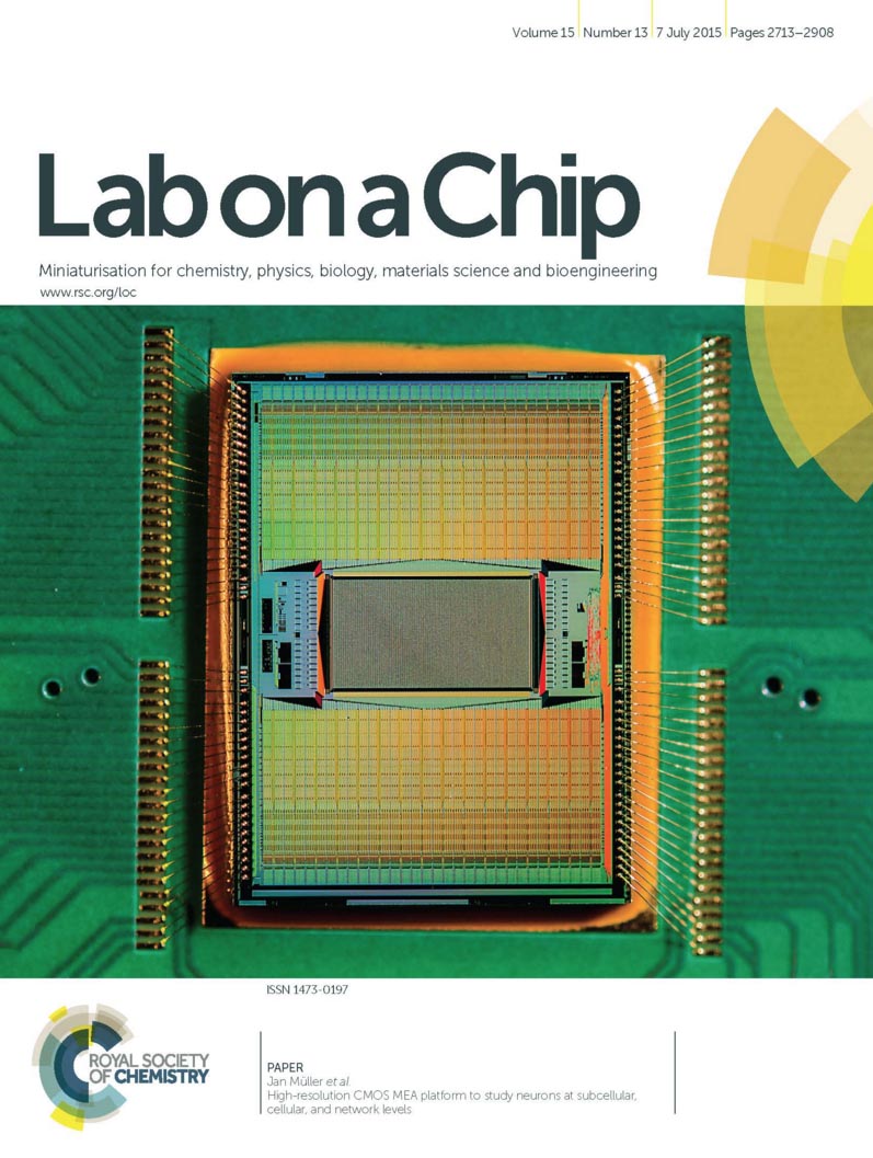
title = {High-resolution CMOS MEA platform to study neurons at subcellular, cellular, and network levels},
author = {Jan Müller and Marco Ballini and Paolo Livi and Yihui Chen and Milos Radivojevic and Amir Shadmani and Vijay Viswam and Ian L Jones and Michele Fiscella and Roland Diggelmann and Alexander Stettler and Urs Frey and Douglas J Bakkum and Andreas Hierlemann},
url = {http://pubs.rsc.org/en/Content/ArticleLanding/2015/LC/C5LC00133A#!divAbstract},
doi = {10.1039/C5LC00133A},
issn = {1473-0197},
year = {2015},
date = {2015-07-07},
journal = {Lab Chip},
volume = {15},
number = {13},
pages = {2767-2780},
publisher = {Royal Society of Chemistry},
abstract = {Studies on information processing and learning properties of neuronal networks would benefit from simultaneous and parallel access to the activity of a large fraction of all neurons in such networks. Here, we present a CMOS-based device, capable of simultaneously recording the electrical activity of over a thousand cells in in vitro neuronal networks. The device provides sufficiently high spatiotemporal resolution to enable, at the same time, access to neuronal preparations on subcellular, cellular, and network level. The key feature is a rapidly reconfigurable array of 26 400 microelectrodes arranged at low pitch (17.5 um) within a large overall sensing area (3.85 × 2.10 mm2). An arbitrary subset of the electrodes can be simultaneously connected to 1024 low-noise readout channels as well as 32 stimulation units. Each electrode or electrode subset can be used to electrically stimulate or record the signals of virtually any neuron on the array. We demonstrate the applicability and potential of this device for various different experimental paradigms: large-scale recordings from whole networks of neurons as well as investigations of axonal properties of individual neurons.},
keywords = {MaxOne, Neuronal Networks},
pubstate = {published},
tppubtype = {article}
}

title = {A network comprising short and long noncoding RNAs and RNA helicase controls mouse retina architecture.},
author = {Jacek Krol and Ilona Krol and Claudia Patricia Patino Alvarez and Michele Fiscella and Andreas Hierlemann and Botond Roska and Witold Filipowicz},
url = {https://www.nature.com/articles/ncomms8305},
doi = {10.1038/ncomms8305},
issn = {2041-1723},
year = {2015},
date = {2015-06-04},
journal = {Nature Communications},
volume = {6},
pages = {7305},
publisher = {Nature Publishing Group},
abstract = {Brain regions, such as the cortex and retina, are composed of layers of uniform thickness. The molecular mechanism that controls this uniformity is not well understood. Here we show that during mouse postnatal development the timed expression of Rncr4, a retina-specific long noncoding RNA, regulates the similarly timed processing of pri-miR-183/96/182, which is repressed at an earlier developmental stage by RNA helicase Ddx3x. Shifting the timing of mature miR-183/96/182 accumulation or interfering with Ddx3x expression leads to the disorganization of retinal architecture, with the photoreceptor layer being most affected. We identify Crb1, a component of the adhesion belt between glial and photoreceptor cells, as a link between Rncr4-regulated miRNA metabolism and uniform retina layering. Our results suggest that the precise timing of glia-neuron interaction controlled by noncoding RNAs and Ddx3x is important for the even distribution of cells across layers.},
keywords = {MaxOne, Retina},
pubstate = {published},
tppubtype = {article}
}

title = {Chronic Co-Variation of Neural Network Configuration and Activity in Mature Dissociated Cultures},
author = {Satoru Okawa and Takeshi Mita and Douglas J Bakkum and Urs Frey and Andreas Hierlemann and Ryohei Kanzaki and Hirokazu Takahashi},
url = {http://onlinelibrary.wiley.com/doi/10.1002/ecj.11736/abstract;jsessionid=791557FA80CF36B1F78DC538C1618924.f03t02},
doi = {10.1002/ecj.11736},
issn = {1942-9541},
year = {2015},
date = {2015-04-08},
journal = {Electronics and Communications in Japan},
volume = {98},
number = {5},
pages = {34-42},
abstract = {Spatiotemporal neural patterns depend on the physical structure of neural circuits. Neural plasticity can thus be associated with changes in the circuit structure. For example, newborn neurons migrate toward existing, already matured, neural networks in order to participate in neural computation. In the present study, we have conducted two experiments to investigate how neural migration is associated with the development of neural activity in primary dissociated cultures of neuronal cells. In Experiment 1, using a mature culture, a high-density CMOS microelectrode array was used to continuously monitor neural migration and activity for more than two weeks. Consequently, we found that even in mature neuronal cultures neurons moved 2.0 ± 1.0 mum a day and that the moving distance was negatively correlated with their firing rate, suggesting that neurons featuring low firing rates tend to migrate actively. In Experiment 2 using a co-culture of mature and immature neurons, we found that immature neurons moved more actively than matured neurons to achieve functional connections to other neurons. These findings suggest that neurons with low firing rates as well as newborn neurons actively migrate in order to establish their connections and function in a neuronal network.},
keywords = {ETH-CMOS-MEA, Neuronal Networks},
pubstate = {published},
tppubtype = {article}
}

title = {Recording large extracellular spikes in microchannels along many axonal sites from individual neurons},
author = {Marta K Lewandowska and Douglas J Bakkum and Santiago B Rompani and Andreas Hierlemann},
url = {http://journals.plos.org/plosone/article?id=10.1371/journal.pone.0118514},
doi = {10.1371/journal.pone.0118514},
issn = {19326203},
year = {2015},
date = {2015-03-03},
journal = {PLoS ONE},
volume = {10},
number = {3},
pages = {1-24},
abstract = {The numerous connections between neuronal cell bodies, made by their dendrites and axons, are vital for information processing in the brain. While dendrites and synapses have been extensively studied, axons have remained elusive to a large extent. We present a novel platform to study axonal physiology and information processing based on combining an 11,011-electrode high-density complementary metal-oxide semiconductor microelectrode array with a poly(dimethylsiloxane) channel device, which isolates axons from somas and, importantly, significantly amplifies recorded axonal signals. The combination of the microelectrode array with recording and stimulation capability with the microfluidic isolation channels permitted us to study axonal signal behavior at great detail. The device, featuring two culture chambers with over 30 channels spanning in between, enabled long-term recording of single spikes from isolated axons with signal amplitudes of 100 uV up to 2 mV. Propagating signals along axons could be recorded with 10 to 50 electrodes per channel. We (i) describe the performance and capabilities of our device for axonal electrophysiology, and (ii) present novel data on axonal signals facilitated by the device. Spontaneous action potentials with characteristic shapes propagated from somas along axons between the two compartments, and these unique shapes could be used to identify individual axons within channels that contained many axonal branches. Stimulation through the electrode array facilitated the identification of somas and their respective axons, enabling interfacing with different compartments of a single cell. Complex spike shapes observed in channels were traced back to single cells, and we show that more complicated spike shapes originate from a linear superposition of multiple axonal signals rather than signal distortion by the channels.},
keywords = {ETH-CMOS-MEA, Neuronal Networks},
pubstate = {published},
tppubtype = {article}
}
title = {Complexity Optimization and High-Throughput Low-Latency Hardware Implementation of a Multi-Electrode Spike-Sorting Algorithm},
author = {Jelena Dragas and David Jäckel and Andreas Hierlemann and Felix Franke},
url = {http://ieeexplore.ieee.org/document/6955847/},
doi = {10.1109/TNSRE.2014.2370510},
issn = {15344320},
year = {2015},
date = {2015-03-01},
journal = {IEEE Transactions on Neural Systems and Rehabilitation Engineering},
volume = {23},
number = {2},
pages = {149--158},
abstract = {Reliable real-time low-latency spike sorting with large data throughput is essential for studies of neural network dynamics and for brain-machine interfaces (BMIs), in which the stimulation of neural networks is based on the networks' most recent activity. However, the majority of existing multi-electrode spike-sorting algorithms are unsuited for processing high quantities of simultaneously recorded data. Recording from large neuronal networks using large high-density electrode sets (thousands of electrodes) imposes high demands on the data-processing hardware regarding computational complexity and data transmission bandwidth; this, in turn, entails demanding requirements in terms of chip area, memory resources and processing latency. This paper presents computational complexity optimisation techniques, which facilitate the use of spike-sorting algorithms in large multi-electrode-based recording systems. The techniques are then applied to a previously published algorithm, on its own, unsuited for large electrode set recordings. Further, a real-time low-latency high-performance VLSI hardware architecture of the modified algorithm is presented, featuring a folded structure capable of processing the activity of hundreds of neurons simultaneously. The hardware is reconfigurable “on-the-fly” and adaptable to the non-stationarities of neuronal recordings. By transmitting exclusively spike time stamps and/or spike waveforms, its real-time processing offers the possibility of data bandwidth and data storage reduction.},
keywords = {Spike Sorting},
pubstate = {published},
tppubtype = {article}
}

title = {Revealing neuronal function through microelectrode array recordings},
author = {Marie Engelene J Obien and Kosmas Deligkaris and Torsten Bullmann and Douglas J Bakkum and Urs Frey},
url = {https://www.frontiersin.org/articles/10.3389/fnins.2014.00423/full},
doi = {10.3389/fnins.2014.00423},
issn = {1662453X},
year = {2015},
date = {2015-01-06},
journal = {Frontiers in Neuroscience},
volume = {9},
pages = {423},
abstract = {Microelectrode arrays and microprobes have been widely utilized to measure neuronal activity, both in vitro and in vivo. The key advantage is the capability to record and stimulate neurons at multiple sites simultaneously. However, unlike the single-cell or single-channel resolution of intracellular recording, microelectrodes detect signals from all possible sources around every sensor. Here, we review the current understanding of microelectrode signals and the techniques for analyzing them. We introduce the ongoing advancements in microelectrode technology, with focus on achieving higher resolution and quality of recordings by means of monolithic integration with on-chip circuitry. We show how recent advanced microelectrode array measurement methods facilitate the understanding of single neurons as well as network function.},
keywords = {MEA Technology, Review},
pubstate = {published},
tppubtype = {article}
}
2014
title = {A 1024-channel CMOS microelectrode array with 26,400 electrodes for recording and stimulation of electrogenic cells in vitro},
author = {Marco Ballini and Jan Müller and Paolo Livi and Yihui Chen and Urs Frey and Alexander Stettler and Amir Shadmani and Vijay Viswam and Ian L Jones and David Jäckel and Milos Radivojevic and Marta K Lewandowska and Wei Gong and Michele Fiscella and Douglas J Bakkum and Flavio Heer and Andreas Hierlemann},
url = {http://ieeexplore.ieee.org/document/6923484/},
doi = {10.1109/JSSC.2014.2359219},
issn = {00189200},
year = {2014},
date = {2014-10-14},
journal = {IEEE Journal of Solid-State Circuits},
volume = {49},
number = {11},
pages = {2705-2719},
abstract = {To advance our understanding of the functioning of neuronal ensembles, systems are needed to enable simultaneous recording from a large number of individual neurons at high spa-tiotemporal resolution and good signal-to-noise ratio. Moreover, stimulation capability is highly desirable for investigating, for example, plasticity and learning processes. Here, we present a microelectrode array (MEA) system on a single CMOS die for in vitro recording and stimulation. The system incorporates 26,400 platinum electrodes, fabricated by in-house post-processing, over a large sensing area (3.85 2.10 mm) with sub-cellular spatial resolution (pitch of 17.5 µm). Owing to an area and power efficient implementation, we were able to integrate 1024 readout channels on chip to record extracellular signals from a user-specified selection of electrodes. These channels feature noise values of 2.4 µV in the action-potential band (300 Hz–10 kHz) and 5.4 µV in the local-field-potential band (1 Hz–300 Hz), and provide programmable gain (up to 78 dB) to accommodate various biological preparations. Amplified and filtered signals are digitized by 10 bit parallel single-slope ADCs at 20 kSamples/s. The system also includes 32 stimulation units, which can elicit neural spikes through either current or voltage pulses. The chip consumes only 75 mW in total, which obviates the need of active cooling even for sensitive cell cultures.},
keywords = {MaxOne, MEA Technology},
pubstate = {published},
tppubtype = {article}
}
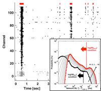
title = {Parameters for burst detection},
author = {Douglas J Bakkum and Milos Radivojevic and Urs Frey and Felix Franke and Andreas Hierlemann and Hirokazu Takahashi},
url = {http://journal.frontiersin.org/article/10.3389/fncom.2013.00193/abstract},
doi = {10.3389/fncom.2013.00193},
issn = {1662-5188},
year = {2014},
date = {2014-01-13},
journal = {Frontiers in Computational Neuroscience},
volume = {7},
pages = {1-12},
abstract = {Bursts of action potentials within neurons and throughout networks are believed to serve roles in how neurons handle and store information, both in vivo and in vitro. Accurate detection of burst occurrences and durations are therefore crucial for many studies. A number of algorithms have been proposed to do so, but a standard method has not been adopted. This is due, in part, to many algorithms requiring the adjustment of multiple ad-hoc parameters and further post-hoc criteria in order to produce satisfactory results. Here, we broadly catalog existing approaches and present a new approach requiring the selection of only a single parameter: the number of spikes N comprising the smallest burst to consider. A burst was identified if N spikes occurred in less than T ms, where the threshold T was automatically determined from observing a probability distribution of inter-spike-intervals. Performance was compared vs. different classes of detectors on data gathered from in vitro neuronal networks grown over microelectrode arrays. Our approach offered a number of useful features including: a simple implementation, no need for ad-hoc or post-hoc criteria, and precise assignment of burst boundary time points. Unlike existing approaches, detection was not biased toward larger bursts, allowing identification and analysis of a greater range of neuronal and network dynamics.},
keywords = {Data Analysis, ETH-CMOS-MEA},
pubstate = {published},
tppubtype = {article}
}