 日本語
日本語 繁體中文
繁體中文 简体中文
简体中文 English
Publications
Selected Publications

High-resolution CMOS MEA platform to study neurons at subcellular, cellular, and network levels

Presenting measurements of neuronal preparations with a novel CMOS-based microelectrode array at high-spatiotemporal-resolution on subcellular, cellular, and network level.
J. Müller, M. Ballini, P. Livi, Y. Chen, M. Radivojevic, A. Shadmani, V. Viswam, I. L. Jones, M. Fiscella, R. Diggelmann, A. Stettler, U. Frey, D. J. Bakkum, and A. Hierlemann, “High-resolution CMOS MEA platform to study neurons at subcellular, cellular, and network levels,” Lab Chip, vol. 15, no. 13, pp. 2767–2780, May 2015.
Revealing Neuronal Function through Microelectrode Array Recordings

Reviewing the current understanding of microelectrode signals and the techniques for analyzing them, with focus on the ongoing advancements in microelectrode technology (in vivo and in vitro) and recent advanced microelectrode array measurement methods that facilitate the understanding of single neurons and network function.
M. E. J. Obien, K. Deligkaris, T. Bullmann, D. J. Bakkum, and U. Frey, “Revealing Neuronal Function through Microelectrode Array Recordings,” Front. Neurosci., 8:423, Jan 2015.

A 1024-Channel CMOS Microelectrode Array With 26,400 Electrodes for Recording and Stimulation of Electrogenic Cells In Vitro
A high-resolution CMOS-based microelectrode array featuring 1,024 low-noise readout channels, 26,400 electrodes at a density of 3,265 electrodes per mm2, including on-chip 10bit ADCs and consuming only 75 mW.
M. Ballini, J. Muller, P. Livi, Y. Chen, U. Frey, A. Stettler, A. Shadmani, V. Viswam, I. L. Jones, D. Jackel, M. Radivojevic, M. K. Lewandowska, W. Gong, M. Fiscella, D. J. Bakkum, F. Heer, and A. Hierlemann, “A 1024-Channel CMOS Microelectrode Array With 26,400 Electrodes for Recording and Stimulation of Electrogenic Cells In Vitro,” IEEE Journal of Solid-State Circuits, vol. 49, no. 11, pp. 2705-2719, 2014.

Tracking axonal action potential propagation on a high-density microelectrode array across hundreds of sites
Demonstrating a method to electrically visualize action potential propagation on axons and revealing
large variations in velocity.
D. J. Bakkum, U. Frey, M. Radivojevic, T. L. Russell, J. Muller, M. Fiscella, H. Takahashi, and A. Hierlemann, “Tracking axonal action potential propagation on a high-density microelectrode array across hundreds of sites,” Nature Communications, 4:2181, Jul 2013.

Microelectronic System for High-Resolution Mapping of Extracellular Electric Fields Applied to Brain Slices
Recording and modeling extracellular action potentials of Purkinje cells at subcellular resolution.
U. Frey, U. Egert, F. Heer, S. Hafizovic, and A. Hierlemann, “Microelectronic System for High-Resolution Mapping of Extracellular Electric Fields Applied to Brain Slices,” Biosensors and Bioelectronics, vol. 24, no. 7, pp. 2191-2198, 2009.

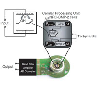
Modulation of Cardiomyocyte Electrical Properties Using Regulated Bone Morphogenetic Protein-2 Expression
Controlling BMP-2 expression to modulate the electrophysiological properties of cardiomyocytes using an HD-MEA for detailed monitoring.
C. D. Sanchez-Bustamante, U. Frey, J. M. Kelm, A. Hierlemann, and M. Fussenegger,
“Modulation of Cardiomyocyte Electrical Properties Using Regulated Bone Morphogenetic Protein-2 Expression,” Tissue Engineering Part A, vol. 14, no. 12, pp. 1969-1988, 2008.
All Publications
| 1. | Lee, Jihyun; Gänswein, Tobias; Ulusan, Hasan; Emmenegger, Vishalini; Saguner, Ardan M; Duru, Firat; and Hierlemann, Andreas: Repeated and On-Demand Intracellular Recordings of Cardiomyocytes Derived from Human Induced Pluripotent Stem Cells. In: ACS Sensors, 2022. (Type: Journal Article | Abstract | Links | BibTeX) @article{Lee2022, title = {Repeated and On-Demand Intracellular Recordings of Cardiomyocytes Derived from Human Induced Pluripotent Stem Cells}, author = {Jihyun Lee and Tobias Gänswein and Hasan Ulusan and Vishalini Emmenegger and Ardan M. Saguner and Firat Duru and and Andreas Hierlemann}, url = {https://pubs.acs.org/doi/10.1021/acssensors.2c01678}, doi = {https://doi.org/10.1021/acssensors.2c01678}, year = {2022}, date = {2022-09-27}, journal = {ACS Sensors}, abstract = {Pharmaceutical compounds may have cardiotoxic properties, triggering potentially life-threatening arrhythmi- as. To investigate proarrhythmic effects of drugs, the patch clamp technique has been used as the gold standard for charac- terizing the electrophysiology of cardiomyocytes in vitro. However, the applicability of this technology for drug screening is limited, as it is complex to use and features low throughput. Recent studies have demonstrated that 3D-nanostructured electrodes enable to obtain intracellular signals from many cardiomyocytes in parallel; however, the tedious electrode fab- rication and limited measurement duration still remain major issues for cardiotoxicity testing. Here, we demonstrate how porous Pt-black electrodes, arranged in high-density microelectrode arrays, can be used to record intracellular-like signals of cardiomyocytes at large-scale repeatedly over an extended period of time. The developed technique, which yields highly parallelized electroporations by using stimulation voltages around 1 Volt peak-to-peak amplitude, enabled intracellular-like recordings at high success rates without causing significant alteration in key electrophysiological features. In a proof of concept study, we investigated electrophysiological modulations induced by two clinically applied drugs, nifedipine and quinidine. As the obtained results were in good agreement with previously published data, we are confident that the devel- oped technique has the potential to be routinely used in in vitro platforms for cardiotoxicity screening.}, keywords = {}, pubstate = {published}, tppubtype = {article} } Pharmaceutical compounds may have cardiotoxic properties, triggering potentially life-threatening arrhythmi- as. To investigate proarrhythmic effects of drugs, the patch clamp technique has been used as the gold standard for charac- terizing the electrophysiology of cardiomyocytes in vitro. However, the applicability of this technology for drug screening is limited, as it is complex to use and features low throughput. Recent studies have demonstrated that 3D-nanostructured electrodes enable to obtain intracellular signals from many cardiomyocytes in parallel; however, the tedious electrode fab- rication and limited measurement duration still remain major issues for cardiotoxicity testing. Here, we demonstrate how porous Pt-black electrodes, arranged in high-density microelectrode arrays, can be used to record intracellular-like signals of cardiomyocytes at large-scale repeatedly over an extended period of time. The developed technique, which yields highly parallelized electroporations by using stimulation voltages around 1 Volt peak-to-peak amplitude, enabled intracellular-like recordings at high success rates without causing significant alteration in key electrophysiological features. In a proof of concept study, we investigated electrophysiological modulations induced by two clinically applied drugs, nifedipine and quinidine. As the obtained results were in good agreement with previously published data, we are confident that the devel- oped technique has the potential to be routinely used in in vitro platforms for cardiotoxicity screening. |
| 2. | Weber, Wilfried; Luzi, Stefan; Karlsson, Maria; Sanchez-Bustamante, Carlota Diaz; Frey, Urs; Hierlemann, Andreas; Fussenegger, Martin: A synthetic mammalian electro-genetic transcription circuit. In: Nucleic Acids Research, 37 (4), pp. 1-8, 2009, ISSN: 03051048. (Type: Journal Article | Abstract | Links | BibTeX) @article{Weber2009, title = {A synthetic mammalian electro-genetic transcription circuit}, author = {Wilfried Weber and Stefan Luzi and Maria Karlsson and Carlota Diaz Sanchez-Bustamante and Urs Frey and Andreas Hierlemann and Martin Fussenegger}, url = {https://academic.oup.com/nar/article-lookup/doi/10.1093/nar/gkp014}, doi = {10.1093/nar/gkp014}, issn = {03051048}, year = {2009}, date = {2009-02-03}, journal = {Nucleic Acids Research}, volume = {37}, number = {4}, pages = {1-8}, abstract = {Electric signal processing has evolved to manage rapid information transfer in neuronal networks and muscular contraction in multicellular organisms and controls the most sophisticated man-built devices. Using a synthetic biology approach to assemble electronic parts with genetic control units engineered into mammalian cells, we designed an electric power-adjustable transcription control circuit able to integrate the intensity of a direct current over time, to translate the amplitude or frequency of an alternating current into an adjustable genetic readout or to modulate the beating frequency of primary heart cells. Successful miniaturization of the electro-genetic devices may pave the way for the design of novel hybrid electrogenetic implants assembled from electronic and genetic parts.}, keywords = {}, pubstate = {published}, tppubtype = {article} } Electric signal processing has evolved to manage rapid information transfer in neuronal networks and muscular contraction in multicellular organisms and controls the most sophisticated man-built devices. Using a synthetic biology approach to assemble electronic parts with genetic control units engineered into mammalian cells, we designed an electric power-adjustable transcription control circuit able to integrate the intensity of a direct current over time, to translate the amplitude or frequency of an alternating current into an adjustable genetic readout or to modulate the beating frequency of primary heart cells. Successful miniaturization of the electro-genetic devices may pave the way for the design of novel hybrid electrogenetic implants assembled from electronic and genetic parts. |
| 3. | Sanchez-Bustamante, Carlota Diaz; Frey, Urs; Kelm, Jens M; Hierlemann, Andreas; Fussenegger, Martin: Modulation of cardiomyocyte electrical properties using regulated bone morphogenetic protein-2 expression.. In: Tissue Engineering. Part A, 14 (12), pp. 1969-1988, 2008, ISSN: 1937-3341. (Type: Journal Article | Abstract | Links | BibTeX) @article{Sanchez-Bustamante2008, title = {Modulation of cardiomyocyte electrical properties using regulated bone morphogenetic protein-2 expression.}, author = {Carlota Diaz Sanchez-Bustamante and Urs Frey and Jens M Kelm and Andreas Hierlemann and Martin Fussenegger}, url = {http://online.liebertpub.com/doi/abs/10.1089/ten.tea.2007.0302?url_ver=Z39.88-2003&rfr_id=ori%3Arid%3Acrossref.org&rfr_dat=cr_pub%3Dpubmed}, doi = {10.1089/ten.tea.2007.0302}, issn = {1937-3341}, year = {2008}, date = {2008-11-19}, journal = {Tissue Engineering. Part A}, volume = {14}, number = {12}, pages = {1969-1988}, abstract = {Because cardiomyocytes lose their ability to divide after birth, any subsequent cell loss or dysfunction results in pathologic cardiac rhythm initiation or impulse conduction. Strategies to restore and control the electrophysiological activity of the heart may, therefore, greatly affect the regeneration of cardiac tissue functionality. Using lentivirus-derived particles to regulate the bone morphogenetic protein-2 (BMP-2) gene expression in a pristinamycin- or gaseous acetaldehyde-inducible manner, we demonstrated the adjustment of cardiomyocyte electrophysiological characteristics. Complementary metal oxide semiconductor-based high-density microelectrode arrays (HD-MEAs) were used to monitor the electrophysiological activity of neonatal rat cardiomyocytes (NRCs) cultured as monolayers (NRCml) or as microtissues (NRCmt). NRCmt more closely resembled heart tissue physiology than did NRCml and could be conveniently monitored using HD-MEAs because of their ability to detect low-signal events and to sub-select the region of interest, namely, areas where the microtissues were placed. Cardiomyocyte-forming microtissues, transduced using lentiviral vectors encoding BMP-2, were capable of restoring myocardial microtissue electrical activity. We also engineered NRCmt to functionally couple within a cardiomyocyte monolayer, thus showing pacemaker-like activity upon local regulation of transgenic BMP-2 expression. The controlled expression of therapeutic transgenes represents a crucial advance for clinical interventions and gene-function analysis.}, keywords = {}, pubstate = {published}, tppubtype = {article} } Because cardiomyocytes lose their ability to divide after birth, any subsequent cell loss or dysfunction results in pathologic cardiac rhythm initiation or impulse conduction. Strategies to restore and control the electrophysiological activity of the heart may, therefore, greatly affect the regeneration of cardiac tissue functionality. Using lentivirus-derived particles to regulate the bone morphogenetic protein-2 (BMP-2) gene expression in a pristinamycin- or gaseous acetaldehyde-inducible manner, we demonstrated the adjustment of cardiomyocyte electrophysiological characteristics. Complementary metal oxide semiconductor-based high-density microelectrode arrays (HD-MEAs) were used to monitor the electrophysiological activity of neonatal rat cardiomyocytes (NRCs) cultured as monolayers (NRCml) or as microtissues (NRCmt). NRCmt more closely resembled heart tissue physiology than did NRCml and could be conveniently monitored using HD-MEAs because of their ability to detect low-signal events and to sub-select the region of interest, namely, areas where the microtissues were placed. Cardiomyocyte-forming microtissues, transduced using lentiviral vectors encoding BMP-2, were capable of restoring myocardial microtissue electrical activity. We also engineered NRCmt to functionally couple within a cardiomyocyte monolayer, thus showing pacemaker-like activity upon local regulation of transgenic BMP-2 expression. The controlled expression of therapeutic transgenes represents a crucial advance for clinical interventions and gene-function analysis. |
| 4. | Greve, Frauke; Lichtenberg, Jan; Kirstein, Kay Uwe; Frey, Urs; Perriard, Jean Claude; Hierlemann, Andreas: A perforated CMOS microchip platform for immobilization and activity monitoring of electrogenic cells. In: Journal of Micromechanics and Microengineering, 17 (3), pp. 462-471, 2007, ISSN: 0960-1317. (Type: Journal Article | Abstract | Links | BibTeX) @article{Greve2007, title = {A perforated CMOS microchip platform for immobilization and activity monitoring of electrogenic cells}, author = {Frauke Greve and Jan Lichtenberg and Kay Uwe Kirstein and Urs Frey and Jean Claude Perriard and Andreas Hierlemann}, url = {http://iopscience.iop.org/article/10.1088/0960-1317/17/3/007/}, doi = {10.1088/0960-1317/17/3/007}, issn = {0960-1317}, year = {2007}, date = {2007-01-30}, journal = {Journal of Micromechanics and Microengineering}, volume = {17}, number = {3}, pages = {462-471}, abstract = {CMOS-based microelectrode systems offer decisive advantages over conventional micro-electrode arrays, which include the possibility to perform on-chip signal conditioning or to efficiently use larger numbers of electrodes to obtain statistically relevant data, e.g., in pharmacological drug screening. A larger number of electrodes can only be realized with the help of on-chip multiplexing and readout schemes, which require integrated electronics. Another fundamental issue in performing high-fidelity recordings from electrogenic cells is a good electrical coupling between the cells and the microelectrodes, in particular, since the recorded extracellular signals are in the range of only 10–1000 µV. In this paper we present the first CMOS microelectrode system with integrated micromechanical cell-placement features fabricated in a commercial CMOS process with subsequent post-CMOS bulk micromachining. This new microdevice aims at enabling the precise placement of single cells in the center of the electrodes to ensure an efficient use of the available electrodes, even for low-density cell cultures. Small through-chip holes have been generated at the metal-electrode sites by using a combination of bulk micromachining and reactive-ion etching. These holes act as orifices so that cell immobilization can be achieved by means of pneumatic anchoring. The chip additionally hosts integrated circuitry, i.e., multiplexers to select the respective readout electrodes, an amplifier with selectable gain (2×, 10×, 100×), and a high-pass filter (100 Hz cut-off). In this paper we show that electrical signals from most of the electrodes can be recorded, even in low-density cultures of neonatal rat cardiomyocytes, by using perforated metal electrodes and by applying a small underpressure from the backside of the chip. The measurements evidenced that, in most cases, about 90% of the electrodes were covered with single cells, approximately 4% were covered with more than one cell due to clustering and approximately 6% were not covered with any cell, mostly as a consequence of orifice clogging. After 4 days of culturing, the cells were still in place on the electrodes so that the cell electrical activity could be measured using the on-chip circuitry. Measured signal amplitudes were in the range of 500–700 µV, while the input-referred noise of the readout was below 15 µVrms (100 Hz–4 kHz bandwidth). We report on the development and fabrication of this new cell-biological tool and present first results collected during the characterization and evaluation of the chip. The recordings of electrical potentials of neonatal rat cardiomyocytes after several days in vitro, which, on the one hand, were conventionally cultured (no pneumatic anchoring) and, on the other hand, were anchored and immobilized, will be detailed.}, keywords = {}, pubstate = {published}, tppubtype = {article} } CMOS-based microelectrode systems offer decisive advantages over conventional micro-electrode arrays, which include the possibility to perform on-chip signal conditioning or to efficiently use larger numbers of electrodes to obtain statistically relevant data, e.g., in pharmacological drug screening. A larger number of electrodes can only be realized with the help of on-chip multiplexing and readout schemes, which require integrated electronics. Another fundamental issue in performing high-fidelity recordings from electrogenic cells is a good electrical coupling between the cells and the microelectrodes, in particular, since the recorded extracellular signals are in the range of only 10–1000 µV. In this paper we present the first CMOS microelectrode system with integrated micromechanical cell-placement features fabricated in a commercial CMOS process with subsequent post-CMOS bulk micromachining. This new microdevice aims at enabling the precise placement of single cells in the center of the electrodes to ensure an efficient use of the available electrodes, even for low-density cell cultures. Small through-chip holes have been generated at the metal-electrode sites by using a combination of bulk micromachining and reactive-ion etching. These holes act as orifices so that cell immobilization can be achieved by means of pneumatic anchoring. The chip additionally hosts integrated circuitry, i.e., multiplexers to select the respective readout electrodes, an amplifier with selectable gain (2×, 10×, 100×), and a high-pass filter (100 Hz cut-off). In this paper we show that electrical signals from most of the electrodes can be recorded, even in low-density cultures of neonatal rat cardiomyocytes, by using perforated metal electrodes and by applying a small underpressure from the backside of the chip. The measurements evidenced that, in most cases, about 90% of the electrodes were covered with single cells, approximately 4% were covered with more than one cell due to clustering and approximately 6% were not covered with any cell, mostly as a consequence of orifice clogging. After 4 days of culturing, the cells were still in place on the electrodes so that the cell electrical activity could be measured using the on-chip circuitry. Measured signal amplitudes were in the range of 500–700 µV, while the input-referred noise of the readout was below 15 µVrms (100 Hz–4 kHz bandwidth). We report on the development and fabrication of this new cell-biological tool and present first results collected during the characterization and evaluation of the chip. The recordings of electrical potentials of neonatal rat cardiomyocytes after several days in vitro, which, on the one hand, were conventionally cultured (no pneumatic anchoring) and, on the other hand, were anchored and immobilized, will be detailed. |
| 5. | Heer, Flavio; Hafizovic, Sadik; Ugniwenko, T; Frey, Urs; Franks, Wendy; Perriard, Evelyne; Perriard, Jean Claude; Blau, Axel; Ziegler, Christiane; Hierlemann, Andreas: Single-chip microelectronic system to interface with living cells. In: Biosensors & Bioelectronics, 22 (11), pp. 2546-2553, 2006, ISSN: 0956-5663. (Type: Journal Article | Abstract | Links | BibTeX) @article{Hierlemann2006, title = {Single-chip microelectronic system to interface with living cells}, author = {Flavio Heer and Sadik Hafizovic and T Ugniwenko and Urs Frey and Wendy Franks and Evelyne Perriard and Jean Claude Perriard and Axel Blau and Christiane Ziegler and Andreas Hierlemann}, url = {http://www.sciencedirect.com/science/article/pii/S0956566306004891?via%3Dihub}, doi = {10.1016/j.bios.2006.10.003}, issn = {0956-5663}, year = {2006}, date = {2006-11-13}, journal = {Biosensors & Bioelectronics}, volume = {22}, number = {11}, pages = {2546-2553}, abstract = {A high degree of connectivity and the coordinated electrical activity of neural cells or networks are believed to be the reason that the brain is capable of highly sophisticated information processing. Likewise, the effectiveness of an animal heart largely depends on such coordinated cell activity. To advance our understanding of these complex biological systems, high spatiotemporal-resolution techniques to monitor the cell electrical activity and an ideally seamless interaction between cells and recording devices are desired. Here we present a monolithic microsystem in complementary metal oxide semiconductor (CMOS) technology that provides bidirectional communication (stimulation and recording) between standard electronics technology and cultured electrogenic cells. The microchip can be directly used as a substrate for cell culturing, it features circuitry units per electrode for stimulation and immediate cell signal treatment, and it provides on-chip signal transformation as well as a digital interface so that a very fast, almost real-time interaction (2ms loop time from event recognition to, e.g., a defined stimulation) is possible at remarkable signal quality. The corresponding spontaneous and stimulated electrical activity recordings with neuronal and cardiac cell cultures will be presented. The system can be used to, e.g., study the development of neural networks, reveal the effects of neuronal plasticity and study cellular or network activity in response to pharmacological treatments.}, keywords = {}, pubstate = {published}, tppubtype = {article} } A high degree of connectivity and the coordinated electrical activity of neural cells or networks are believed to be the reason that the brain is capable of highly sophisticated information processing. Likewise, the effectiveness of an animal heart largely depends on such coordinated cell activity. To advance our understanding of these complex biological systems, high spatiotemporal-resolution techniques to monitor the cell electrical activity and an ideally seamless interaction between cells and recording devices are desired. Here we present a monolithic microsystem in complementary metal oxide semiconductor (CMOS) technology that provides bidirectional communication (stimulation and recording) between standard electronics technology and cultured electrogenic cells. The microchip can be directly used as a substrate for cell culturing, it features circuitry units per electrode for stimulation and immediate cell signal treatment, and it provides on-chip signal transformation as well as a digital interface so that a very fast, almost real-time interaction (2ms loop time from event recognition to, e.g., a defined stimulation) is possible at remarkable signal quality. The corresponding spontaneous and stimulated electrical activity recordings with neuronal and cardiac cell cultures will be presented. The system can be used to, e.g., study the development of neural networks, reveal the effects of neuronal plasticity and study cellular or network activity in response to pharmacological treatments. |
| 6. | Franks, Wendy; Tosatti, Samuele; Heer, Flavio; Seif, Philipp; Textor, Marcus; Hierlemann, Andreas: Patterned cell adhesion by self-assembled structures for use with a CMOS cell-based biosensor. In: Biosensors & Bioelectronics, 22 (7), pp. 1426-1433, 2006, ISSN: 0956-5663. (Type: Journal Article | Abstract | Links | BibTeX) @article{Hierlemann2006b, title = {Patterned cell adhesion by self-assembled structures for use with a CMOS cell-based biosensor}, author = {Wendy Franks and Samuele Tosatti and Flavio Heer and Philipp Seif and Marcus Textor and Andreas Hierlemann}, url = {http://www.sciencedirect.com/science/article/pii/S095656630600282X?via%3Dihub}, doi = {10.1016/j.bios.2006.06.031}, issn = {0956-5663}, year = {2006}, date = {2006-10-19}, journal = {Biosensors & Bioelectronics}, volume = {22}, number = {7}, pages = {1426-1433}, abstract = {A strategy for patterned cell adhesion based on chemical surface modification is presented. To confine cell adhesion to specific locations, an engineered surface for high-contrast protein adsorption and, hence, cell attachment has been developed. Surface functionalization is based on selective molecular-assembly patterning (SMAP). An amine-terminated self-assembled monolayer is used to define areas of cell adhesion. A protein-repellent grafted copolymer, poly(l-lysine)-graft-poly(ethylene glycol) (PLL-g-PEG), is used to render the surrounding silicon dioxide resistant to protein adsorption. X-ray photoelectron spectroscopy, scanning ellipsometry and fluorescence microscopy techniques were used to monitor the individual steps of the patterning process. Successful guided growth using these layers is demonstrated with primary neonatal rat cardiomyocytes, up to 4 days in vitro, and with the HL-1 cardiomyocyte cell line, up to 7 days in vitro. The advantage of the presented method is that high-resolution engineered surfaces can be realized using a simple, cost-effective, dip-and-rinse process. The technique has been developed for application on a CMOS cell-based biosensor, which comprises an array of microelectrodes to extracellularly record electrical activity from cardiomyocytes.}, keywords = {}, pubstate = {published}, tppubtype = {article} } A strategy for patterned cell adhesion based on chemical surface modification is presented. To confine cell adhesion to specific locations, an engineered surface for high-contrast protein adsorption and, hence, cell attachment has been developed. Surface functionalization is based on selective molecular-assembly patterning (SMAP). An amine-terminated self-assembled monolayer is used to define areas of cell adhesion. A protein-repellent grafted copolymer, poly(l-lysine)-graft-poly(ethylene glycol) (PLL-g-PEG), is used to render the surrounding silicon dioxide resistant to protein adsorption. X-ray photoelectron spectroscopy, scanning ellipsometry and fluorescence microscopy techniques were used to monitor the individual steps of the patterning process. Successful guided growth using these layers is demonstrated with primary neonatal rat cardiomyocytes, up to 4 days in vitro, and with the HL-1 cardiomyocyte cell line, up to 7 days in vitro. The advantage of the presented method is that high-resolution engineered surfaces can be realized using a simple, cost-effective, dip-and-rinse process. The technique has been developed for application on a CMOS cell-based biosensor, which comprises an array of microelectrodes to extracellularly record electrical activity from cardiomyocytes. |
| 7. | Linder, Vincent; Koster, Sander; Franks, Wendy; Kraus, Tobias; Verpoorte, Elisabeth; Heer, Flavio; Hierlemann, Andreas; de Rooij, Nico F: Microfluidics/CMOS orthogonal capabilities for cell biology. In: Biomedical Microdevices, 8 (2), pp. 159-166, 2006, ISSN: 1572-8781. (Type: Journal Article | Abstract | Links | BibTeX) @article{Linder2006, title = {Microfluidics/CMOS orthogonal capabilities for cell biology}, author = {Vincent Linder and Sander Koster and Wendy Franks and Tobias Kraus and Elisabeth Verpoorte and Flavio Heer and Andreas Hierlemann and Nico F de Rooij}, url = {https://link.springer.com/article/10.1007%2Fs10544-006-7711-9}, doi = {10.1007/s10544-006-7711-9}, issn = {1572-8781}, year = {2006}, date = {2006-06-01}, journal = {Biomedical Microdevices}, volume = {8}, number = {2}, pages = {159-166}, abstract = {The study of individual cells and cellular networks can greatly benefit from the capabilities of microfabricated devices for the stimulation and the recording of electrical cellular events. In this contribution, we describe the development of a device, which combines capabilities for both electrical and pharmacological cell stimulation, and the subsequent recording of electrical cellular activity. The device combines the unique advantages of integrated circuitry (CMOS technology) for signal processing and microfluidics for drug delivery. Both techniques are ideally suited to study electrogenic mammalian cells, because feature sizes are of the same order as the cell diameter, ∼50 mum. Despite these attractive features, we observe a size mismatch between microfluidic devices, with bulky fluidic connections to the outside world, and highly miniaturized CMOS chips. To overcome this problem, we developed a microfluidic flow cell that accommodates a small CMOS chip. We simulated the performances of a flow cell based on a 3-D microfluidic system, and then fabricated the device to experimentally verify the nutrient delivery and localized drug delivery performance. The flow-cell has a constant nutrient flow, and six drug inlets that can individually deliver a drug to the cells. The experimental analysis of the nutrient and drug flow mass transfer properties in the flowcell are in good agreement with our simulations. For an experimental proof-of-principle, we successfully delivered, in a spatially resolved manner, a `drug' to a culture of HL-1 cardiac myocytes.}, keywords = {}, pubstate = {published}, tppubtype = {article} } The study of individual cells and cellular networks can greatly benefit from the capabilities of microfabricated devices for the stimulation and the recording of electrical cellular events. In this contribution, we describe the development of a device, which combines capabilities for both electrical and pharmacological cell stimulation, and the subsequent recording of electrical cellular activity. The device combines the unique advantages of integrated circuitry (CMOS technology) for signal processing and microfluidics for drug delivery. Both techniques are ideally suited to study electrogenic mammalian cells, because feature sizes are of the same order as the cell diameter, ∼50 mum. Despite these attractive features, we observe a size mismatch between microfluidic devices, with bulky fluidic connections to the outside world, and highly miniaturized CMOS chips. To overcome this problem, we developed a microfluidic flow cell that accommodates a small CMOS chip. We simulated the performances of a flow cell based on a 3-D microfluidic system, and then fabricated the device to experimentally verify the nutrient delivery and localized drug delivery performance. The flow-cell has a constant nutrient flow, and six drug inlets that can individually deliver a drug to the cells. The experimental analysis of the nutrient and drug flow mass transfer properties in the flowcell are in good agreement with our simulations. For an experimental proof-of-principle, we successfully delivered, in a spatially resolved manner, a `drug' to a culture of HL-1 cardiac myocytes. |
| 8. | Kraus, Tobias; Verpoorte, Elisabeth; Linder, Vincent; Franks, Wendy; Hierlemann, Andreas; Heer, Flavio; Hafizovic, Sadik; Fujii, Teruo; de Rooij, Nico F; Koster, Sander: Characterization of a microfluidic dispensing system for localised stimulation of cellular networks. In: Lab Chip, 6 (2), pp. 218-229, 2006. (Type: Journal Article | Abstract | Links | BibTeX) @article{Koster2006, title = {Characterization of a microfluidic dispensing system for localised stimulation of cellular networks}, author = {Tobias Kraus and Elisabeth Verpoorte and Vincent Linder and Wendy Franks and Andreas Hierlemann and Flavio Heer and Sadik Hafizovic and Teruo Fujii and Nico F de Rooij and Sander Koster}, url = {http://pubs.rsc.org/en/Content/ArticleLanding/2006/LC/b511768b#!divAbstract}, doi = {10.1039/B511768B}, year = {2006}, date = {2006-01-04}, journal = {Lab Chip}, volume = {6}, number = {2}, pages = {218-229}, publisher = {The Royal Society of Chemistry}, abstract = {We present a 3-D microfluidic device designed for localized drug delivery to cellular networks. The device features a flow cell comprising a main channel for nutrient delivery as well as multiple channels for drug delivery. This device is one key component of a larger, fully integrated system now under development, based upon a microelectrode array (MEA) with on-chip CMOS circuitry for recording and stimulation of electrogenic cells (e.g. neurons, cardiomyocytes). As a critical system unit, the microfluidics must be carefully designed and characterized to ensure that candidate drugs are delivered to specific regions of the culture at known concentrations. Furthermore, microfluidic design and functionality is dictated by the size, geometry, and material/electrical characteristics of the CMOS MEA. Therefore, this paper reports on the design considerations and fabrication of the flow cell, including theoretical and experimental analysis of the mass transfer properties of the nutrient and drug flows, which are in good agreement with one another. To demonstrate proof of concept, the flow cell was mounted on a dummy CMOS chip, which had been plated with HL-1 cardiomyocytes. A test chemical compound was delivered to the cell culture in a spatially resolved manner. Envisioned applications of this stand-alone system include simultaneous toxicological testing of multiple compounds and chemical stimulation of natural neural networks for neuroscience investigations}, keywords = {}, pubstate = {published}, tppubtype = {article} } We present a 3-D microfluidic device designed for localized drug delivery to cellular networks. The device features a flow cell comprising a main channel for nutrient delivery as well as multiple channels for drug delivery. This device is one key component of a larger, fully integrated system now under development, based upon a microelectrode array (MEA) with on-chip CMOS circuitry for recording and stimulation of electrogenic cells (e.g. neurons, cardiomyocytes). As a critical system unit, the microfluidics must be carefully designed and characterized to ensure that candidate drugs are delivered to specific regions of the culture at known concentrations. Furthermore, microfluidic design and functionality is dictated by the size, geometry, and material/electrical characteristics of the CMOS MEA. Therefore, this paper reports on the design considerations and fabrication of the flow cell, including theoretical and experimental analysis of the mass transfer properties of the nutrient and drug flows, which are in good agreement with one another. To demonstrate proof of concept, the flow cell was mounted on a dummy CMOS chip, which had been plated with HL-1 cardiomyocytes. A test chemical compound was delivered to the cell culture in a spatially resolved manner. Envisioned applications of this stand-alone system include simultaneous toxicological testing of multiple compounds and chemical stimulation of natural neural networks for neuroscience investigations |
Lee, Jihyun; Gänswein, Tobias; Ulusan, Hasan; Emmenegger, Vishalini; Saguner, Ardan M; Duru, Firat; and Hierlemann, Andreas Repeated and On-Demand Intracellular Recordings of Cardiomyocytes Derived from Human Induced Pluripotent Stem Cells Journal Article ACS Sensors, 2022. Abstract | Links | BibTeX | タグ: Cardiomyocytes, CMOS, HD-MEA, MaxOne, MEA Technology Weber, Wilfried; Luzi, Stefan; Karlsson, Maria; Sanchez-Bustamante, Carlota Diaz; Frey, Urs; Hierlemann, Andreas; Fussenegger, Martin A synthetic mammalian electro-genetic transcription circuit Journal Article Nucleic Acids Research, 37 (4), pp. 1-8, 2009, ISSN: 03051048. Abstract | Links | BibTeX | タグ: Cardiomyocytes, ETH-CMOS-MEA Sanchez-Bustamante, Carlota Diaz; Frey, Urs; Kelm, Jens M; Hierlemann, Andreas; Fussenegger, Martin Modulation of cardiomyocyte electrical properties using regulated bone morphogenetic protein-2 expression. Journal Article Tissue Engineering. Part A, 14 (12), pp. 1969-1988, 2008, ISSN: 1937-3341. Abstract | Links | BibTeX | タグ: Cardiomyocytes, ETH-CMOS-MEA Greve, Frauke; Lichtenberg, Jan; Kirstein, Kay Uwe; Frey, Urs; Perriard, Jean Claude; Hierlemann, Andreas A perforated CMOS microchip platform for immobilization and activity monitoring of electrogenic cells Journal Article Journal of Micromechanics and Microengineering, 17 (3), pp. 462-471, 2007, ISSN: 0960-1317. Abstract | Links | BibTeX | タグ: 2D Neuronal Culture, Cardiomyocytes, ETH-CMOS-MEA, MEA Technology Heer, Flavio; Hafizovic, Sadik; Ugniwenko, T; Frey, Urs; Franks, Wendy; Perriard, Evelyne; Perriard, Jean Claude; Blau, Axel; Ziegler, Christiane; Hierlemann, Andreas Single-chip microelectronic system to interface with living cells Journal Article Biosensors & Bioelectronics, 22 (11), pp. 2546-2553, 2006, ISSN: 0956-5663. Abstract | Links | BibTeX | タグ: Cardiomyocytes, ETH-CMOS-MEA, Neuronal Networks Franks, Wendy; Tosatti, Samuele; Heer, Flavio; Seif, Philipp; Textor, Marcus; Hierlemann, Andreas Patterned cell adhesion by self-assembled structures for use with a CMOS cell-based biosensor Journal Article Biosensors & Bioelectronics, 22 (7), pp. 1426-1433, 2006, ISSN: 0956-5663. Abstract | Links | BibTeX | タグ: Cardiomyocytes, ETH-CMOS-MEA Linder, Vincent; Koster, Sander; Franks, Wendy; Kraus, Tobias; Verpoorte, Elisabeth; Heer, Flavio; Hierlemann, Andreas; de Rooij, Nico F Microfluidics/CMOS orthogonal capabilities for cell biology Journal Article Biomedical Microdevices, 8 (2), pp. 159-166, 2006, ISSN: 1572-8781. Abstract | Links | BibTeX | タグ: Cardiomyocytes, ETH-CMOS-MEA Kraus, Tobias; Verpoorte, Elisabeth; Linder, Vincent; Franks, Wendy; Hierlemann, Andreas; Heer, Flavio; Hafizovic, Sadik; Fujii, Teruo; de Rooij, Nico F; Koster, Sander Characterization of a microfluidic dispensing system for localised stimulation of cellular networks Journal Article Lab Chip, 6 (2), pp. 218-229, 2006. Abstract | Links | BibTeX | タグ: Cardiomyocytes, ETH-CMOS-MEA
2022

title = {Repeated and On-Demand Intracellular Recordings of Cardiomyocytes Derived from Human Induced Pluripotent Stem Cells},
author = {Jihyun Lee and Tobias Gänswein and Hasan Ulusan and Vishalini Emmenegger and Ardan M. Saguner and Firat Duru and and Andreas Hierlemann},
url = {https://pubs.acs.org/doi/10.1021/acssensors.2c01678},
doi = {https://doi.org/10.1021/acssensors.2c01678},
year = {2022},
date = {2022-09-27},
journal = {ACS Sensors},
abstract = {Pharmaceutical compounds may have cardiotoxic properties, triggering potentially life-threatening arrhythmi- as. To investigate proarrhythmic effects of drugs, the patch clamp technique has been used as the gold standard for charac- terizing the electrophysiology of cardiomyocytes in vitro. However, the applicability of this technology for drug screening is limited, as it is complex to use and features low throughput. Recent studies have demonstrated that 3D-nanostructured electrodes enable to obtain intracellular signals from many cardiomyocytes in parallel; however, the tedious electrode fab- rication and limited measurement duration still remain major issues for cardiotoxicity testing. Here, we demonstrate how porous Pt-black electrodes, arranged in high-density microelectrode arrays, can be used to record intracellular-like signals of cardiomyocytes at large-scale repeatedly over an extended period of time. The developed technique, which yields highly parallelized electroporations by using stimulation voltages around 1 Volt peak-to-peak amplitude, enabled intracellular-like recordings at high success rates without causing significant alteration in key electrophysiological features. In a proof of concept study, we investigated electrophysiological modulations induced by two clinically applied drugs, nifedipine and quinidine. As the obtained results were in good agreement with previously published data, we are confident that the devel- oped technique has the potential to be routinely used in in vitro platforms for cardiotoxicity screening.},
keywords = {Cardiomyocytes, CMOS, HD-MEA, MaxOne, MEA Technology},
pubstate = {published},
tppubtype = {article}
}
2009
title = {A synthetic mammalian electro-genetic transcription circuit},
author = {Wilfried Weber and Stefan Luzi and Maria Karlsson and Carlota Diaz Sanchez-Bustamante and Urs Frey and Andreas Hierlemann and Martin Fussenegger},
url = {https://academic.oup.com/nar/article-lookup/doi/10.1093/nar/gkp014},
doi = {10.1093/nar/gkp014},
issn = {03051048},
year = {2009},
date = {2009-02-03},
journal = {Nucleic Acids Research},
volume = {37},
number = {4},
pages = {1-8},
abstract = {Electric signal processing has evolved to manage rapid information transfer in neuronal networks and muscular contraction in multicellular organisms and controls the most sophisticated man-built devices. Using a synthetic biology approach to assemble electronic parts with genetic control units engineered into mammalian cells, we designed an electric power-adjustable transcription control circuit able to integrate the intensity of a direct current over time, to translate the amplitude or frequency of an alternating current into an adjustable genetic readout or to modulate the beating frequency of primary heart cells. Successful miniaturization of the electro-genetic devices may pave the way for the design of novel hybrid electrogenetic implants assembled from electronic and genetic parts.},
keywords = {Cardiomyocytes, ETH-CMOS-MEA},
pubstate = {published},
tppubtype = {article}
}
2008
title = {Modulation of cardiomyocyte electrical properties using regulated bone morphogenetic protein-2 expression.},
author = {Carlota Diaz Sanchez-Bustamante and Urs Frey and Jens M Kelm and Andreas Hierlemann and Martin Fussenegger},
url = {http://online.liebertpub.com/doi/abs/10.1089/ten.tea.2007.0302?url_ver=Z39.88-2003&rfr_id=ori%3Arid%3Acrossref.org&rfr_dat=cr_pub%3Dpubmed},
doi = {10.1089/ten.tea.2007.0302},
issn = {1937-3341},
year = {2008},
date = {2008-11-19},
journal = {Tissue Engineering. Part A},
volume = {14},
number = {12},
pages = {1969-1988},
abstract = {Because cardiomyocytes lose their ability to divide after birth, any subsequent cell loss or dysfunction results in pathologic cardiac rhythm initiation or impulse conduction. Strategies to restore and control the electrophysiological activity of the heart may, therefore, greatly affect the regeneration of cardiac tissue functionality. Using lentivirus-derived particles to regulate the bone morphogenetic protein-2 (BMP-2) gene expression in a pristinamycin- or gaseous acetaldehyde-inducible manner, we demonstrated the adjustment of cardiomyocyte electrophysiological characteristics. Complementary metal oxide semiconductor-based high-density microelectrode arrays (HD-MEAs) were used to monitor the electrophysiological activity of neonatal rat cardiomyocytes (NRCs) cultured as monolayers (NRCml) or as microtissues (NRCmt). NRCmt more closely resembled heart tissue physiology than did NRCml and could be conveniently monitored using HD-MEAs because of their ability to detect low-signal events and to sub-select the region of interest, namely, areas where the microtissues were placed. Cardiomyocyte-forming microtissues, transduced using lentiviral vectors encoding BMP-2, were capable of restoring myocardial microtissue electrical activity. We also engineered NRCmt to functionally couple within a cardiomyocyte monolayer, thus showing pacemaker-like activity upon local regulation of transgenic BMP-2 expression. The controlled expression of therapeutic transgenes represents a crucial advance for clinical interventions and gene-function analysis.},
keywords = {Cardiomyocytes, ETH-CMOS-MEA},
pubstate = {published},
tppubtype = {article}
}
2007
title = {A perforated CMOS microchip platform for immobilization and activity monitoring of electrogenic cells},
author = {Frauke Greve and Jan Lichtenberg and Kay Uwe Kirstein and Urs Frey and Jean Claude Perriard and Andreas Hierlemann},
url = {http://iopscience.iop.org/article/10.1088/0960-1317/17/3/007/},
doi = {10.1088/0960-1317/17/3/007},
issn = {0960-1317},
year = {2007},
date = {2007-01-30},
journal = {Journal of Micromechanics and Microengineering},
volume = {17},
number = {3},
pages = {462-471},
abstract = {CMOS-based microelectrode systems offer decisive advantages over conventional micro-electrode arrays, which include the possibility to perform on-chip signal conditioning or to efficiently use larger numbers of electrodes to obtain statistically relevant data, e.g., in pharmacological drug screening. A larger number of electrodes can only be realized with the help of on-chip multiplexing and readout schemes, which require integrated electronics. Another fundamental issue in performing high-fidelity recordings from electrogenic cells is a good electrical coupling between the cells and the microelectrodes, in particular, since the recorded extracellular signals are in the range of only 10–1000 µV. In this paper we present the first CMOS microelectrode system with integrated micromechanical cell-placement features fabricated in a commercial CMOS process with subsequent post-CMOS bulk micromachining. This new microdevice aims at enabling the precise placement of single cells in the center of the electrodes to ensure an efficient use of the available electrodes, even for low-density cell cultures. Small through-chip holes have been generated at the metal-electrode sites by using a combination of bulk micromachining and reactive-ion etching. These holes act as orifices so that cell immobilization can be achieved by means of pneumatic anchoring. The chip additionally hosts integrated circuitry, i.e., multiplexers to select the respective readout electrodes, an amplifier with selectable gain (2×, 10×, 100×), and a high-pass filter (100 Hz cut-off). In this paper we show that electrical signals from most of the electrodes can be recorded, even in low-density cultures of neonatal rat cardiomyocytes, by using perforated metal electrodes and by applying a small underpressure from the backside of the chip. The measurements evidenced that, in most cases, about 90% of the electrodes were covered with single cells, approximately 4% were covered with more than one cell due to clustering and approximately 6% were not covered with any cell, mostly as a consequence of orifice clogging. After 4 days of culturing, the cells were still in place on the electrodes so that the cell electrical activity could be measured using the on-chip circuitry. Measured signal amplitudes were in the range of 500–700 µV, while the input-referred noise of the readout was below 15 µVrms (100 Hz–4 kHz bandwidth). We report on the development and fabrication of this new cell-biological tool and present first results collected during the characterization and evaluation of the chip. The recordings of electrical potentials of neonatal rat cardiomyocytes after several days in vitro, which, on the one hand, were conventionally cultured (no pneumatic anchoring) and, on the other hand, were anchored and immobilized, will be detailed.},
keywords = {2D Neuronal Culture, Cardiomyocytes, ETH-CMOS-MEA, MEA Technology},
pubstate = {published},
tppubtype = {article}
}
2006
title = {Single-chip microelectronic system to interface with living cells},
author = {Flavio Heer and Sadik Hafizovic and T Ugniwenko and Urs Frey and Wendy Franks and Evelyne Perriard and Jean Claude Perriard and Axel Blau and Christiane Ziegler and Andreas Hierlemann},
url = {http://www.sciencedirect.com/science/article/pii/S0956566306004891?via%3Dihub},
doi = {10.1016/j.bios.2006.10.003},
issn = {0956-5663},
year = {2006},
date = {2006-11-13},
journal = {Biosensors & Bioelectronics},
volume = {22},
number = {11},
pages = {2546-2553},
abstract = {A high degree of connectivity and the coordinated electrical activity of neural cells or networks are believed to be the reason that the brain is capable of highly sophisticated information processing. Likewise, the effectiveness of an animal heart largely depends on such coordinated cell activity. To advance our understanding of these complex biological systems, high spatiotemporal-resolution techniques to monitor the cell electrical activity and an ideally seamless interaction between cells and recording devices are desired. Here we present a monolithic microsystem in complementary metal oxide semiconductor (CMOS) technology that provides bidirectional communication (stimulation and recording) between standard electronics technology and cultured electrogenic cells. The microchip can be directly used as a substrate for cell culturing, it features circuitry units per electrode for stimulation and immediate cell signal treatment, and it provides on-chip signal transformation as well as a digital interface so that a very fast, almost real-time interaction (2ms loop time from event recognition to, e.g., a defined stimulation) is possible at remarkable signal quality. The corresponding spontaneous and stimulated electrical activity recordings with neuronal and cardiac cell cultures will be presented. The system can be used to, e.g., study the development of neural networks, reveal the effects of neuronal plasticity and study cellular or network activity in response to pharmacological treatments.},
keywords = {Cardiomyocytes, ETH-CMOS-MEA, Neuronal Networks},
pubstate = {published},
tppubtype = {article}
}
title = {Patterned cell adhesion by self-assembled structures for use with a CMOS cell-based biosensor},
author = {Wendy Franks and Samuele Tosatti and Flavio Heer and Philipp Seif and Marcus Textor and Andreas Hierlemann},
url = {http://www.sciencedirect.com/science/article/pii/S095656630600282X?via%3Dihub},
doi = {10.1016/j.bios.2006.06.031},
issn = {0956-5663},
year = {2006},
date = {2006-10-19},
journal = {Biosensors & Bioelectronics},
volume = {22},
number = {7},
pages = {1426-1433},
abstract = {A strategy for patterned cell adhesion based on chemical surface modification is presented. To confine cell adhesion to specific locations, an engineered surface for high-contrast protein adsorption and, hence, cell attachment has been developed. Surface functionalization is based on selective molecular-assembly patterning (SMAP). An amine-terminated self-assembled monolayer is used to define areas of cell adhesion. A protein-repellent grafted copolymer, poly(l-lysine)-graft-poly(ethylene glycol) (PLL-g-PEG), is used to render the surrounding silicon dioxide resistant to protein adsorption. X-ray photoelectron spectroscopy, scanning ellipsometry and fluorescence microscopy techniques were used to monitor the individual steps of the patterning process. Successful guided growth using these layers is demonstrated with primary neonatal rat cardiomyocytes, up to 4 days in vitro, and with the HL-1 cardiomyocyte cell line, up to 7 days in vitro. The advantage of the presented method is that high-resolution engineered surfaces can be realized using a simple, cost-effective, dip-and-rinse process. The technique has been developed for application on a CMOS cell-based biosensor, which comprises an array of microelectrodes to extracellularly record electrical activity from cardiomyocytes.},
keywords = {Cardiomyocytes, ETH-CMOS-MEA},
pubstate = {published},
tppubtype = {article}
}
title = {Microfluidics/CMOS orthogonal capabilities for cell biology},
author = {Vincent Linder and Sander Koster and Wendy Franks and Tobias Kraus and Elisabeth Verpoorte and Flavio Heer and Andreas Hierlemann and Nico F de Rooij},
url = {https://link.springer.com/article/10.1007%2Fs10544-006-7711-9},
doi = {10.1007/s10544-006-7711-9},
issn = {1572-8781},
year = {2006},
date = {2006-06-01},
journal = {Biomedical Microdevices},
volume = {8},
number = {2},
pages = {159-166},
abstract = {The study of individual cells and cellular networks can greatly benefit from the capabilities of microfabricated devices for the stimulation and the recording of electrical cellular events. In this contribution, we describe the development of a device, which combines capabilities for both electrical and pharmacological cell stimulation, and the subsequent recording of electrical cellular activity. The device combines the unique advantages of integrated circuitry (CMOS technology) for signal processing and microfluidics for drug delivery. Both techniques are ideally suited to study electrogenic mammalian cells, because feature sizes are of the same order as the cell diameter, ∼50 mum. Despite these attractive features, we observe a size mismatch between microfluidic devices, with bulky fluidic connections to the outside world, and highly miniaturized CMOS chips. To overcome this problem, we developed a microfluidic flow cell that accommodates a small CMOS chip. We simulated the performances of a flow cell based on a 3-D microfluidic system, and then fabricated the device to experimentally verify the nutrient delivery and localized drug delivery performance. The flow-cell has a constant nutrient flow, and six drug inlets that can individually deliver a drug to the cells. The experimental analysis of the nutrient and drug flow mass transfer properties in the flowcell are in good agreement with our simulations. For an experimental proof-of-principle, we successfully delivered, in a spatially resolved manner, a `drug' to a culture of HL-1 cardiac myocytes.},
keywords = {Cardiomyocytes, ETH-CMOS-MEA},
pubstate = {published},
tppubtype = {article}
}
title = {Characterization of a microfluidic dispensing system for localised stimulation of cellular networks},
author = {Tobias Kraus and Elisabeth Verpoorte and Vincent Linder and Wendy Franks and Andreas Hierlemann and Flavio Heer and Sadik Hafizovic and Teruo Fujii and Nico F de Rooij and Sander Koster},
url = {http://pubs.rsc.org/en/Content/ArticleLanding/2006/LC/b511768b#!divAbstract},
doi = {10.1039/B511768B},
year = {2006},
date = {2006-01-04},
journal = {Lab Chip},
volume = {6},
number = {2},
pages = {218-229},
publisher = {The Royal Society of Chemistry},
abstract = {We present a 3-D microfluidic device designed for localized drug delivery to cellular networks. The device features a flow cell comprising a main channel for nutrient delivery as well as multiple channels for drug delivery. This device is one key component of a larger, fully integrated system now under development, based upon a microelectrode array (MEA) with on-chip CMOS circuitry for recording and stimulation of electrogenic cells (e.g. neurons, cardiomyocytes). As a critical system unit, the microfluidics must be carefully designed and characterized to ensure that candidate drugs are delivered to specific regions of the culture at known concentrations. Furthermore, microfluidic design and functionality is dictated by the size, geometry, and material/electrical characteristics of the CMOS MEA. Therefore, this paper reports on the design considerations and fabrication of the flow cell, including theoretical and experimental analysis of the mass transfer properties of the nutrient and drug flows, which are in good agreement with one another. To demonstrate proof of concept, the flow cell was mounted on a dummy CMOS chip, which had been plated with HL-1 cardiomyocytes. A test chemical compound was delivered to the cell culture in a spatially resolved manner. Envisioned applications of this stand-alone system include simultaneous toxicological testing of multiple compounds and chemical stimulation of natural neural networks for neuroscience investigations},
keywords = {Cardiomyocytes, ETH-CMOS-MEA},
pubstate = {published},
tppubtype = {article}
}