2nd In-Vitro 2D & 3D Neuronal Networks Summit
MaxWell User Meeting 2022
April 6
| Schedule in CEST | click here to convert to your timezone | |
|---|---|---|
| 08:00 - 08:40 | Registration | |
| 08:40 - 08:45 | Opening Remarks | Dr. Marie Obien | MaxWell Biosystems, Switzerland |
| 08:45 - 09:00 | Welcome Address | Dr. Urs Frey | MaxWell Biosystems, Switzerland |
| 09:00 - 09:30 | Scientific Talk | Human neural networks with sparse TDP-43 pathology reveal NPTX2 misregulation in ALS/FTLD Dr. Marián Hruška-Plocháň | Polymenidou Lab, University of Zurich, Switzerland Human cellular models of neurodegeneration require reproducibility and longevity. To explore the TDP-43 pathologies, we generated iPSC-derived, colony morphology neural stem cells (iCoMoNSCs) that differentiated into a self-organized multicellular system, which matured into long-lived functional neural networks. Overexpression of TDP-43 in neurons led to progressive fragmentation and aggregation, resulting in loss of function and neurotoxicity. The strongest RNA target revealed by scRNA-seq encoded for NPTX2, which was misaccumulated in ALS and FTLD patient neurons with TDP-43 pathology.
|
| 09:30 - 10:00 | Scientific Talk | High resolution spatiotemporal evaluation of rat sensory axons Dr. Kenta Shimba | Jimbo Lab, University of Tokyo, Japan Axons are considered to play an important role in the computational functions in central and peripheral nervous system. Although electrophysiological methods have been used to characterize conduction properties, spatial and temporal resolution has been limited. We have been studying relationships between axonal structure and conduction properties using MaxOne system. In my presentation, I will talk about recent advances in our study on rat sensory axons.
|
| 10:00 - 10:30 | Scientific Talk | Leveraging neurons-on-a-chip technology to model neurodevelopmental disorders Prof. Dr. Nael Nadif Kasri | Radboud University Medical Centre, Netherlands Recent progress in human genetics has led to the identification of hundreds of genes associated with neurodevelopmental disorders. In his talk, Nael Nadif Kasri will discuss his strategy to link genetic deficits observed in patients to neuronal network measurements. He combines induced pluripotent stem cell-derived neurons (excitatory and inhibitory) with micro-electrode arrays (MEAs) and transcriptomics analysis (MEA-seq) to unravel the pathomechanism underlying specific syndromes and identify genotype-phenotype correlations. Specifically he will discuss the latest results related to mutations in SETD1A, a gene that has been associated to schizophrenia and developmental delay.
|
| 10:30 - 10:50 | Coffee Break | |
| 10:50 - 11:20 | Scientific Talk | A high-efficient protocol to derive functional caudal spinal cord motor neurons from hPSCs Prof. Dr. Naihe Jing | Institute of Biochemistry and Cell Biology, China In vitro motor neuron differentiation from hPSCs remains inefficient and lacks of functional maturation. We established a protocol to differentiate hPSCs into human spinal cord neural progenitor cells (hSCNPCs) through human neuromesodermal progenitors. The hSCNPCs can be passaged over 40 times in vitro, and can be further differentiated into posterior spinal cord motor neurons. Both patchclamp and HD-MEA analysis showed that the hSCNPCs differentiated spinal motor neurons have had mature and functional electrophysiological properties.
|
| 11:20 - 11:50 | Roundtable | |
| 11:50 - 12:10 | Product Presentation | AxonTracking Assay: high-resolution and high-throughput mapping of propagating action potentials Dr. David Jäckel | MaxWell Biosystems, Switzerland |
| 12:10 - 12:25 | Short Scientific Talk | Novel approaches for characterizing human iPSC-derived neuronal networks Prof. Dr. Bastian Hengerer | Boehringer Ingelheim International GmbH, Germany Routine use of human iPSC-derived neuron cultures for pharmacological studies in pharma research is hampered by large inter-assay variability in standard assay formats. The complexity of readouts with multi electrode assays offers an alternative approach which enables exploring the functional connectivity of neuronal (and non-neuronal) cells in the culture. The AxonTracking Assay adds an additional level of details to the analyses and permits to study neuronal functions with single cell resolution.
|
| 12:25 - 12:40 | Open Discussion | |
| 12:40 - 12:45 | Closing Remarks | Dr. Marie Obien | MaxWell Biosystems, Switzerland |
| 12:45 - 14:45 | Lunch Poster Session Meet and Greet Networking Session Activities and Quizzes Product Exhibition |
|
| 14:45 - 14:50 | Opening Remarks | Dr. Marie Obien | MaxWell Biosystems, Switzerland |
| 14:50 - 15:20 | MxW Presentation | |
| 15:20 - 15:50 | Scientific Talk | MEAs as key to bottom-up neuroscience Prof. Dr. Janos Vörös | ETH Zurich, Switzerland Bottom-up neuroscience attempts to address fundamental questions by creating and studying well-defined small networks of neurons. In this talk, the basic concepts of guiding axons with microstructures will be introduced to achieve oriented connections between the networks. MEAs enable interaction with such networks. Repeated 4 Hz electrical stimulation induces stable network responses revealing interesting non-linear effects in the input-output relationship. The approach is also compatible with human iPSC-derived neurons and spheroids.
|
| 15:50 - 16:20 | Scientific Talk | Human brain organoids reveal asynchronous development of cortical neuron classes as a shared feature of autism risk genes Dr. Bruna Paulsen | Arlotta Lab, Harvard University, USA Genetic risk for autism spectrum disorder is associated with mutations in hundreds of genes; however, neurodevelopmental abnormalities resulting from these mutations remain unclear. Here, we will describe how human brain organoids have been recently used to uncover shared cell type-specific neurodevelopmental abnormalities among three autism risk genes. Our data show a convergent phenotype of asynchronous development of two main cortical neuronal lineages and suggests that a shared clinical pathology may derive from higher-order processes of neuronal differentiation and circuit wiring.
|
| 16:20 - 16:40 | Coffee Break | |
| 16:40 - 16:55 | Short Scientific Talk | The iCOUNT: a novel tool to study cell division history in mice and in human brain organoids Dr. Annina Denoth-Lippuner | Jessberger Lab, University of Zurich, Switzerland Neural stem cells (NSCs) generate neurons and glia cells throughout life. However, how many times they divide and whether increased divisions affect NSCs and their progeny remains unknown. Thus, we created an inducible cell division counter, the iCOUNT. Using the iCOUNT in mice and in human cortical organoids revealed molecular changes occurring with increased cell division history and manipulating them was able to enhance NSC proliferation capacity. In the future, we plan to investigate the electrophysiological properties of neurons derived from early and late NSC divisions in cortical, hippocampal, and glia-enriched human brain organoids.
|
| 16:55 - 17:10 | Short Scientific Talk | Multimodal remote monitoring and analysis of human neural cells in vitro using brain organoids Dr. Jess Sevetson | Haussler-Salama Lab, UC Santa Cruz, USA The mechanisms behind the emergence of human cognitive abilities is one of the most fundamental questions in neuroscience, yet one of the hardest to answer. The Braingeneers are an interdisciplinary collaboration of scientists using iPSC-derived cortical organoids in conjunction with cutting-edge techniques in engineering and machine learning to study human brain development and function. We aim to use in vitro systems neuroscience to advance therapeutic discoveries. I will present our recent advances and emerging methodologies for incorporating the MaxOne system into our analysis.
|
| 17:10 - 17:40 | Scientific Talk | Part I: Brain Organoids Model Tauopathy Part II: The Brain Organoid: A Free Running Encoder Prof. Dr. Kenneth Kosik | UC Santa Barbara, USA |
| 17:40 - 18:10 | Roundtable | |
| 18:10 - 18:15 | Closing Remarks | Dr. Marie Obien | MaxWell Biosystems, Switzerland |
| 18:15 - 19:00 | Networking Session |
April 7
| Schedule in CEST | click here to convert to your timezone | |
|---|---|---|
| 08:00 - 09:00 | Registration | |
| 09:00 - 09:05 | Opening Remarks | Dr. Marie Obien | MaxWell Biosystems, Switzerland |
| 09:05 - 09:15 | Welcome Address | Dr. Urs Frey | MaxWell Biosystems, Switzerland |
| 09:15 - 09:45 | Scientific Talk | Functional imaging of brain organoids using high-density microelectrode arrays Dr. Manuel Schröter | Hierlemann Lab, ETH Zurich, Switzerland High-density microelectrode arrays (HD-MEAs) represent an attractive analysis platform to perform functional studies of human cerebral organoids (hCOs) at the subcellular, cellular and network scale. We used HD-MEAs to infer electrophysiological recordings from sliced hCOs and probed their neuronal dynamics pharmacologically. I’ll present results on how the obtained recordings can be spike-sorted and further studied across scales (e.g. by tracking neurons over time, quantifying their axonal action-potential velocities, and by inferring functional connectivity).
|
| 09:45 - 10:15 | Scientific Talk | Structured real-time closed-loop stimulation drives goal-directed activity changes in cortical neural cultures Dr. Brett Kagan | Cortical Labs, Australia Living organisms require feedback about the outcome of their actions for to learn. To investigate how biological neural networks (BNNs) learn, it is critical provide similar feedback mechanisms to the culture. This work presents the DishBrain system, a real-time method of providing electrophysiological stimulation and recording to embody BNNs in a closed-loop virtual environment. Through this method goal-directed activity changes can be observed in simulated gameplay, where BNNs show evidence of learning to perform a task with only sparse information about the outcome of their own activity.
|
| 10:15 - 10:35 | Coffee Break | |
| 10:35 - 11:05 | Scientific Talk | Evaluation of multi-well microelectrode arrays (MEA) for neurotoxicity screening in a 3D human brain iPSC-derived model using a chemical training set Dr. David Pamies | Zurich Fontanellaz Lab, University of Lausanne, Switzerland The use of Multielectrode arrays (MEAs) have been proposed as an in vitro neurotoxicity screening method and a recent study demonstrated high reproducibility and reliability of MEA measurements across five laboratories. However, most of the work has been done in 2D cortical rat cultures. The development of new primary human cell culture technologies such as 3D culture in combination with iPSC derived models promise to generate more relevant human physiological systems. In order to internal validate the model, we have performed a set of chemical experiments by using the MaxTwo platform using various concentrations and different time points.
|
| 11:05 - 11:35 | Roundtable | |
| 11:35 - 11:40 | Intro to Session | Dr. Marie Obien | MaxWell Biosystems, Switzerland |
| 11:40 - 11:55 | Short Scientific Talk | Analysis of cultured hippocampal neuronal activity Dr. Nobuyoshi Matsumoto | Ikegaya Lab, University of Tokyo, Japan The reward system refers to a group of neural structures associated with a pleasant state responsible for associative learning, which are partially supported by the hippocampus. Neural pathways connecting within the reward system include dopaminergic neurons. We are currently interested in how dopamine per se affects hippocampal neurons; thus, we recorded the neural activity of the primary hippocampal culture using the MaxOne system while dopamine was cumulatively added to the culture. In this talk, I will briefly show preliminary results of our ongoing pharmacological experiments.
|
| 11:55 - 12:10 | Short Scientific Talk | Engineered biological neural networks on high density CMOS microelectrode arrays Jens Duru | Vörös Lab, ETH Zurich, Switzerland How the brain processes and stores information remains an unsolved question, due to its high complexity. Further progress in neuroscience requires a new approach, in which circuits of only a few neurons are assembled in vitro, providing a platform with low complexity. In this “bottom-up neuroscience” approach, we make use of PDMS microstructures that confine and direct the growth of neurons and neurites. We show that we can maintain engineered neural networks of primary rat neurons on CMOS MEAs for several weeks in vitro and make recordings across the whole extent of the microstructure. Furthermore, we are able to stimulate the networks and track the signal propagation with unprecedented detail.
|
| 12:10 - 12:25 | Short Scientific Talk | Patterning cortical neurons on high-density MEAs Prof. Dr. Hideaki Yamamoto | Tohoku University, Japan Cell patterning technology is opening new applications of cultured neuronal networks. I will briefly share the issues that we faced when making patterns of cortical neurons on high-density MEAs and discuss how the Flat Chip could possibly solve the issues. |
| 12:25 - 12:30 | Closing Remarks | Dr. Marie Obien | MaxWell Biosystems, Switzerland |
| 12:30 - 14:00 | Lunch Poster Session Meet and Greet Networking Session Activities and Quizzes Product Exhibition |
|
| 14:00 - 14:05 | Opening Remarks | Dr. Marie Obien | MaxWell Biosystems, Switzerland |
| 14:05 - 14:35 | Scientific Talk | Probing synaptic connectivity and functions by combined Ca2+ imaging and micro-electrode array recordings Xiaohan Xue | Hierlemann Lab, ETH Zurich, Switzerland I will introduce a novel method that - for the first time - enables researchers to simultaneously observe presynaptic neuron spiking and postsynaptic activity of dendritic spines and to localize the respective synapse within the dendritic tree of the postsynaptic neuron. Thereby, it is possible to unambiguously identify monosynaptic connections. We developed the method and the corresponding open-source analysis pipeline, which also evaluates the reliability of the mapped synaptic connections.
|
| 14:35 - 15:05 | Scientific Talk | Electrophysiological characterization of neurons modeling neurological diseases using high-density microelectrode arrays Dr. Dario Magnani | Charles River Laboratories, United Kingdom In this work, we studied the electrophysiology of healthy and disease lines (ALS and HD) of glutamatergic neurons generated via forward programming (bit.bio, Cambridge, UK), using high-density microelectrode arrays (HD-MEAs, MaxWell Biosystems, Zurich, Switzerland). We extracted electrophysiological metrics such as neuron firing rate, action potential amplitude, and interspike interval over time, as well characterising the extension of the axonal arborization of single neurons and computed the axonal conduction velocity. We found significant differences in the maturation of ALS and HD versus healthy neurons.
|
| 15:05 - 15:35 | Scientific Talk | Investigating neural connectivity and function of disease-associated CASK loss-of-function mutations in human induced neurons Danny McSweeney | Pak Lab, University of Massachusetts Amherst, USA Loss-of-function mutations in calcium/calmodulin-dependent serine protein kinase (CASK) cause severe developmental phenotypes, including microcephaly with pontine and cerebellar hypoplasia, X-linked intellectual disability, and autism. Using CRISPR-engineered CASK KO human embryonic stem cells differentiated into cortical excitatory neurons, we investigated gene expression changes, characterized neurite and synaptic morphology, and assayed synaptic transmission using multi-electrode arrays and whole-cell patch-clamp electrophysiology. In this talk, I will highlight the main findings correlating gene expression, morphology, and function.
|
| 15:35 - 15:55 | Coffee Break | |
| 15:55 - 16:25 | Scientific Talk | Establishing human iPSC-neural and glial co-culture systems as a model for seizurogenic compound testing and disease modeling Dr. Katherine Czysz | FUJIFILM Cellular Dynamics, USA Due to their translatability in the drug development process, human induced pluripotent stem cell (iPSC)-derived neural models are an important cellular tool for detecting neurotoxic compounds and modelling disease. In this study we establish a multi-electrode array (MEA) assay for neural co-cultures using iCell iPSC-derived excitatory neurons and glial cells. We demonstrate its ability to model seizurogenic activity following addition of seizure promoting compounds. We also present data using MEA to detect subtle differences in electrical activity between healthy vs. disease iPSC-neural co-cultures.
|
| 16:25 - 16:55 | Scientific Talk | Functional phenotyping of neurodevelopmental and psychiatric disorders with human iPSC-derived neurons Dr. Maria Sundberg | Sahin Lab, Boston Children’s Hospital, USA Copy number variations of the genetic region of 16p11.2 leads to severe developmental deficits, intellectual disability, autism, and schizophrenia. 16p11.2 region contains 29 protein coding genes and most of them are expressed in the brain. Here we have studied how 16p11.2 deletion affects cortical development in these neurodevelopmental and psychiatric disorders using iPSC derived cortical neurons. Our results suggest that the 16p11.2 deletion disturbs cortical development causing network hyperactivation and axonal deficits. These preliminary results will in the future facilitate unveiling specific molecular pathways that may be targeted to treat these disorders.
|
| 16:55 - 17:25 | Roundtable | |
| 17:25 - 17:30 | Closing Remarks | Dr. Marie Obien | MaxWell Biosystems, Switzerland |
| 18:00 - 21:00 | MxW Evening Reception & Office Tour | Restaurant CUBE, Albisriederstrasse 245, 8047 Zürich & MaxWell Biosystems |
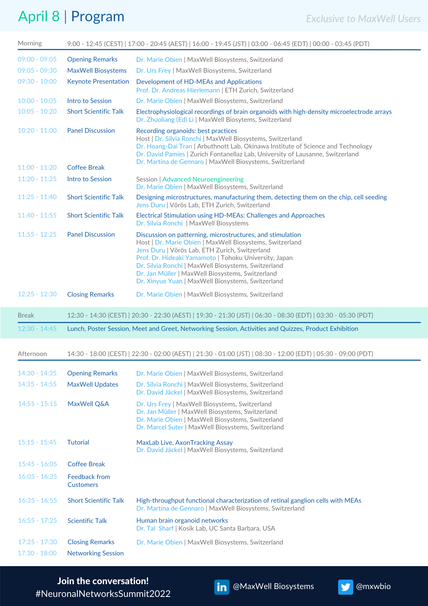
April 8


 English


