 English
 繁體中文
繁體中文 简体中文
简体中文 日本語
日本語
Discover over 100 publications featuring our technology
All Publications
Fenton, Timothy A; Haouchine, Olivia Y; Hallam, Elizabeth L; Smith, Emily M; Jackson, Kiya C; Rahbarian, Darlene; Canales, Cesar; Adhikari, Anna; Nord, Alexander S; Ben-Shalom, Roy; Silverman, Jill L Hyperexcitability and translational phenotypes in a preclinical mouse model of SYNGAP1-Related Intellectual Disability Journal Article Research Square, 2024. Abstract | Links | BibTeX | Tags: Activity Scan Assay, HD-MEA, MaxOne, MEA Metrics, MEA Technology, Network Assay, Primary Neuronal Cell Culture, Spike Sorting Kasuba, Krishna Chaitanya; Buccino, Alessio Paolo; Bartram, Julian; Gaub, Benjamin M; Fauser, Felix J; Ronchi, Silvia; Kumar, Sreedhar Saseendran; Geissler, Sydney; Nava, Michele M; Hierlemann, Andreas; Müller, Daniel J Nature Nanotechnology, 2024. Abstract | Links | BibTeX | Tags: Action Potential, Brain Slice, HD-MEA, MaxOne, Primary Neuronal Cell Culture, Spike Sorting Hruska-Plochan, Marian; Wiersma, Vera I; Betz, Katharina M; Mallona, Izaskun; Ronchi, Silvia; Maniecka, Zuzanna; Hock, Eva-Maria; Tantardini, Elena; Laferriere, Florent; Sahadevan, Sonu; Hoop, Vanessa; Delvendahl, Igor; Pérez-Berlanga, Manuela; Gatta, Beatrice; Panatta, Martina; van der Bourg, Alexander; Bohaciakova, Dasa; Sharma, Puneet; Vos, Laura De; Frontzek, Karl; Aguzzi, Adriano; Lashley, Tammaryn; Robinson, Mark D; Karayannis, Theofanis; Mueller, Martin; Hierlemann, Andreas; Polymenidou, Magdalini A model of human neural networks reveals NPTX2 pathology in ALS and FTLD Journal Article Nature, 2024. Abstract | Links | BibTeX | Tags: Activity Scan Assay, HD-MEA, IPSC, MaxOne, MEA Technology, Network Assay, Primary Neuronal Cell Culture, Spike Sorting Hornauer, Philipp; Prack, Gustavo; Anastasi, Nadia; Ronchi, Silvia; Kim, Taehoon; Donner, Christian; Fiscella, Michele; Borgwardt, Karsten; Taylor, Verdon; Jagasia, Ravi; Roqueiro, Damian; Hierlemann, Andreas; Schröter, Manuel DeePhys: A machine learning–assisted platform for electrophysiological phenotyping of human neuronal networks Journal Article Stem Cell Reports, 2024. Abstract | Links | BibTeX | Tags: 2D Neuronal Culture, HD-MEA, IPSC, MaxOne, MEA Technology, Spike Sorting Elliott, Matthew A T; Schweiger, Hunter E; Robbins, Ash; Vera-Choqqueccota, Samira; Ehrlich, Drew; Hernandez, Sebastian; Voitiuk, Kateryna; Geng, Jinghui; Sevetson, Jess L; Core, Cordero; Rosen, Yohei M; Teodorescu, Mircea; Wagner, Nico O; Haussler, David; Mostajo-Radji, Mohammed A Internet-Connected Cortical Organoids for Project-Based Stem Cell and Neuroscience Education Journal Article eNeuro, 2023. Abstract | Links | BibTeX | Tags: 3D Culture, Activity Scan Assay, HD-MEA, IPSC, MaxOne, MEA Technology, Organoids, Spike Sorting, Stimulation Kelley, Matt R; Chipman, Laura B; Asano, Shoh; Knott, Matthew; Howard, Samantha T; Berg, Allison P bioRxiv, 2023. Abstract | Links | BibTeX | Tags: Activity Scan Assay, Axon Tracking Assay, HD-MEA, IPSC, MaxTwo, MEA Technology, Network Assay, Spike Sorting Bartram, Julian; Franke, Felix; Kumar, Sreedhar Saseendran; Buccino, Alessio Paolo; Xue, Xiaohan; Gänswein, Tobias; Schröter, Manuel; Kim, Taehoon; Kasuba, Krishna Chaitanya; Hierlemann, Andreas Parallel reconstruction of the excitatory and inhibitory inputs received by single neurons reveals the synaptic basis of recurrent spiking Journal Article eLife, 2023. Abstract | Links | BibTeX | Tags: HD-MEA, MaxOne, MEA Metrics, MEA Technology, Modeling, Primary Neuronal Cell Culture, Spike Sorting Duru, Jens; Maurer, Benedikt; Doran, Ciara Giles; Jelitto, Robert; Küchler, Joël; Ihle, Stephan J; Ruff, Tobias; John, Robert; Genocchi, Barbara; Vörös, János Investigation of the input-output relationship of engineered neural networks using high-density microelectrode arrays Journal Article SSRN, 2023. Abstract | Links | BibTeX | Tags: HD-MEA, MaxOne, MEA Technology, Primary Neuronal Cell Culture, Spike Sorting, Stimulation Bartram, Julian; Franke, Felix; Kumar, Sreedhar Saseendran; Buccino, Alessio Paolo; Xue, Xiaohan; Gänswein, Tobias; Schröter, Manuel; Kim, Taehoon; Kasuba, Krishna Chaitanya; Hierlemann, Andreas Parallel reconstruction of the excitatory and inhibitory inputs received by single neurons reveals the synaptic basis of recurrent spiking Journal Article bioRxiv, 2023. Abstract | Links | BibTeX | Tags: CMOS, HD-MEA, Modeling, Primary Neuronal Cell Culture, Spike Sorting Habibey, Rouhollah; Striebel, Johannes; Schmieder, Felix; Czarske, Jürgen; Busskamp, Volker Long-term morphological and functional dynamics of human stem cell-derived neuronal networks on high-density micro-electrode arrays Journal Article Frontiers in Neuroscience, 2022. Abstract | Links | BibTeX | Tags: 2D Neuronal Culture, CMOS, HD-MEA, IPSC, MaxOne, MEA Metrics, Modeling, Spike Sorting Diggelmann, Roland; Fiscella, Michele; Hierlemann, Andreas; Franke, Felix 26th Annual Computational Neuroscience Meeting (CNS2017) Antwerp, Belgium , 2017. Abstract | Links | BibTeX | Tags: Spike Sorting Dragas, Jelena; Jäckel, David; Hierlemann, Andreas; Franke, Felix Complexity Optimization and High-Throughput Low-Latency Hardware Implementation of a Multi-Electrode Spike-Sorting Algorithm Journal Article IEEE Transactions on Neural Systems and Rehabilitation Engineering, 23 (2), pp. 149–158, 2015, ISSN: 15344320. Abstract | Links | BibTeX | Tags: Spike Sorting Franke, Felix; Jackel, David; Dragas, Jelena; Muller, Jan; Radivojevic, Milos; Bakkum, Douglas J; Hierlemann, Andreas Frontiers in Neural Circuits, 6 , pp. 105, 2012, ISSN: 1662-5110. Abstract | Links | BibTeX | Tags: Neuronal Networks, Review, Spike Sorting Jäckel, David; Frey, Urs; Fiscella, Michele; Franke, Felix; Hierlemann, Andreas Applicability of independent component analysis on high-density microelectrode array recordings Journal Article Journal of Neurophysiology, 108 (1), pp. 334-348, 2012, ISSN: 0022-3077. Abstract | Links | BibTeX | Tags: ETH-CMOS-MEA, Spike Sorting
2024
![]()
title = {Hyperexcitability and translational phenotypes in a preclinical mouse model of SYNGAP1-Related Intellectual Disability},
author = {Timothy A Fenton and Olivia Y Haouchine and Elizabeth L Hallam and Emily M Smith and Kiya C Jackson and Darlene Rahbarian and Cesar Canales and Anna Adhikari and Alexander S Nord and Roy Ben-Shalom and Jill L Silverman},
url = {https://www.researchsquare.com/article/rs-4067746/v1},
doi = {10.21203/rs.3.rs-4067746/v1},
year = {2024},
date = {2024-03-19},
journal = {Research Square},
abstract = {Disruption of SYNGAP1 directly causes a genetically identifiable neurodevelopmental disorder (NDD) called SYNGAP1-related intellectual disability (SRID). Without functional SynGAP1 protein, individuals are developmentally delayed and have prominent features of intellectual disability, motor impairments, and epilepsy. Over the past two decades, there have been numerous discoveries indicting the critical role of Syngap1. Several rodent models with a loss of Syngap1 have been engineered identifying precise roles in neuronal structure and function, as well as key biochemical pathways key for synapse integrity. Homozygous loss of Syngap1 is lethal. Heterozygous mutations of Syngap1 result in a broad range of behavioral phenotypes. Our in vivo functional data, using the mouse model from the Huganir laboratory, corroborated earlier reported behaviors including robust hyperactivity and deficits in learning and memory in young adults. In extension, we report impairments in slow wave sleep, a critical component of the domain of sleep. We characterized Syngap1+/- mice by using neurophysiology collected with wireless, telemetric electroencephalography (EEG). Syngap1+/- mice also exhibited elevated spiking events and spike trains, in addition to elevated power, most notably in the delta frequency band. For the first time, we illustrated how primary neurons from Syngap1+/- mice function and display increased network firing activity, greater bursts, and shorter inter-burst intervals between peaks by employing high density microelectrode arrays (HD-MEA). Our reported data bridge in-vitro electrophysiological neuronal activity and function with in vivo neurophysiological brain activity and function. These data elucidate quantitative, translational biomarkers in vivo and in vitro that can be utilized for the development of and efficacy assessment of targeted treatments for SRID.},
keywords = {Activity Scan Assay, HD-MEA, MaxOne, MEA Metrics, MEA Technology, Network Assay, Primary Neuronal Cell Culture, Spike Sorting},
pubstate = {published},
tppubtype = {article}
}
![]()
title = {Mechanical stimulation and electrophysiological monitoring at subcellular resolution reveals differential mechanosensation of neurons within networks},
author = {Krishna Chaitanya Kasuba and Alessio Paolo Buccino and Julian Bartram and Benjamin M. Gaub and Felix J. Fauser and Silvia Ronchi and Sreedhar Saseendran Kumar and Sydney Geissler and Michele M. Nava and Andreas Hierlemann and Daniel J. Müller },
url = {https://www.nature.com/articles/s41565-024-01609-1},
doi = {10.1038/s41565-024-01609-1},
year = {2024},
date = {2024-02-20},
journal = {Nature Nanotechnology},
abstract = {A growing consensus that the brain is a mechanosensitive organ is driving the need for tools that mechanically stimulate and simultaneously record the electrophysiological response of neurons within neuronal networks. Here we introduce a synchronized combination of atomic force microscopy, high-density microelectrode array and fluorescence microscopy to monitor neuronal networks and to mechanically characterize and stimulate individual neurons at piconewton force sensitivity and nanometre precision while monitoring their electrophysiological activity at subcellular spatial and millisecond temporal resolution. No correlation is found between mechanical stiffness and electrophysiological activity of neuronal compartments. Furthermore, spontaneously active neurons show exceptional functional resilience to static mechanical compression of their soma. However, application of fast transient (∼500 ms) mechanical stimuli to the neuronal soma can evoke action potentials, which depend on the anchoring of neuronal membrane and actin cytoskeleton. Neurons show higher responsivity, including bursts of action potentials, to slower transient mechanical stimuli (∼60 s). Moreover, transient and repetitive application of the same compression modulates the neuronal firing rate. Seemingly, neuronal networks can differentiate and respond to specific characteristics of mechanical stimulation. Ultimately, the developed multiparametric tool opens the door to explore manifold nanomechanobiological responses of neuronal systems and new ways of mechanical control.},
keywords = {Action Potential, Brain Slice, HD-MEA, MaxOne, Primary Neuronal Cell Culture, Spike Sorting},
pubstate = {published},
tppubtype = {article}
}
![]()
title = {A model of human neural networks reveals NPTX2 pathology in ALS and FTLD},
author = {Marian Hruska-Plochan and Vera I. Wiersma and Katharina M. Betz and Izaskun Mallona and Silvia Ronchi and Zuzanna Maniecka and Eva-Maria Hock and Elena Tantardini and Florent Laferriere and Sonu Sahadevan and Vanessa Hoop and Igor Delvendahl and Manuela Pérez-Berlanga and Beatrice Gatta and Martina Panatta and Alexander van der Bourg and Dasa Bohaciakova and Puneet Sharma and Laura De Vos and Karl Frontzek and Adriano Aguzzi and Tammaryn Lashley and Mark D. Robinson and Theofanis Karayannis and Martin Mueller and Andreas Hierlemann and Magdalini Polymenidou },
url = {https://www.nature.com/articles/s41586-024-07042-7},
doi = {10.1038/s41586-024-07042-7},
year = {2024},
date = {2024-02-14},
journal = {Nature},
abstract = {Human cellular models of neurodegeneration require reproducibility and longevity, which is necessary for simulating age-dependent diseases. Such systems are particularly needed for TDP-43 proteinopathies1, which involve human-specific mechanisms that cannot be directly studied in animal models. Here, to explore the emergence and consequences of TDP-43 pathologies, we generated induced pluripotent stem cell-derived, colony morphology neural stem cells (iCoMoNSCs) via manual selection of neural precursors. Single-cell transcriptomics and comparison to independent neural stem cells showed that iCoMoNSCs are uniquely homogenous and self-renewing. Differentiated iCoMoNSCs formed a self-organized multicellular system consisting of synaptically connected and electrophysiologically active neurons, which matured into long-lived functional networks (which we designate iNets). Neuronal and glial maturation in iNets was similar to that of cortical organoids. Overexpression of wild-type TDP-43 in a minority of neurons within iNets led to progressive fragmentation and aggregation of the protein, resulting in a partial loss of function and neurotoxicity. Single-cell transcriptomics revealed a novel set of misregulated RNA targets in TDP-43-overexpressing neurons and in patients with TDP-43 proteinopathies exhibiting a loss of nuclear TDP-43. The strongest misregulated target encoded the synaptic protein NPTX2, the levels of which are controlled by TDP-43 binding on its 3′ untranslated region. When NPTX2 was overexpressed in iNets, it exhibited neurotoxicity, whereas correcting NPTX2 misregulation partially rescued neurons from TDP-43-induced neurodegeneration. Notably, NPTX2 was consistently misaccumulated in neurons from patients with amyotrophic lateral sclerosis and frontotemporal lobar degeneration with TDP-43 pathology. Our work directly links TDP-43 misregulation and NPTX2 accumulation, thereby revealing a TDP-43-dependent pathway of neurotoxicity.},
keywords = {Activity Scan Assay, HD-MEA, IPSC, MaxOne, MEA Technology, Network Assay, Primary Neuronal Cell Culture, Spike Sorting},
pubstate = {published},
tppubtype = {article}
}

title = {DeePhys: A machine learning–assisted platform for electrophysiological phenotyping of human neuronal networks},
author = {Philipp Hornauer and Gustavo Prack and Nadia Anastasi and Silvia Ronchi and Taehoon Kim and Christian Donner and Michele Fiscella and Karsten Borgwardt and Verdon Taylor and Ravi Jagasia and Damian Roqueiro and Andreas Hierlemann and Manuel Schröter},
url = {https://www.sciencedirect.com/science/article/pii/S2213671123005015},
doi = {10.1016/j.stemcr.2023.12.008},
year = {2024},
date = {2024-01-25},
journal = {Stem Cell Reports},
abstract = {Reproducible functional assays to study in vitro neuronal networks represent an important cornerstone in the quest to develop physiologically relevant cellular models of human diseases. Here, we introduce DeePhys, a MATLAB-based analysis tool for data-driven functional phenotyping of in vitro neuronal cultures recorded by high-density microelectrode arrays. DeePhys is a modular workflow that offers a range of techniques to extract features from spike-sorted data, allowing for the examination of functional phenotypes both at the individual cell and network levels, as well as across development. In addition, DeePhys incorporates the capability to integrate novel features and to use machine-learning-assisted approaches, which facilitates a comprehensive evaluation of pharmacological interventions. To illustrate its practical application, we apply DeePhys to human induced pluripotent stem cell–derived dopaminergic neurons obtained from both patients and healthy individuals and showcase how DeePhys enables phenotypic screenings.},
keywords = {2D Neuronal Culture, HD-MEA, IPSC, MaxOne, MEA Technology, Spike Sorting},
pubstate = {published},
tppubtype = {article}
}
2023
![]()
title = {Internet-Connected Cortical Organoids for Project-Based Stem Cell and Neuroscience Education},
author = {Matthew A. T. Elliott and Hunter E. Schweiger and Ash Robbins and Samira Vera-Choqqueccota and Drew Ehrlich and Sebastian Hernandez and Kateryna Voitiuk and Jinghui Geng and Jess L. Sevetson and Cordero Core and Yohei M. Rosen and Mircea Teodorescu and Nico O. Wagner and David Haussler and Mohammed A. Mostajo-Radji},
url = {https://www.eneuro.org/lookup/doi/10.1523/ENEURO.0308-23.2023},
doi = {10.1523/ENEURO.0308-23.2023},
year = {2023},
date = {2023-11-28},
journal = {eNeuro},
abstract = {The introduction of Internet-connected technologies to the classroom has the potential to revolutionize STEM education by allowing students to perform experiments in complex models that are unattainable in traditional teaching laboratories. By connecting laboratory equipment to the cloud, we introduce students to experimentation in pluripotent stem cell (PSC)-derived cortical organoids in two different settings: using microscopy to monitor organoid growth in an introductory tissue culture course and using high-density (HD) multielectrode arrays (MEAs) to perform neuronal stimulation and recording in an advanced neuroscience mathematics course. We demonstrate that this approach develops interest in stem cell and neuroscience in the students of both courses. All together, we propose cloud technologies as an effective and scalable approach for complex project-based university training.},
keywords = {3D Culture, Activity Scan Assay, HD-MEA, IPSC, MaxOne, MEA Technology, Organoids, Spike Sorting, Stimulation},
pubstate = {published},
tppubtype = {article}
}
![]()
title = {Potentiating NaV1.1 in Dravet syndrome patient iPSC-derived GABAergic neurons increases neuronal firing frequency and decreases network synchrony},
author = {Matt R Kelley and Laura B Chipman and Shoh Asano and Matthew Knott and Samantha T Howard and Allison P Berg},
url = {https://www.biorxiv.org/content/10.1101/2023.09.28.559990v1},
doi = {10.1101/2023.09.28.559990},
year = {2023},
date = {2023-09-29},
journal = {bioRxiv},
abstract = {Dravet syndrome is a developmental and epileptic encephalopathy characterized by seizures, behavioral abnormalities, developmental deficits, and elevated risk of sudden unexpected death in epilepsy (SUDEP). Most patient cases are caused by de novo loss-of-function mutations in the gene SCN1A, causing a haploinsufficiency of the alpha subunit of the voltage-gated sodium channel NaV1.1. Within the brain, NaV1.1 is primarily localized to the axons of inhibitory neurons, and decreased NaV1.1 function is hypothesized to reduce GABAergic inhibitory neurotransmission within the brain, driving neuronal network hyperexcitability and subsequent pathology. We have developed a human in vitro model of Dravet syndrome using differentiated neurons derived from patient iPSC and enriched for GABA expressing neurons. Neurons were plated on high definition multielectrode arrays (HD-MEAs), permitting recordings from the same cultures over the 7-weeks duration of study at the network, single cell, and subcellular resolution. Using this capability, we characterized the features of axonal morphology and physiology. Neurons developed increased spiking activity and synchronous network bursting. Recordings were processed through a spike sorting pipeline for curation of single unit activity and to assess the effects of pharmacological treatments. At 7-weeks, the application of the GABAAR receptor agonist muscimol eliminated network bursting, indicating the presence of GABAergic neurotransmission. To identify the role of NaV1.1 on neuronal and network activity, cultures were treated with a dose-response of the NaV1.1 potentiator δ-theraphotoxin-Hm1a. This resulted in a strong increase in firing rates of putative GABAergic neurons, an increase in the intraburst firing rate, and eliminated network bursting. These results validate that potentiation of NaV1.1 in Dravet patient iPSC-derived neurons results in decreased firing synchrony in neuronal networks through increased GABAergic neuron activity and support the use of human neurons and HD-MEAs as viable high-throughput electrophysiological platform to enable therapeutic discovery.},
keywords = {Activity Scan Assay, Axon Tracking Assay, HD-MEA, IPSC, MaxTwo, MEA Technology, Network Assay, Spike Sorting},
pubstate = {published},
tppubtype = {article}
}

title = {Parallel reconstruction of the excitatory and inhibitory inputs received by single neurons reveals the synaptic basis of recurrent spiking},
author = {Julian Bartram and Felix Franke and Sreedhar Saseendran Kumar and Alessio Paolo Buccino and Xiaohan Xue and Tobias Gänswein and Manuel Schröter and Taehoon Kim and Krishna Chaitanya Kasuba and Andreas Hierlemann},
url = {https://elifesciences.org/reviewed-preprints/86820},
doi = {10.7554/eLife.86820},
year = {2023},
date = {2023-05-17},
journal = {eLife},
abstract = {Self-sustained recurrent activity in cortical networks is thought to be important for multiple crucial processes, including circuit development and homeostasis. Yet, the precise relationship between the synaptic input patterns and the spiking output of individual neurons remains largely unresolved. Here, we developed, validated and applied a novel in vitro experimental platform and analytical procedures that provide – for individual neurons – simultaneous excitatory and inhibitory synaptic activity estimates during recurrent network activity. Our approach combines whole-network high-density microelectrode array (HD-MEA) recordings from rat neuronal cultures with patch clamping and enables a comprehensive mapping and characterization of active incoming connections to single postsynaptic neurons. We found that, during network states with excitation(E)-inhibition(I) balance, postsynaptic spiking coincided precisely with the maxima of fast fluctuations in the input E/I ratio. These spike-associated E/I ratio escalations were largely due to a rapid bidirectional change in synaptic inhibition that was modulated by the network-activity level. Our approach also uncovered the underlying circuit architecture and we show that individual neurons received a few key inhibitory connections – often from special hub neurons – that were instrumental in controlling postsynaptic spiking. Balanced network theory predicts dynamical regimes governed by small and rapid input fluctuation and featuring a fast neuronal responsiveness. Our findings – obtained in self-organized neuronal cultures – suggest that the emergence of these favorable regimes and associated network architectures is an inherent property of cortical networks in general.},
keywords = {HD-MEA, MaxOne, MEA Metrics, MEA Technology, Modeling, Primary Neuronal Cell Culture, Spike Sorting},
pubstate = {published},
tppubtype = {article}
}

title = {Investigation of the input-output relationship of engineered neural networks using high-density microelectrode arrays},
author = {Jens Duru and Benedikt Maurer and Ciara Giles Doran and Robert Jelitto and Joël Küchler and Stephan J. Ihle and Tobias Ruff and Robert John and Barbara Genocchi and János Vörös},
url = {https://www.ssrn.com/abstract=4427959},
doi = {DOI: 10.2139/ssrn.4427959},
year = {2023},
date = {2023-04-24},
journal = {SSRN},
abstract = {Bottom-up neuroscience utilizes small, engineered biological neural networks to study neuronal activity in systems of reduced complexity. We present a platform that establishes up to six independent networks formed by primary rat neurons on planar complementary metal–oxide–semiconductor (CMOS) microelectrode arrays (MEAs). We introduce an approach that allows repetitive stimulation and recording of network activity at any of the over 700 electrodes underlying a network. We demonstrate that the continuous application of a repetitive super-threshold stimulus yields a reproducible network answer within a 15 ms post-stimulus window. This response can be tracked with high spatiotemporal resolution across the whole extent of the network. Moreover, we show that the location of the stimulation plays a significant role in the networks’ early response to the stimulus. By applying a stimulation pattern to all network-underlying electrodes in sequence, the sensitivity of the whole network to the stimulus can be visualized. We demonstrate that microchannels reduce the voltage stimulation threshold and induce the strongest network response. By varying the stimulation amplitude and frequency we reveal discrete network transition points. Finally, we introduce vector fields to follow stimulation-induced spike propagation pathways within the network. Overall we show that our defined neural networks on CMOS MEAs enable us to elicit highly reproducible activity patterns that can be precisely modulated by stimulation amplitude, stimulation frequency and the site of stimulation.},
keywords = {HD-MEA, MaxOne, MEA Technology, Primary Neuronal Cell Culture, Spike Sorting, Stimulation},
pubstate = {published},
tppubtype = {article}
}
![]()
title = {Parallel reconstruction of the excitatory and inhibitory inputs received by single neurons reveals the synaptic basis of recurrent spiking},
author = {Julian Bartram and Felix Franke and Sreedhar Saseendran Kumar and Alessio Paolo Buccino and Xiaohan Xue and Tobias Gänswein and Manuel Schröter and Taehoon Kim and Krishna Chaitanya Kasuba and Andreas Hierlemann},
url = {https://www.biorxiv.org/content/10.1101/2023.01.06.523018v2},
doi = {https://doi.org/10.1101/2023.01.06.523018},
year = {2023},
date = {2023-01-08},
journal = {bioRxiv},
abstract = {Self-sustained recurrent activity in cortical networks is thought to be important for multiple crucial processes, including circuit development and homeostasis. However, the precise relationship between synaptic input patterns and spiking output of individual neurons remains unresolved during spontaneous network activity. Here, using whole-network high-density microelectrode array (HD-MEA) recordings and patch clamping, we developed a novel experimental approach and analytical tools that provide a comprehensive long-term input-output characterization of individual neurons in cortical cell cultures. We found that, during in vivo-like network activity with excitation(E)-inhibition(I) balance, postsynaptic spiking coincided with the maxima of rapid, network state-dependent fluctuations in the input E/I ratio. Our approach also uncovered the underlying circuit architecture and we identified a few key inhibitory inputs – often from special hub neurons – that were instrumental in mediating these E/I ratio changes. Balanced network theory predicts dynamical regimes governed by input fluctuation and featuring a fast neuronal responsiveness. Our findings – obtained in self-organized neuronal cultures – suggest that the emergence of these favorable regimes and associated network architectures is an inherent property of all cortical networks.},
keywords = {CMOS, HD-MEA, Modeling, Primary Neuronal Cell Culture, Spike Sorting},
pubstate = {published},
tppubtype = {article}
}
2022

title = {Long-term morphological and functional dynamics of human stem cell-derived neuronal networks on high-density micro-electrode arrays},
author = {Rouhollah Habibey and Johannes Striebel and Felix Schmieder and Jürgen Czarske and Volker Busskamp},
url = {https://www.frontiersin.org/articles/10.3389/fnins.2022.951964/full},
doi = {10.3389/fnins.2022.951964},
year = {2022},
date = {2022-10-04},
journal = {Frontiers in Neuroscience},
abstract = {Comprehensive electrophysiological characterizations of human induced pluripotent stem cell (hiPSC)-derived neuronal networks are essential to determine to what extent these in vitro models recapitulate the functional features of in vivo neuronal circuits. High-density micro-electrode arrays (HD-MEAs) offer non-invasive recording with the best spatial and temporal resolution possible to date. For 3 months, we tracked the morphology and activity features of developing networks derived from a transgenic hiPSC line in which neurogenesis is inducible by neurogenic transcription factor overexpression. Our morphological data revealed large-scale structural changes from homogeneously distributed neurons in the first month to the formation of neuronal clusters over time. This led to a constant shift in position of neuronal cells and clusters on HD-MEAs and corresponding changes in spatial distribution of the network activity maps. Network activity appeared as scarce action potentials (APs), evolved as local bursts with longer duration and changed to network-wide synchronized bursts with higher frequencies but shorter duration over time, resembling the emerging burst features found in the developing human brain. Instantaneous firing rate data indicated that the fraction of fast spiking neurons (150–600 Hz) increases sharply after 63 days post induction (dpi). Inhibition of glutamatergic synapses erased burst features from network activity profiles and confirmed the presence of mature excitatory neurotransmission. The application of GABAergic receptor antagonists profoundly changed the bursting profile of the network at 120 dpi. This indicated a GABAergic switch from excitatory to inhibitory neurotransmission during circuit development and maturation. Our results suggested that an emerging GABAergic system at older culture ages is involved in regulating spontaneous network bursts. In conclusion, our data showed that long-term and continuous microscopy and electrophysiology readouts are crucial for a meaningful characterization of morphological and functional maturation in stem cell-derived human networks. Most importantly, assessing the level and duration of functional maturation is key to subject these human neuronal circuits on HD-MEAs for basic and biomedical applications.},
keywords = {2D Neuronal Culture, CMOS, HD-MEA, IPSC, MaxOne, MEA Metrics, Modeling, Spike Sorting},
pubstate = {published},
tppubtype = {article}
}
2017

title = {Pre-whitening as a means to improve dimensionality reduction and simplify clustering in spike-sorters for multi-electrode recordings},
author = {Roland Diggelmann and Michele Fiscella and Andreas Hierlemann and Felix Franke},
url = {https://bmcneurosci.biomedcentral.com/articles/10.1186/s12868-017-0371-2},
year = {2017},
date = {2017-07-15},
address = {Antwerp, Belgium },
organization = {26th Annual Computational Neuroscience Meeting (CNS2017)},
abstract = {Spike sorting is the process to extract single neuronal activity from extracellular recordings. It makes use of the fact that spikes from a single neuron feature highly similar waveforms, whereas spikes from different neurons have different waveforms. Clustering algorithms are used to find groups of similar spikes that putatively originated from the same neuron. However, since spike waveforms especially in multi-electrode recordings can have a high dimensionality, their dimensionality needs to be reduced before clustering. Principal component analysis (PCA) is one of the most commonly employed dimensionality reduction methods for this purpose [1]. It reduces the dimensions to those where the variance of the data was highest, presumably those along which the waveforms of separate neurons differ most strongly, However, if the noise is not uniform in all dimensions, high variability can also mean high noise, which would render a dimension useless for discrimination. We, therefore, propose an additional pre-whitening step before PCA and discuss two beneficial effects on the subsequent clustering. We illustrate these effects by using spikes from retinal ganglion cells recorded with high-density multi-electrode arrays (HD-MEA).},
keywords = {Spike Sorting},
pubstate = {published},
tppubtype = {conference}
}
2015

title = {Complexity Optimization and High-Throughput Low-Latency Hardware Implementation of a Multi-Electrode Spike-Sorting Algorithm},
author = {Jelena Dragas and David Jäckel and Andreas Hierlemann and Felix Franke},
url = {http://ieeexplore.ieee.org/document/6955847/},
doi = {10.1109/TNSRE.2014.2370510},
issn = {15344320},
year = {2015},
date = {2015-03-01},
journal = {IEEE Transactions on Neural Systems and Rehabilitation Engineering},
volume = {23},
number = {2},
pages = {149--158},
abstract = {Reliable real-time low-latency spike sorting with large data throughput is essential for studies of neural network dynamics and for brain-machine interfaces (BMIs), in which the stimulation of neural networks is based on the networks' most recent activity. However, the majority of existing multi-electrode spike-sorting algorithms are unsuited for processing high quantities of simultaneously recorded data. Recording from large neuronal networks using large high-density electrode sets (thousands of electrodes) imposes high demands on the data-processing hardware regarding computational complexity and data transmission bandwidth; this, in turn, entails demanding requirements in terms of chip area, memory resources and processing latency. This paper presents computational complexity optimisation techniques, which facilitate the use of spike-sorting algorithms in large multi-electrode-based recording systems. The techniques are then applied to a previously published algorithm, on its own, unsuited for large electrode set recordings. Further, a real-time low-latency high-performance VLSI hardware architecture of the modified algorithm is presented, featuring a folded structure capable of processing the activity of hundreds of neurons simultaneously. The hardware is reconfigurable “on-the-fly” and adaptable to the non-stationarities of neuronal recordings. By transmitting exclusively spike time stamps and/or spike waveforms, its real-time processing offers the possibility of data bandwidth and data storage reduction.},
keywords = {Spike Sorting},
pubstate = {published},
tppubtype = {article}
}
2012

title = {High-density microelectrode array recordings and real-time spike sorting for closed-loop experiments: an emerging technology to study neural plasticity},
author = {Felix Franke and David Jackel and Jelena Dragas and Jan Muller and Milos Radivojevic and Douglas J Bakkum and Andreas Hierlemann},
url = {https://www.frontiersin.org/article/10.3389/fncir.2012.00105},
doi = {10.3389/fncir.2012.00105},
issn = {1662-5110},
year = {2012},
date = {2012-12-20},
journal = {Frontiers in Neural Circuits},
volume = {6},
pages = {105},
abstract = {Understanding plasticity of neural networks is a key to comprehending their development and function. A powerful technique to study neural plasticity includes recording and control of pre- and postsynaptic neural activity, e.g., by using simultaneous intracellular recording and stimulation of several neurons. Intracellular recording is, however, a demanding technique and has its limitations in that only a small number of neurons can be stimulated and recorded from at the same time. Extracellular techniques offer the possibility to simultaneously record from larger numbers of neurons with relative ease, at the expenses of increased efforts to sort out single neuronal activities from the recorded mixture, which is a time consuming and error prone step, referred to as spike sorting. In this mini-review, we describe recent technological developments in two separate fields, namely CMOS-based high-density microelectrode arrays, which also allow for extracellular stimulation of neurons, and real-time spike sorting. We argue that these techniques, when combined, will provide a powerful tool to study plasticity in neural networks consisting of several thousand neurons in vitro.},
keywords = {Neuronal Networks, Review, Spike Sorting},
pubstate = {published},
tppubtype = {article}
}
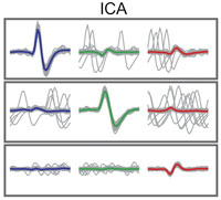
title = {Applicability of independent component analysis on high-density microelectrode array recordings},
author = {David Jäckel and Urs Frey and Michele Fiscella and Felix Franke and Andreas Hierlemann},
url = {http://jn.physiology.org/cgi/doi/10.1152/jn.01106.2011},
doi = {10.1152/jn.01106.2011},
issn = {0022-3077},
year = {2012},
date = {2012-04-04},
journal = {Journal of Neurophysiology},
volume = {108},
number = {1},
pages = {334-348},
abstract = {Emerging complementary metal oxide semiconductor (CMOS)-based, high-density microelectrode array (HD-MEA) devices provide high spatial resolution at subcellular level and a large number of readout channels. These devices allow for simultaneous recording of extracellular activity of a large number of neurons with every neuron being detected by multiple electrodes. To analyze the recorded signals, spiking events have to be assigned to individual neurons, a process referred to as "spike sorting." For a set of observed signals, which constitute a linear mixture of a set of source signals, independent component (IC) analysis (ICA) can be used to demix blindly the data and extract the individual source signals. This technique offers great potential to alleviate the problem of spike sorting in HD-MEA recordings, as it represents an unsupervised method to separate the neuronal sources. The separated sources or ICs then constitute estimates of single-neuron signals, and threshold detection on the ICs yields the sorted spike times. However, it is unknown to what extent extracellular neuronal recordings meet the requirements of ICA. In this paper, we evaluate the applicability of ICA to spike sorting of HD-MEA recordings. The analysis of extracellular neuronal signals, recorded at high spatiotemporal resolution, reveals that the recorded data cannot be modeled as a purely linear mixture. As a consequence, ICA fails to separate completely the neuronal signals and cannot be used as a stand-alone method for spike sorting in HD-MEA recordings. We assessed the demixing performance of ICA using simulated data sets and found that the performance strongly depends on neuronal density and spike amplitude. Furthermore, we show how postprocessing techniques can be used to overcome the most severe limitations of ICA. In combination with these postprocessing techniques, ICA represents a viable method to facilitate rapid spike sorting of multidimensional neuronal recordings.},
keywords = {ETH-CMOS-MEA, Spike Sorting},
pubstate = {published},
tppubtype = {article}
}
Selected Publications

High-resolution CMOS MEA platform to study neurons at subcellular, cellular, and network levels

Presenting measurements of neuronal preparations with a novel CMOS-based microelectrode array at high-spatiotemporal-resolution on subcellular, cellular, and network level.
J. Müller, M. Ballini, P. Livi, Y. Chen, M. Radivojevic, A. Shadmani, V. Viswam, I. L. Jones, M. Fiscella, R. Diggelmann, A. Stettler, U. Frey, D. J. Bakkum, and A. Hierlemann, “High-resolution CMOS MEA platform to study neurons at subcellular, cellular, and network levels,” Lab Chip, vol. 15, no. 13, pp. 2767–2780, May 2015.
Revealing Neuronal Function through Microelectrode Array Recordings

Reviewing the current understanding of microelectrode signals and the techniques for analyzing them, with focus on the ongoing advancements in microelectrode technology (in vivo and in vitro) and recent advanced microelectrode array measurement methods that facilitate the understanding of single neurons and network function.
M. E. J. Obien, K. Deligkaris, T. Bullmann, D. J. Bakkum, and U. Frey, “Revealing Neuronal Function through Microelectrode Array Recordings,” Front. Neurosci., 8:423, Jan 2015.

A 1024-Channel CMOS Microelectrode Array With 26,400 Electrodes for Recording and Stimulation of Electrogenic Cells In Vitro
A high-resolution CMOS-based microelectrode array featuring 1,024 low-noise readout channels, 26,400 electrodes at a density of 3,265 electrodes per mm2, including on-chip 10bit ADCs and consuming only 75 mW.
M. Ballini, J. Muller, P. Livi, Y. Chen, U. Frey, A. Stettler, A. Shadmani, V. Viswam, I. L. Jones, D. Jackel, M. Radivojevic, M. K. Lewandowska, W. Gong, M. Fiscella, D. J. Bakkum, F. Heer, and A. Hierlemann, “A 1024-Channel CMOS Microelectrode Array With 26,400 Electrodes for Recording and Stimulation of Electrogenic Cells In Vitro,” IEEE Journal of Solid-State Circuits, vol. 49, no. 11, pp. 2705-2719, 2014.

Tracking axonal action potential propagation on a high-density microelectrode array across hundreds of sites
Demonstrating a method to electrically visualize action potential propagation on axons and revealing
large variations in velocity.
D. J. Bakkum, U. Frey, M. Radivojevic, T. L. Russell, J. Muller, M. Fiscella, H. Takahashi, and A. Hierlemann, “Tracking axonal action potential propagation on a high-density microelectrode array across hundreds of sites,” Nature Communications, 4:2181, Jul 2013.

Microelectronic System for High-Resolution Mapping of Extracellular Electric Fields Applied to Brain Slices
Recording and modeling extracellular action potentials of Purkinje cells at subcellular resolution.
U. Frey, U. Egert, F. Heer, S. Hafizovic, and A. Hierlemann, “Microelectronic System for High-Resolution Mapping of Extracellular Electric Fields Applied to Brain Slices,” Biosensors and Bioelectronics, vol. 24, no. 7, pp. 2191-2198, 2009.

Modulation of Cardiomyocyte Electrical Properties Using Regulated Bone Morphogenetic Protein-2 Expression
Controlling BMP-2 expression to modulate the electrophysiological properties of cardiomyocytes using an HD-MEA for detailed monitoring.
C. D. Sanchez-Bustamante, U. Frey, J. M. Kelm, A. Hierlemann, and M. Fussenegger,
“Modulation of Cardiomyocyte Electrical Properties Using Regulated Bone Morphogenetic Protein-2 Expression,” Tissue Engineering Part A, vol. 14, no. 12, pp. 1969-1988, 2008.