Modeling Pain-Related Nociceptive Networks with HD-MEA and Microfluidics
Date
Type
Tags
"The high quality of the HD-MEA signal was essential. Furthermore, the high density is great to map the whole circuit and allows us to place the microstructure anywhere we want. The fact that you have low noise at the electrode level not only facilitates things with the PDMS, but also recording from axons, which have a very low amplitude."
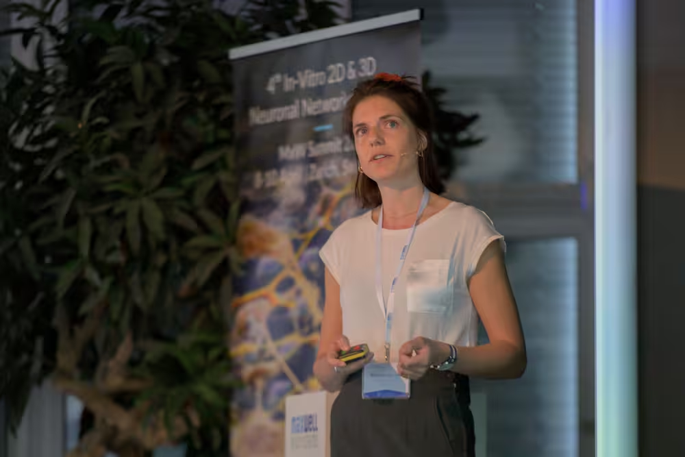
With the MPS World Summit currently bringing together the global microphysiological systems community in Washington DC, we took the opportunity to look back at one of the highlights of last year’s event: having Dr. Blandine Clement join us at our MaxWell Innovation Showcase “Unraveling Neural Complexity: Next-Generation Electrophysiology for Functional Characterization of 2D, 3D, and Microfluidic-Based Neuronal Models”.

For some of us, Blandine is a familiar face from her time at MaxWell Biosystems, where she contributed to the development of MaxLab Live even before starting her PhD. Others have met her more recently at conferences around the world, where she brings both scientific insight and a touch of humor to every conversation.
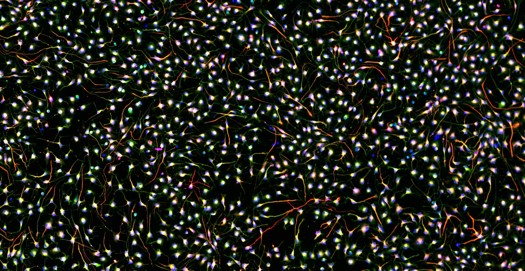
Having seen her present at multiple events, including our 4th In-Vitro 2D & 3D Neuronal Networks Summit in 2024, we have come to appreciate her work on PDMS multicompartment models for studying peripheral nervous system pain. Combining iPSC-derived neurons and keratinocytes, her research is as visually striking as it is impactful, often brought to life through vivid fluorescent imaging on the MaxOne+ chip.
Here, we took the opportunity to reflect with Blandine on her journey, from MaxWell team member to valued technology user.
In this
conversation
Hi Blandine, to start, could you tell us how you first entered the field of bioengineering?
I wanted to become a medical doctor first. I enjoyed scientific disciplines in school, and with time I realized that I was very interested in the research behind health and medicine progress. That is what brought me to biomedical engineering, this perfect combination between engineering and medical applications that allows you to have an impact.
At what point did your interest in medicine evolve into a focus on neuroscience?
I cannot recollect exactly when and how I became so interested in neuroscience, but my interest in the brain came quite early actually. I do remember that when I was fifteen, I visited a museum as part of a school exchange and in the museum shop, a book captured my attention. It had one hundred questions about the brain, just general knowledge, and fun facts. I bought it and still see it as a symbolic turning point toward my path in neuroscience (and I still have it on my bookshelf!). Later, I got fascinated by an interview describing the work on spinal cord injury done at EPFL, which determined my decision to study there.
Did that naturally lead you to focus on pain?
The path was not that straight forward actually. During my master thesis, I did a ten-month exchange in Boston, focusing on neurorehabilitation, and working on exoskeletons for stroke patients. However, although the project was very rewarding clinically, I felt that I was missing a more fundamental approach to studying the nervous system. I wanted to know more about the mechanisms. Then I discovered in vitro neuroscience through MaxWell Biosystems, which brought this complete shift in how to study the nervous system.
Is that what guided you toward the bottom-up neuroscience approach developed in the Vörös Lab?
Yes, exactly. And in this bottom-up neuroscience you need to find your niche. When I joined, most of my colleagues were working on plasticity and memory. The pain research branch was started by Dr. Tobias Ruff, a PostDoc in the lab, who applied for a grant on the effects of mechanical stress on nerves. Following this, he initiated some work with rat dorsal root ganglion neurons, provided by a collaborator. This project was ongoing when I joined the lab and that’s how I found, as Janos likes to refer to it, “my little square of grass”.
With such a broad spectrum of research in the Vörös Lab, what would you say unifies it all?
The overarching goal is to develop new interfaces that enable precise interactive measurement of living cells, or biological and chemical entities. The research focuses on integrating microfabricated technology, surface chemistry, and electronics to create sensitive platforms and biosensors for studying cellular behavior. Applications are very diverse, spanning neuroscience, diagnostics, mechanobiology….
Looking back at your PhD, could you expand on your projects and share a few of the most important lessons you took from that experience?
The first lesson was learning to swim in the dark. Figuring out how to leverage the use of microfluidics and HD-MEAs to extract meaningful information that can be useful for patients and clinicians. From there, the next step was to model the interaction between multiple cell types, to develop physiologically relevant microphysiological systems. I tried to put some glial cells and skin cells in the model. All these steps with the goal to better mimic the peripheral nervous system and understand pain nociceptive mechanisms.

You mentioned that your project involved microfluidics and HD-MEAs. Could you elaborate on the role of the HD-MEA in this context?
The HD-MEA had an essential role in the project, since it’s the sensing part of the platform. For us the high quality of the signal was essential, because the microchannels amplify the signal from the neurons but can also amplify noise. The high density is great to map the whole circuit and allows us to place the microstructure anywhere we want. Being able to stimulate in a controlled way was amazing too.
If you had to choose just one feature of the technology to keep, which would it be?
The flat electrodes. It facilitates PDMS attachment so much, which results in an easy lab protocol. At the end of the day, an easy protocol is all we wish for. The more difficult one is, the more risk of failure. And as I mentioned, the great quality of the signal is essential too. The fact that at the electrode level you have low noise not only facilitates things with the PDMS, but also recording from axons, which have a very low amplitude, and both things were crucial for my project.

Let’s switch gears for a moment. Having worked at MaxWell before your PhD and having used the technology as a researcher, what do you appreciate most about the experience?
Wow! A deep question. Well, it was very easy throughout my PhD for me to reach out to all MaxWellers. It has always been a very welcoming environment. Since we are based in Zurich too, I enjoy stopping by the office and catching up with those that were there during my time or meeting new faces. It is always funny how most people were aware I used to work there and now being a user of the PDMS approach. Jokes aside, there’s a certain familiarity and such a strong community inside and out. Now, as a user, I also value the quality of support greatly. I hope the Support Team reads this! 😊
Thank you very much for your time, Blandine. It was a pleasure talking with you and hearing about your journey.
Thank you for the opportunity!
Thank You
We would like to sincerely thank Dr. Blandine Clément for taking the time to share her journey and research with us. From her early experience at MaxWell Biosystems to her PhD work at ETH Zürich, we greatly appreciate her continued collaboration and contributions to the advancement of in vitro neuroscience and neuroengineering.
Discover More
Interested in learning more about Dr. Blandine Clément’s work on nociceptive networks, microfluidics, and HD-MEA electrophysiology? Explore her most recent publications below.
If you are interested in more details, please contact us via info@mxwbio.com or schedule a call with one of our experts.
Related Resources
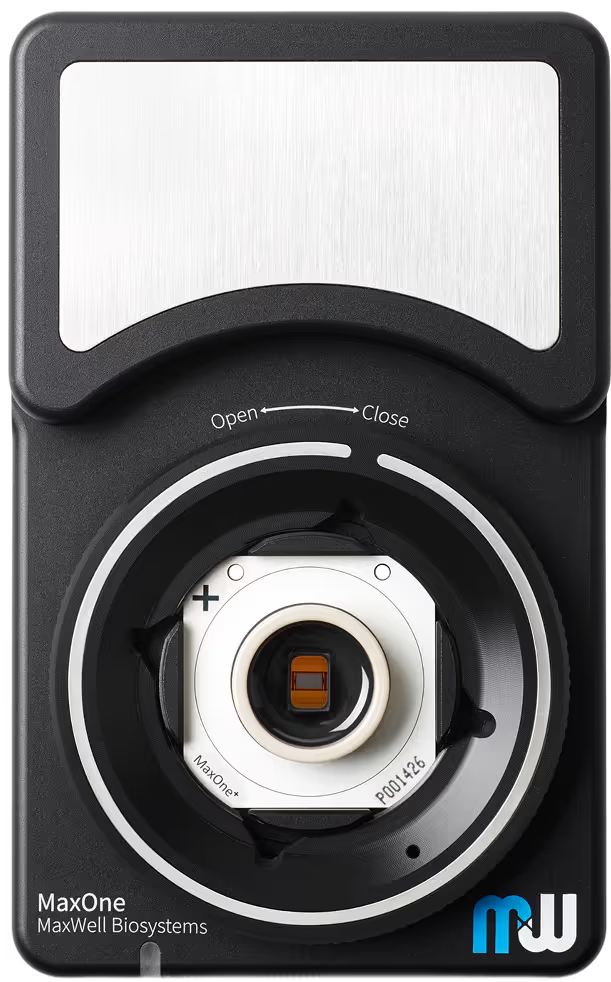
MaxOne
Versatility and functionality in one compact device
Our Technology
Multi-/Micro-electrode Arrays
Microphysiological Systems
Neuronal Cell Cultures