Open Source Hardware & Software Modules for Multimodal Electrophysiology Experiments with Prof. Dr. Mircea Teodorescu & Kateryna Voitiuk
(ミルチャ・テオドレスク教授とカテリーナ・ヴォイティウク氏による多様な電子生理学的実験におけるオープンソースハードウェア及びソフトモジュール)
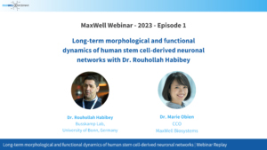
Long-term morphological and functional dynamics of human stem cell-derived neuronal networks with Dr. Rouhollah Habibey
(ルホラ・ハビベイ博士によるヒト幹細胞由来ニューロンネットワークの長期的形態・機能ダイナミクス)
What’s your cell story?
Characterizing the activity of human iPSC derived-neurons in 2D and 3D cultures at high resolution
(2Dおよび3D培養におけるヒトiPSC由来ニューロンの活動の高解像度特性化) 2021年11月11日
Dr. Silvia Ronchi
Scientific Application Specialist at MaxWell Biosystems
Giulio Zorzi
Product Manager & Application Engineer at MaxWell Biosystems
Abstract
Alongside our participation at the SfN Annual Meeting 2021, we will host two sessions presenting you replays of a Case Study presentation based on a paper published at Advanced Biology and our MaxTwo showcase. Both will be followed by a Live Q&A session. During these sessions, we will introduce our technology, products and applications and allow all attendees to ask questions to our team during the live Q&A session.
These sessions will be hosted by Dr. Marie Obien who will briefly introduce the company, technology, products and applications. The case study is presented by Dr. Silvia Ronchi, which is entitled, “Electrophysiological phenotype characterization of human iPSC derived-neuronal cell lines by means of high-density microelectrode arrays”, highlighting how the neuronal activity in 2D samples can be easily captured, label-free, at single-cell resolution by using MaxWell Biosystems’ high-density microelectrode array (HD-MEA) platforms. Giulio Zorzi showcases MaxTwo, a powerful system to characterize the function of human iPSC-derived neurons that can help you advance your research in different applications. The presentations will be followed by a live Q&A session.
Overall, the presentations will provide an overview on how HD-MEA technology can efficiently advance research in 2D and 3Dhuman derived from induced pluripotent stems cells (hiPSCs) brain models, as promising tools for investigating development, disease progression, and to test drug toxicity/efficacy in-vitro, and accelerate drug development for neurodegenerative diseases.
Presenting MaxTwo: A powerful system to characterize the function of human iPSC-derived neurons
(MaxTwo: ヒトiPS細胞由来神経細胞の機能を特徴付ける強力なシステム) September 29, 2021 | 10am / 5pm (CET), 1am / 8am (PDT), 4am / 11am (EDT)
2021年9月29日午後5時 / 30日午前0時 (日本時間)
Dr. Marie Obien
VP Marketing and Sales at MaxWell Biosystems
Giulio Zorzi
Product Manager & Application Engineer at MaxWell Biosystems
Abstract
In our live broadcast from our laboratory here in Zurich, Switzerland, we will:
Showcase how to use the MaxTwo System
Present how MaxTwo can help you advance your research in different applications
Engage the audience and answer questions throughout the session
Phenotyping of neurodevelopmental and psychiatric disorders with human iPSC-derived dopaminergic neurons
(ヒトiPS細胞由来ドーパミン性作動ニューロンによる神経発達障害と精神障害の表現型) July 8, 2021 | 5pm (CET), 8am (PST), 11am (EDT)
2021年7月9日 0am (日本時間)
Dr. Marie Obien
VP Marketing and Sales at MaxWell Biosystems
Speaker
Speaker
Dr. Maria Sundberg
Research Fellow, Group of Prof. Sahin, Boston Children’s Hospital
Hannah Pinson
PhD Candidate, Group of Prof. Ginis, Vrije Universiteit Brussel & Visitor in Prof. Tegmark group, MIT
Abstract
In recent years, the genetic causes of autism and schizophrenia have been studied intensively. In addition to monogenic deficits, deletions or duplications of specific chromosomal loci have also been associated with neurodevelopmental and psychiatric disorders. One of these regions is 16p11.2, which contains 29 protein coding genes, most of which are also expressed in the brain. Clinical studies have shown that deletion of 16p11.2 leads to severe developmental deficits, intellectual disability, and autism. On the other hand, patients with duplication of 16p11.2 locus have an increased risk of developing schizophrenia, bipolar disorder, depression and autism. Deficits in the dopamine signaling can cause behavioral problems and deficits in social interactions in the patients with autism and schizophrenia.
In this webinar, our speakers will:
Speak about how they studied the effects of 16p11.2 copy number variations on dopamine signaling by differentiating human iPSCs, with either a 16p11.2 duplication or deletion, into dopaminergic (DA) neurons in vitro and how they characterized molecular and functional phenotypes of these neurons compared to healthy control neurons.
Present their assessment of network activity using MaxOne, MaxWell’s high-density micro-electrode array (MEA) platform and give a short introduction to the data analysis pipeline used in this study. They will show a brief overview of the detection of synchronised sensors and bursting patterns, including a link to the used code.
Discuss their results: They detected that the cells carrying 16p11.2 deletion had an increased soma size, increased synaptic marker expression, and hyperactive DA-neuron networks compared to healthy control DA-neurons. Increased RhoA expression was also detected in the 16p11.2 deletion DA-neurons. Treatment of the neurons with a specific RhoA pathway inhibitor, Rhosin, rescued the network hyperactivation and the abnormal morphological development of the DA-neurons with 16p11.2 deletion. These results show that inhibition of RhoA pathway can serve as a potential therapeutic target for neurodevelopmental and neuropsychiatric disorders associated with 16p11.2 deletion.
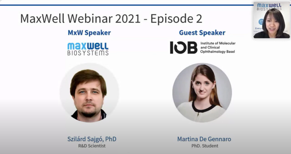
SpikeInterface, a Unified Framework for Spike Sorting (スパイクインターフェイス、スパイクソーティングのための統合フレームワーク) March 25, 2021 | 5pm (CET), 8am (PST), 11am (EDT)
Dr. Marie Obien
VP Marketing and Sales at MaxWell Biosystems
Speaker
Speaker
Dr. Szilard Sajgo
Application Specialist at MaxWell Biosystems
Dr. Alessio Buccino
Postdoctoral Fellow at ETH Zürich
Abstract
Understanding how assemblies of neurons encode information requires recording of large populations of cells. In recent years, high-density multi-electrode arrays (HD-MEAs) have been developed to record simultaneously from thousands of electrodes. Each electrode records from multiple surrounding neurons at the same time. In order to assign electrical signals recorded by HD-MEAs to individual neurons, a critical step called spike sorting needs to be performed. During this step, the extracellular action potentials originating from hundreds to thousands of neurons need to be disentangled from the background noise and from each other.
We had an introduction to spike sorting in a webinar earlier this year (link to the replay) and are happy to host this new webinar as a follow-up.
Dr. Szilárd Sajgó, R&D scientist at MaxWell Biosystems, will:
Explain the basic concept of spike sorting
Highlight how HD-MEAs facilitate reliable spike clustering.
This is followed by Dr. Alessio Buccino, ETH Postdoctoral Fellow in the group of Prof. Hierlemann, who will:
Present SpikeInterface, a Python framework designed to unify preexisting spike sorting technologies into a single codebase and to facilitate straightforward comparison and adoption of different approaches.
Explain how, with a few lines of code, researchers can reproducibly run, compare, and benchmark most modern spike sorting algorithms; pre-process, post-process, and visualize extracellular datasets; validate, curate, and export sorting outputs; and more.
Demonstrate the use of SpikeInterface on real and simulated datasets to reduce the burden of manual curation and to more comprehensively benchmark automated spike sorters.
Morphological, functional & transcriptomic correlation of retinal organoids to the human retina(網膜オルガノイド とヒトの網膜との形態学的、機能的、トランスクリプトームの相関関係について) February 18, 2021 | 5pm (CET), 8am (PST), 11am (EDT)
2021年2月19日午前1時(日本時間)
Genetic disorders of the human retina cause visual impairment to millions of people worldwide. The retina is a well characterized tissue at the back of the eye, and is a potential target for visual restoration therapies. Light-sensitive human retinal organoids recapitulate cell-types, circuitry and transcriptomic profiles of the human retina, offering a relevant tool for translational studies.
In this webinar, Dr. Szilárd Sajgó, our R&D scientist and an expert in retina research will:
Introduce the different analyses of retinal function that can be performed using high-density microelecrode arrays (HD-MEAs),
Explain how the efficacy of visual restoration therapies can now be easily assessed using HD-MEAs.
Demonstrate how the generation of a library of 285,441 single-cell transcriptomes from light-responsive human retinae and retinal organoids at different time points allows the comparison of developmental rates and genomic expression profiles,
Explain how electrophysiological assessments, such as HD-MEA and calcium imaging, ensure the collection of transcriptomes from functional, light-responsive human retinae ex-vivo,
Discuss how this research is allowing to define cellular targets for disease mechanisms investigation in organoids and targeted repair in the human retina.
Fast and Accurate Spike Sorting for Thousands of Channels(何千ものチャネルに対する迅速かつ正確なスパイソーティング)
January 14, 2021 | 5pm (CET), 8am (PST), 11am (EDT)
2021年1月15日午前1時(日本時間)
Understanding how assemblies of neurons encode information requires recording of large populations of cells. In recent years, high-density multi-electrode arrays (HD-MEAs) and silicon probes have been developed to record simultaneously from thousands of electrodes. Each electrode records from multiple surrounding neurons at the same time. In order to assign electrical signals recorded by HD-MEAs to individual neurons, a critical step called spike sorting needs to be performed . During this step, the extracellular action potentials originating from hundreds to thousands of neurons need to be disentangled from the background noise and from each other.
In this webinar, our speakers will:
explain the basic concept of spike sorting and highlight how HD-MEAs facilitate reliable spike clustering.
present a fast and accurate spike sorting algorithm, validated with in vivo and in vitro ground truth experiments.
explain how the SpyKING CIRCUS software is optimized for solving temporally overlapping spikes in large-scale extracellular recordings.
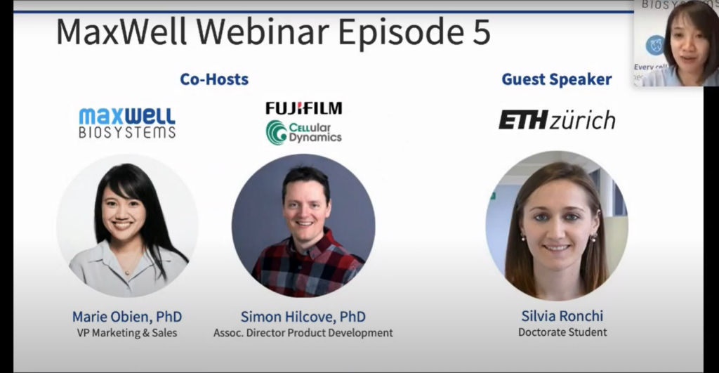
Co-hosted with FUJIFILM Cellular Dynamics:
Electrophysiological Phenotype Characterization of Human iPSC-Derived Neuronal Cell Lines by Means of High-Density Microelectrode Arrays(高密度微小電極アレイによるヒトiPS細胞由来神経細胞株の電気生理学的表現型の特徴付け) October 15, 2020 | 5pm (CET), 8am (PST), 11am (EDT)
2020年10月16日 | 午前0時(日本時間)
Marie Obien, Ph.D.
VP Marketing and Sales at MaxWell Biosystems
Simon Hilcove, Ph.D.
Assoc. Director Product Development at FUJIFILM Cellular Dynamics
Speaker
Silvia Ronchi
Doctorate Student at ETH Zürich
Abstract:
Recent advances in the field of cellular reprogramming have opened a route to study the fundamental mechanisms underlying common neurological disorders. High-density microelectrode-arrays (HD-MEAs) provide unprecedented means to study neuronal physiology at different scales, ranging from network through single-neuron to subcellular features.
In this webinar, we will:
Introduce how HD-MEAs was used to characterize and compare human induced-pluripotent-stem-cell (iPSC)-derived dopaminergic and motor neurons, including isogenic neuronal lines modeling Parkinson’s disease and amyotrophic lateral sclerosis in-vitro.
Present the metrics used for phenotype characterization and drug testing.
Demonstrate the ability to detect drug effects with HD-MEA.
Label-free functional characterization of 3D organoids at single-cell resolution(単一細胞解像度での3Dオルガノイドのラベルフリー機能的評価) September 10, 2020 | 5pm (CET), 8am (PST), 11am (EDT)
2020年9月11日 | 午前0時(日本時間)
Dr. Marie Obien
VP Marketing and Sales at MaxWell Biosystems
Dr. Szilard Sajgo
Application Specialist at MaxWell Biosystems
Abstract
Human organoids that originate from human induced pluripotent stem cells (h-iPSCs) are emerging as promising tools for investigating development and disease progression. In order to adopt human organoids for rapid and cost-effective drug screenings, it is necessary to assess their cell type composition, gene expression patterns and physiological function. The electrical activity of brain, retina or muscle organoids can now be easily captured, label-free, at single-cell resolution by using MaxWell Biosystems’ high-density microelectrode array (HD-MEA) technology.
In this webinar, the speakers will:
Introduce high-resolution functional imaging of organoids with the MaxTwo HD-MEA platform
Present results from organoids modeling different brain compartments
Demonstrate the potential of HD-MEA technology for characterizing the physiological function of human brain organoids and for testing compounds.
Dr. Marie Obien
VP Marketing and Sales at MaxWell Biosystems
Dr. David Jäckel
Senior Product Manager at MaxWell Biosystems
Abstract
The high-density microelectrode arrays (HD-MEAs) high-content electrophysiology platforms MaxOne and MaxTwo allow label-free recording and stimulation of every active cell on a dish at unprecedented spatio-temporal resolution.
A powerful and easy-to-use software interface is key to reveal the full potential of this technology. MaxLab Live is an all-in-one software for live visualization, recording, and analysis of extracellular HD-MEA signals from different biological preparations.
In this webinar, the speakers will:
Introduce you to the MaxLab Live Software and User Interface
Show how to get the visualization of your data in real-time at the level you need – from whole sample network to sub-cellular activity
Present how to run standardized and repeatable experiments with MaxLab Live Assays
Assessing retinal function in health and disease at single-cell resolution
(単一細胞解像度での健康及び疾患の網膜機能評価) June 11, 2020 | 5pm (CET), 8am (PST), 11am (EDT)
2020年6月12日| 午前0時(日本時間)
Dr. Marie Obien
VP Marketing and Sales at MaxWell Biosystems
Dr. Szilard Sajgo
Application Scientist at MaxWell Biosystems
Abstract
Genetic diseases of the human retina cause visual impairment to millions of people worldwide. The retina is a well characterized tissue at the back of the eye, and is a potential target for visual restoration therapies. The efficacy of these therapies can now be easily assessed using high-density microelectrode arrays (HD-MEA). In addition, HD-MEA technology can also be used to assess disease phenotypes, as well as other aspects of visual processing and development.
This webinar will include the following:
Introduction to different analyses of retinal function using HD-MEAs
Showcase of how HD-MEAs can contribute to visual restoration therapies
Presentation of a case study involving FRMD7, a defective gene in human congenital nystagmus
Functional characterization of human iPSC-derived neurons at single-cell resolution
(単一細胞解像度でのヒトiPS細胞由来神経細胞の機能的特性評価) April 23, 2020 | 5pm (CET), 8am (PST), 11am (EDT)
2020年4月24日| 午前0時(日本時間)
Dr. Marie Obien
VP Marketing and Sales at MaxWell Biosystems
Dr. Michele Fiscella
VP Scientific Affairs at MaxWell Biosystems
Abstract
Recent developments in induced pluripotent stem cell (iPSC) technology have enabled easier access to human cells in vitro. With increasing availability of human iPSC-derived neurons, both healthy and disease cell lines, screening compounds for neurodegenerative diseases on human cells can potentially be performed in the earlier stages of drug discovery. To accelerate the functional characterization of iPSC-derived neurons and the effect of compounds, reproducible and relevant results are necessary.
In this webinar, the speakers will:
Introduce high-resolution functional imaging of human iPSC-derived neurons
Showcase how to extract functional features of hundreds of cells in a cell culture sample label-free
Discuss electrophysiological parameters for characterizing the differences among several human neuronal cell lines
We use cookies on our website to give you the most relevant experience by remembering your preferences and repeat visits. By clicking “Accept”, you consent to the use of ALL the cookies. Read more about our Privacy Policy
This website uses cookies to improve your experience while you navigate through the website. Out of these, the cookies that are categorized as necessary are stored on your browser as they are essential for the working of basic functionalities of the website. We also use third-party cookies that help us analyze and understand how you use this website. These cookies will be stored in your browser only with your consent. You also have the option to opt-out of these cookies. But opting out of some of these cookies may affect your browsing experience.
 日本語
日本語 繁體中文
繁體中文 简体中文
简体中文 English

 Open Source Hardware & Software Modules for Multimodal Electrophysiology Experiments with Prof. Dr. Mircea Teodorescu & Kateryna Voitiuk
 Open Source Hardware & Software Modules for Multimodal Electrophysiology Experiments with Prof. Dr. Mircea Teodorescu & Kateryna Voitiuk