 日本語
日本語 繁體中文
繁體中文 简体中文
简体中文 English
網膜応用分野のためのMaxOne 
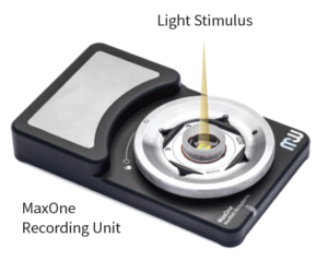
MaxOneは視覚研究に理想的です。
MaxOneと光刺激セットアップを併用することで、すべての科学者は生体外で網膜神経節細胞の機能を研究することが可能です。

この高解像度を保って全ての網膜神経節細胞の活動を記録することを可能にします。
- 26,400個の電極
- 8平方mmのセンサーエリア
- 1平方mm辺り3.265個の電極
このようなことができます
網膜神経節細胞(RGC)の機能を同定
MEA上ですべての網膜神経節細胞のタイプを同定し記録します
MEA上のすべての網膜神経節細胞の光応答はMaxOneで記録および解析ができます。
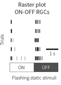
- 閃光する静的な光は異なる網膜神経節細胞の発火特性(ON型、OFF型、あるいはON-OFF型)を明らかにします。

- 方向選択性網膜神経節細胞の応答は移動刺激を用いることによって抽出され得ます。
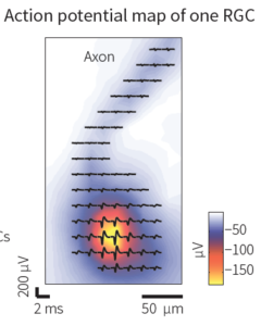
- MaxOneの信号雑音比と高時空間解像度は網膜神経節細胞軸索信号の解析が可能です。
網膜神経節細胞の限定された受容野モザイクを明らかにします
前例の無い解像度で網膜神経節細胞の受容野モザイクを抽出し分析します。
MaxOneは複数の網膜神経節細胞を同時に記録し、移動バー刺激によって活性化した視覚受容野モザイクをとらえます。
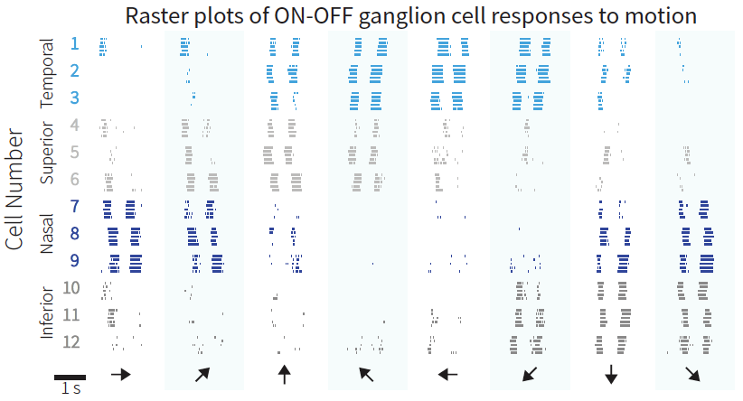
- ON-OFF型方向選択性網膜神経節細胞の発火特性はそれらの好む方向を明らかにします。

- 高解像度受容野モザイクは同型の網膜神経節細胞間でほとんどオーバーラップしません。
スパイクソーティング
MaxOneの高空間解像度は信頼できるスパイクソーティングを容易にします。
複数の電極は網膜神経節細胞よりスパイクを検出します。追加の空間情報はスパイクのクラスタリングの精度を向上させます。
 複数の電極は網膜神経節細胞よりスパイクを検出します。追加の空間情報はスパイクのクラスタリングの精度を向上させます。
* ISI v.: Inter-spike interval violation(発火時間間隔の妨害)
網膜ホルダー
MaxOne組織ホルダーはMEA上の網膜を平坦にすることで、繰り返し安定した実験ができます。

組織ホルダーは灌流溶液下で実験中、MEA上に網膜を押して平らにし固定し続けることができます。
- 3軸マニピュレータが正確なホルダー制御を可能にします。
- 膜もしくは細かいメッシュを使用できます。
- マグネットプレートが灌水チューブ用のスタンドとして役立ちます。
論文 —
網膜の応用分野
Idrees, Saad; Baumann, Matthias-Philipp; Korympidou, Maria M; Schubert, Timm; Kling, Alexandra; Franke, Katrin; Hafed, Ziad M; Franke, Felix; Münch, Thomas A Suppression without inhibition: how retinal computation contributes to saccadic suppression Journal Article Communications Biology, 2022. Idrees, Saad; Münch, Thomas A Different contrast encoding in ON and OFF visual pathways Journal Article BioRxiv, 2020. Idrees, Saad; Baumann, Matthias P; Franke, Felix; Münch, Thomas A; Hafed, Ziad M Perceptual saccadic suppression starts in the retina Journal Article Nature Communications, 11 (1977), 2020. da Drinnenberg Antonia; Franke, Felix; Morikawa Rei Jüttner; Hillier Daniel; Hantz Peter; Hierlemann Andreas; Azeredo Silveira Rava; Roska Botond K; How diverse retinal functions arise from feedback at the first visual synapse Journal Article Neuron, 99 (1), pp. 117-134, 2018. Hillier, Daniel; Fiscella, Michele; Drinnenberg, Antonia; Trenholm, Stuart; Rompani, Santiago B; Raics, Zoltan; Katona, Gergely; Jüttner, Josephine; Hierlemann, Andreas; Rozsa, Balazs; Roska, Botond Causal evidence for retina-dependent and -independent visual motion computations in mouse cortex Journal Article Nature Neuroscience, 20 (7), pp. 960–968, 2017, ISSN: 1097-6256. Franke, Felix; Fiscella, Michele; Sevelev, Maksim; Roska, Botond; Hierlemann, Andreas; Azeredo da Silveira, Rava Structures of Neural Correlation and How They Favor Coding Journal Article Neuron, 89 (2), pp. 409-422, 2016, ISSN: 10974199. Yonehara, Keisuke; Fiscella, Michele; Drinnenberg, Antonia; Esposti, Federico; Trenholm, Stuart; Krol, Jacek; Franke, Felix; Scherf, Brigitte Gross; Kusnyerik, Akos; Müller, Jan; Szabo, Arnold; Jüttner, Josephine; Cordoba, Francisco; Reddy, Ashrithpal Police; Németh, János; Nagy, Zoltán Zsolt; Munier, Francis; Hierlemann, Andreas; Roska, Botond Congenital Nystagmus Gene FRMD7 Is Necessary for Establishing a Neuronal Circuit Asymmetry for Direction Selectivity Journal Article Neuron, 89 (1), pp. 177-193, 2016, ISSN: 10974199. Jones, Ian L; Russell, Thomas L; Farrow, Karl; Fiscella, Michele; Franke, Felix; Müller, Jan; Jäckel, David; Hierlemann, Andreas A method for electrophysiological characterization of hamster retinal ganglion cells using a high-density CMOS microelectrode array Journal Article Frontiers in Neuroscience, 9 , pp. 360, 2015, ISSN: 1662453X. Fiscella, Michele; Franke, Felix; Farrow, Karl; Müller, Jan; Roska, Botond; Azeredo da Silveira, Rava ; Hierlemann, Andreas Visual coding with a population of direction-selective neurons Journal Article Journal of Neurophysiology, 114 (4), pp. 2485-2499, 2015, ISSN: 0022-3077. Krol, Jacek; Krol, Ilona; Alvarez, Claudia Patricia Patino; Fiscella, Michele; Hierlemann, Andreas; Roska, Botond; Filipowicz, Witold A network comprising short and long noncoding RNAs and RNA helicase controls mouse retina architecture. Journal Article Nature Communications, 6 , pp. 7305, 2015, ISSN: 2041-1723. Fiscella, Michele; Farrow, Karl; Jones, Ian L; Jäckel, David; Müller, Jan; Frey, Urs; Bakkum, Douglas J; Hantz, Péter; Roska, Botond; Hierlemann, Andreas Journal of Neuroscience Methods, 211 (1), pp. 103-113, 2012, ISSN: 01650270.![]()
title = {Suppression without inhibition: how retinal computation contributes to saccadic suppression},
author = {Saad Idrees and Matthias-Philipp Baumann and Maria M. Korympidou and Timm Schubert and Alexandra Kling and Katrin Franke and Ziad M. Hafed and Felix Franke and Thomas A. Münch },
url = {https://www.nature.com/articles/s42003-022-03526-2},
year = {2022},
date = {2022-07-12},
journal = {Communications Biology},
abstract = {Visual perception remains stable across saccadic eye movements, despite the concurrent strongly disruptive visual flow. This stability is partially associated with a reduction in visual sensitivity, known as saccadic suppression, which already starts in the retina with reduced ganglion cell sensitivity. However, the retinal circuit mechanisms giving rise to such sup- pression remain unknown. Here, we describe these mechanisms using electrophysiology in mouse, pig, and macaque retina, 2-photon calcium imaging, computational modeling, and human psychophysics. We find that sequential stimuli, like those that naturally occur during saccades, trigger three independent suppressive mechanisms in the retina. The main mechanism is triggered by contrast-reversing sequential stimuli and originates within the receptive field center of ganglion cells. It does not involve inhibition or other known sup- pressive mechanisms like saturation or adaptation. Instead, it relies on temporal filtering of the inherently slow response of cone photoreceptors coupled with downstream non- linearities. Two further mechanisms of suppression are present predominantly in ON ganglion cells and originate in the receptive field surround, highlighting another disparity between ON and OFF ganglion cells. The mechanisms uncovered here likely play a role in shaping the retinal output following eye movements and other natural viewing conditions where sequential stimulation is ubiquitous.},
keywords = {},
pubstate = {published},
tppubtype = {article}
}

title = {Different contrast encoding in ON and OFF visual pathways},
author = {Saad Idrees and Thomas A. Münch },
url = {https://www.biorxiv.org/content/10.1101/2020.11.25.398230v1},
doi = {10.1101/2020.11.25.398230},
year = {2020},
date = {2020-11-26},
journal = {BioRxiv},
abstract = {Subjective visual experience builds on sensory encoding of light reflected by different objects in our environment. Most retinal ganglion cells encode changes in light intensity, quantified as contrast, rather than the absolute intensity. Mathematically, contrast is often defined as a relative change in light intensity. Activity in the visual system and perceptual responses are usually explained with such definitions of contrast. Here, for the first time, we explicitly explored how contrast is actually represented in the visual system. Using mouse retina electrophysiology, we show that response strength of OFF retinal ganglion cells does not represent relative, but absolute changes in light intensity. ON RGC response strength is governed by a combination of absolute and relative change in light intensity. This is true for a wide range of ambient light levels, at least from scotopic to high mesopic regimes. Consequently, light decrements and increments are represented asymmetrically in the retina, which may explain the asymmetries in responses to negative and positive contrast observed throughout the visual system. These findings may help to more thoroughly design and interpret vision science studies where responses are driven by contrast of the visual stimuli.},
keywords = {},
pubstate = {published},
tppubtype = {article}
}

title = {Perceptual saccadic suppression starts in the retina},
author = {Saad Idrees and Matthias P. Baumann and Felix Franke and Thomas A. Münch and Ziad M. Hafed},
url = {https://www.nature.com/articles/s41467-020-15890-w},
doi = {10.1038/s41467-020-15890-w},
year = {2020},
date = {2020-04-24},
journal = {Nature Communications},
volume = {11},
number = {1977},
keywords = {},
pubstate = {published},
tppubtype = {article}
}
title = {How diverse retinal functions arise from feedback at the first visual synapse},
author = {Drinnenberg, Antonia; Franke, Felix; Morikawa, Rei K; Jüttner; Hillier, Daniel; Hantz, Peter; Hierlemann, Andreas; Azeredo da Silveira, Rava; Roska, Botond},
url = {https://www.cell.com/neuron/fulltext/S0896-6273(18)30469-0},
doi = {10.1016/j.neuron.2018.06.001},
year = {2018},
date = {2018-06-21},
journal = {Neuron},
volume = {99},
number = {1},
pages = {117-134},
abstract = {Many brain regions contain local interneurons of distinct types. How does an interneuron type contribute to the input-output transformations of a given brain region? We addressed this question in the mouse retina by chemogenetically perturbing horizontal cells, an interneuron type providing feedback at the first visual synapse, while monitoring the light-driven spiking activity in thousands of ganglion cells, the retinal output neurons. We uncovered six reversible perturbation-induced effects in the response dynamics and response range of ganglion cells. The effects were enhancing or suppressive, occurred in different response epochs, and depended on the ganglion cell type. A computational model of the retinal circuitry reproduced all perturbation-induced effects and led us to assign specific functions to horizontal cells with respect to different ganglion cell types. Our combined experimental and theoretical work reveals how a single interneuron type can differentially shape the dynamical properties of distinct output channels of a brain region.},
keywords = {},
pubstate = {published},
tppubtype = {article}
}
title = {Causal evidence for retina-dependent and -independent visual motion computations in mouse cortex},
author = {Daniel Hillier and Michele Fiscella and Antonia Drinnenberg and Stuart Trenholm and Santiago B Rompani and Zoltan Raics and Gergely Katona and Josephine Jüttner and Andreas Hierlemann and Balazs Rozsa and Botond Roska},
url = {http://www.nature.com/doifinder/10.1038/nn.4566},
doi = {10.1038/nn.4566},
issn = {1097-6256},
year = {2017},
date = {2017-05-22},
journal = {Nature Neuroscience},
volume = {20},
number = {7},
pages = {960--968},
abstract = {How neuronal computations in the sensory periphery contribute to computations in the cortex is not well understood. We examined this question in the context of visual-motion processing in the retina and primary visual cortex (V1) of mice. We disrupted retinal direction selectivity, either exclusively along the horizontal axis using FRMD7 mutants or along all directions by ablating starburst amacrine cells, and monitored neuronal activity in layer 2/3 of V1 during stimulation with visual motion. In control mice, we found an over-representation of cortical cells preferring posterior visual motion, the dominant motion direction an animal experiences when it moves forward. In mice with disrupted retinal direction selectivity, the over-representation of posterior-motion-preferring cortical cells disappeared, and their responses at higher stimulus speeds were reduced. This work reveals the existence of two functionally distinct, sensory-periphery-dependent and -independent computations of visual motion in the cortex.},
keywords = {},
pubstate = {published},
tppubtype = {article}
}

title = {Structures of Neural Correlation and How They Favor Coding},
author = {Felix Franke and Michele Fiscella and Maksim Sevelev and Botond Roska and Andreas Hierlemann and Rava {Azeredo da Silveira}},
url = {http://www.sciencedirect.com/science/article/pii/S0896627315011393?via%3Dihub},
doi = {10.1016/j.neuron.2015.12.037},
issn = {10974199},
year = {2016},
date = {2016-01-20},
journal = {Neuron},
volume = {89},
number = {2},
pages = {409-422},
publisher = {Elsevier Inc.},
abstract = {The neural representation of information suffers from "noise"-the trial-to-trial variability in the response of neurons. The impact of correlated noise upon population coding has been debated, but a direct connection between theory and experiment remains tenuous. Here, we substantiate this connection and propose a refined theoretical picture. Using simultaneous recordings from a population of direction-selective retinal ganglion cells, we demonstrate that coding benefits from noise correlations. The effect is appreciable already in small populations, yet it is a collective phenomenon. Furthermore, the stimulus-dependent structure of correlation is key. We develop simple functional models that capture the stimulus-dependent statistics. We then use them to quantify the performance of population coding, which depends upon interplays of feature sensitivities and noise correlations in the population. Because favorable structures of correlation emerge robustly in circuits with noisy, nonlinear elements, they will arise and benefit coding beyond the confines of retina. Coding in the brain suffers from the variability of neural responses. Using experiment and theory, Franke et al. show that this "noise" comes with a particular structure, which emerges from circuit properties and which counteracts the harmful effect of variability.},
keywords = {},
pubstate = {published},
tppubtype = {article}
}
title = {Congenital Nystagmus Gene FRMD7 Is Necessary for Establishing a Neuronal Circuit Asymmetry for Direction Selectivity},
author = {Keisuke Yonehara and Michele Fiscella and Antonia Drinnenberg and Federico Esposti and Stuart Trenholm and Jacek Krol and Felix Franke and Brigitte Gross Scherf and Akos Kusnyerik and Jan Müller and Arnold Szabo and Josephine Jüttner and Francisco Cordoba and Ashrithpal Police Reddy and János Németh and Zoltán Zsolt Nagy and Francis Munier and Andreas Hierlemann and Botond Roska},
url = {http://www.sciencedirect.com/science/article/pii/S0896627315010387?via%3Dihub},
doi = {10.1016/j.neuron.2015.11.032},
issn = {10974199},
year = {2016},
date = {2016-01-06},
journal = {Neuron},
volume = {89},
number = {1},
pages = {177-193},
abstract = {Neuronal circuit asymmetries are important components of brain circuits, but the molecular pathways leading to their establishment remain unknown. Here we found that the mutation of FRMD7, a gene that is defective in human congenital nystagmus, leads to the selective loss of the horizontal optokinetic reflex in mice, as it does in humans. This is accompanied by the selective loss of horizontal direction selectivity in retinal ganglion cells and the transition from asymmetric to symmetric inhibitory input to horizontal direction-selective ganglion cells. In wild-type retinas, we found FRMD7 specifically expressed in starburst amacrine cells, the interneuron type that provides asymmetric inhibition to direction-selective retinal ganglion cells. This work identifies FRMD7 as a key regulator in establishing a neuronal circuit asymmetry, and it suggests the involvement of a specific inhibitory neuron type in the pathophysiology of a neurological disease.},
keywords = {},
pubstate = {published},
tppubtype = {article}
}

title = {A method for electrophysiological characterization of hamster retinal ganglion cells using a high-density CMOS microelectrode array},
author = {Ian L Jones and Thomas L Russell and Karl Farrow and Michele Fiscella and Felix Franke and Jan Müller and David Jäckel and Andreas Hierlemann},
url = {https://www.frontiersin.org/articles/10.3389/fnins.2015.00360/full},
doi = {10.3389/fnins.2015.00360},
issn = {1662453X},
year = {2015},
date = {2015-10-13},
journal = {Frontiers in Neuroscience},
volume = {9},
pages = {360},
abstract = {Knowledge of neuronal cell types in the mammalian retina is important for the understanding of human retinal disease and the advancement of sight-restoring technology, such as retinal prosthetic devices. A somewhat less utilized animal model for retinal research is the hamster, which has a visual system that is characterized by an area centralis and a wide visual field with a broad binocular component. The hamster retina is optimally suited for recording on the microelectrode array (MEA), because it intrinsically lies flat on the MEA surface and yields robust, large-amplitude signals. However, information in the literature about hamster retinal ganglion cell functional types is scarce. The goal of our work is to develop a method featuring a high-density (HD) Complementary metal-oxide-semiconductor (CMOS) MEA technology along with a sequence of standardized visual stimuli in order to categorize ganglion cells in isolated Syrian Hamster (Mesocricetus auratus) retina. Since the HD-MEA is capable of recording at a higher spatial resolution than most MEA systems (17.5 um electrode pitch), we capitalized on this feature and were able to record from a large proportion of RGCs within a selected region. Secondly, we chose our stimuli so that they could be run during the experiment without intervention or computation steps. The visual stimulus set was designed to activate the receptive fields of most ganglion cells in parallel and to incorporate various visual features to which different cell types respond uniquely. Based on the ganglion cell responses, basic cell properties were determined: direction selectivity, speed tuning, width tuning, transience and latency. These properties were clustered in order to identify ganglion cell types in the hamster retina. Ultimately, we recorded up to a cell density 2780 cells/mm2 at 2 mm (42°) from the optic nerve head. Using 5 parameters extracted from the responses to visual stimuli, we obtained 7 ganglion cell types.},
keywords = {},
pubstate = {published},
tppubtype = {article}
}
title = {Visual coding with a population of direction-selective neurons},
author = {Michele Fiscella and Felix Franke and Karl Farrow and Jan Müller and Botond Roska and Rava {Azeredo da Silveira} and Andreas Hierlemann},
url = {http://jn.physiology.org/lookup/doi/10.1152/jn.00919.2014},
doi = {10.1152/jn.00919.2014},
issn = {0022-3077},
year = {2015},
date = {2015-08-19},
journal = {Journal of Neurophysiology},
volume = {114},
number = {4},
pages = {2485-2499},
abstract = {The brain decodes the visual scene from the action potentials of ∼20 retinal ganglion cell types. Among the retinal ganglion cells, direction-selective ganglion cells (DSGCs) encode motion direction. Several studies have focused on the encoding or decoding of motion direction by recording multiunit activity, mainly in the visual cortex. In this study, we simultaneously recorded from all four types of ON-OFF DSGCs of the rabbit retina using a microelectronics-based high-density microelectrode array (HDMEA) and decoded their concerted activity using probabilistic and linear decoders. Furthermore, we investigated how the modification of stimulus parameters (velocity, size, angle of moving object) and the use of different tuning curve fits influenced decoding precision. Finally, we simulated ON-OFF DSGC activity, based on real data, in order to understand how tuning curve widths and the angular distribution of the cells' preferred directions influence decoding performance. We found that probabilistic decoding strategies outperformed, on average, linear methods and that decoding precision was robust to changes in stimulus parameters such as velocity. The removal of noise correlations among cells, by random shuffling trials, caused a drop in decoding precision. Moreover, we found that tuning curves are broad in order to minimize large errors at the expense of a higher average error, and that the retinal direction-selective system would not substantially benefit, on average, from having more than four types of ON-OFF DSGCs or from a perfect alignment of the cells' preferred directions.},
keywords = {},
pubstate = {published},
tppubtype = {article}
}

title = {A network comprising short and long noncoding RNAs and RNA helicase controls mouse retina architecture.},
author = {Jacek Krol and Ilona Krol and Claudia Patricia Patino Alvarez and Michele Fiscella and Andreas Hierlemann and Botond Roska and Witold Filipowicz},
url = {https://www.nature.com/articles/ncomms8305},
doi = {10.1038/ncomms8305},
issn = {2041-1723},
year = {2015},
date = {2015-06-04},
journal = {Nature Communications},
volume = {6},
pages = {7305},
publisher = {Nature Publishing Group},
abstract = {Brain regions, such as the cortex and retina, are composed of layers of uniform thickness. The molecular mechanism that controls this uniformity is not well understood. Here we show that during mouse postnatal development the timed expression of Rncr4, a retina-specific long noncoding RNA, regulates the similarly timed processing of pri-miR-183/96/182, which is repressed at an earlier developmental stage by RNA helicase Ddx3x. Shifting the timing of mature miR-183/96/182 accumulation or interfering with Ddx3x expression leads to the disorganization of retinal architecture, with the photoreceptor layer being most affected. We identify Crb1, a component of the adhesion belt between glial and photoreceptor cells, as a link between Rncr4-regulated miRNA metabolism and uniform retina layering. Our results suggest that the precise timing of glia-neuron interaction controlled by noncoding RNAs and Ddx3x is important for the even distribution of cells across layers.},
keywords = {},
pubstate = {published},
tppubtype = {article}
}
title = {Recording from defined populations of retinal ganglion cells using a high-density CMOS-integrated microelectrode array with real-time switchable electrode selection},
author = {Michele Fiscella and Karl Farrow and Ian L Jones and David Jäckel and Jan Müller and Urs Frey and Douglas J Bakkum and Péter Hantz and Botond Roska and Andreas Hierlemann},
url = {http://www.sciencedirect.com/science/article/pii/S0165027012003287?via%3Dihub},
doi = {10.1016/j.jneumeth.2012.08.017},
issn = {01650270},
year = {2012},
date = {2012-08-16},
journal = {Journal of Neuroscience Methods},
volume = {211},
number = {1},
pages = {103-113},
publisher = {Elsevier B.V.},
abstract = {In order to understand how retinal circuits encode visual scenes, the neural activity of defined populations of retinal ganglion cells (RGCs) has to be investigated. Here we report on a method for stimulating, detecting, and subsequently targeting defined populations of RGCs. The possibility to select a distinct population of RGCs for extracellular recording enables the design of experiments that can increase our understanding of how these neurons extract precise spatio-temporal features from the visual scene, and how the brain interprets retinal signals. We used light stimulation to elicit a response from physiologically distinct types of RGCs and then utilized the dynamic-configurability capabilities of a microelectronics-based high-density microelectrode array (MEA) to record their synchronous action potentials. The layout characteristics of the MEA made it possible to stimulate and record from multiple, highly overlapping RGCs simultaneously without light-induced artifacts. The high-density of electrodes and the high signal-to-noise ratio of the MEA circuitry allowed for recording of the activity of each RGC on 14 ± 7 electrodes. The spatial features of the electrical activity of each RGC greatly facilitated spike sorting. We were thus able to localize, identify and record from defined RGCs within a region of mouse retina. In addition, we stimulated and recorded from genetically modified RGCs to demonstrate the applicability of optogenetic methods, which introduces an additional feature to target a defined cell type. The developed methodologies can likewise be applied to other neuronal preparations including brain slices or cultured neurons.},
keywords = {},
pubstate = {published},
tppubtype = {article}
}