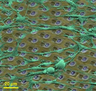
Presenting measurements of neuronal preparations with a novel CMOS-based microelectrode array at high-spatiotemporal-resolution on subcellular, cellular, and network level.
J. Müller, M. Ballini, P. Livi, Y. Chen, M. Radivojevic, A. Shadmani, V. Viswam, I. L. Jones, M. Fiscella, R. Diggelmann, A. Stettler, U. Frey, D. J. Bakkum, and A. Hierlemann, “High-resolution CMOS MEA platform to study neurons at subcellular, cellular, and network levels,” Lab Chip, vol. 15, no. 13, pp. 2767–2780, May 2015.
Reviewing the current understanding of microelectrode signals and the techniques for analyzing them, with focus on the ongoing advancements in microelectrode technology (in vivo and in vitro) and recent advanced microelectrode array measurement methods that facilitate the understanding of single neurons and network function.
M. E. J. Obien, K. Deligkaris, T. Bullmann, D. J. Bakkum, and U. Frey, “Revealing Neuronal Function through Microelectrode Array Recordings,” Front. Neurosci., 8:423, Jan 2015.
A high-resolution CMOS-based microelectrode array featuring 1,024 low-noise readout channels, 26,400 electrodes at a density of 3,265 electrodes per mm2, including on-chip 10bit ADCs and consuming only 75 mW.
M. Ballini, J. Muller, P. Livi, Y. Chen, U. Frey, A. Stettler, A. Shadmani, V. Viswam, I. L. Jones, D. Jackel, M. Radivojevic, M. K. Lewandowska, W. Gong, M. Fiscella, D. J. Bakkum, F. Heer, and A. Hierlemann, “A 1024-Channel CMOS Microelectrode Array With 26,400 Electrodes for Recording and Stimulation of Electrogenic Cells In Vitro,” IEEE Journal of Solid-State Circuits, vol. 49, no. 11, pp. 2705-2719, 2014.
Demonstrating a method to electrically visualize action potential propagation on axons and revealing
large variations in velocity.
D. J. Bakkum, U. Frey, M. Radivojevic, T. L. Russell, J. Muller, M. Fiscella, H. Takahashi, and A. Hierlemann, “Tracking axonal action potential propagation on a high-density microelectrode array across hundreds of sites,” Nature Communications, 4:2181, Jul 2013.
Recording and modeling extracellular action potentials of Purkinje cells at subcellular resolution.
U. Frey, U. Egert, F. Heer, S. Hafizovic, and A. Hierlemann, “Microelectronic System for High-Resolution Mapping of Extracellular Electric Fields Applied to Brain Slices,” Biosensors and Bioelectronics, vol. 24, no. 7, pp. 2191-2198, 2009.
Controlling BMP-2 expression to modulate the electrophysiological properties of cardiomyocytes using an HD-MEA for detailed monitoring.
C. D. Sanchez-Bustamante, U. Frey, J. M. Kelm, A. Hierlemann, and M. Fussenegger,
“Modulation of Cardiomyocyte Electrical Properties Using Regulated Bone Morphogenetic Protein-2 Expression,” Tissue Engineering Part A, vol. 14, no. 12, pp. 1969-1988, 2008.
@article{Bandarabadi2020,
title = {Sleep as a default state of cortical and subcortical networks},
author = {Mojtaba Bandarabadi and Anne Vassalli and Mehdi Tafti},
url = {https://www.sciencedirect.com/science/article/pii/S2468867319301907?via%3Dihub},
doi = {10.1016/j.cophys.2019.12.004},
year = {2020},
date = {2020-06-20},
journal = {Current Opinion in Physiology},
volume = {15},
pages = {60-67},
abstract = {Sleep has been conceptualized as ‘activity-dependent’, hence a response to prior waking experience, and proposed to be ‘the price the brain pays for plasticity during wakefulness’. We here propose that at the level of neuronal networks, particularly those arising from isolated embryonic thalamocortical cells maintained in culture, it represents a default mode of functioning. We show that cell assemblies in ex vivo cultures express powerful sleep specific patterns of oscillatory activity, as well as metabolic and molecular signatures of the sleep state. We summarize recent evidences that support our hypothesis and discuss potential applications of developing ex vivo sleep models to answer open questions in the field.},
keywords = {},
pubstate = {published},
tppubtype = {article}
}
Sleep has been conceptualized as ‘activity-dependent’, hence a response to prior waking experience, and proposed to be ‘the price the brain pays for plasticity during wakefulness’. We here propose that at the level of neuronal networks, particularly those arising from isolated embryonic thalamocortical cells maintained in culture, it represents a default mode of functioning. We show that cell assemblies in ex vivo cultures express powerful sleep specific patterns of oscillatory activity, as well as metabolic and molecular signatures of the sleep state. We summarize recent evidences that support our hypothesis and discuss potential applications of developing ex vivo sleep models to answer open questions in the field.
@article{Obien2015,
title = {Revealing neuronal function through microelectrode array recordings},
author = {Marie Engelene J Obien and Kosmas Deligkaris and Torsten Bullmann and Douglas J Bakkum and Urs Frey},
url = {https://www.frontiersin.org/articles/10.3389/fnins.2014.00423/full},
doi = {10.3389/fnins.2014.00423},
issn = {1662453X},
year = {2015},
date = {2015-01-06},
journal = {Frontiers in Neuroscience},
volume = {9},
pages = {423},
abstract = {Microelectrode arrays and microprobes have been widely utilized to measure neuronal activity, both in vitro and in vivo. The key advantage is the capability to record and stimulate neurons at multiple sites simultaneously. However, unlike the single-cell or single-channel resolution of intracellular recording, microelectrodes detect signals from all possible sources around every sensor. Here, we review the current understanding of microelectrode signals and the techniques for analyzing them. We introduce the ongoing advancements in microelectrode technology, with focus on achieving higher resolution and quality of recordings by means of monolithic integration with on-chip circuitry. We show how recent advanced microelectrode array measurement methods facilitate the understanding of single neurons as well as network function.},
keywords = {},
pubstate = {published},
tppubtype = {article}
}
Microelectrode arrays and microprobes have been widely utilized to measure neuronal activity, both in vitro and in vivo. The key advantage is the capability to record and stimulate neurons at multiple sites simultaneously. However, unlike the single-cell or single-channel resolution of intracellular recording, microelectrodes detect signals from all possible sources around every sensor. Here, we review the current understanding of microelectrode signals and the techniques for analyzing them. We introduce the ongoing advancements in microelectrode technology, with focus on achieving higher resolution and quality of recordings by means of monolithic integration with on-chip circuitry. We show how recent advanced microelectrode array measurement methods facilitate the understanding of single neurons as well as network function.
@article{Hierlemann2012,
title = {High-density microelectrode array recordings and real-time spike sorting for closed-loop experiments: an emerging technology to study neural plasticity},
author = {Felix Franke and David Jackel and Jelena Dragas and Jan Muller and Milos Radivojevic and Douglas J Bakkum and Andreas Hierlemann},
url = {https://www.frontiersin.org/article/10.3389/fncir.2012.00105},
doi = {10.3389/fncir.2012.00105},
issn = {1662-5110},
year = {2012},
date = {2012-12-20},
journal = {Frontiers in Neural Circuits},
volume = {6},
pages = {105},
abstract = {Understanding plasticity of neural networks is a key to comprehending their development and function. A powerful technique to study neural plasticity includes recording and control of pre- and postsynaptic neural activity, e.g., by using simultaneous intracellular recording and stimulation of several neurons. Intracellular recording is, however, a demanding technique and has its limitations in that only a small number of neurons can be stimulated and recorded from at the same time. Extracellular techniques offer the possibility to simultaneously record from larger numbers of neurons with relative ease, at the expenses of increased efforts to sort out single neuronal activities from the recorded mixture, which is a time consuming and error prone step, referred to as spike sorting. In this mini-review, we describe recent technological developments in two separate fields, namely CMOS-based high-density microelectrode arrays, which also allow for extracellular stimulation of neurons, and real-time spike sorting. We argue that these techniques, when combined, will provide a powerful tool to study plasticity in neural networks consisting of several thousand neurons in vitro.},
keywords = {},
pubstate = {published},
tppubtype = {article}
}
Understanding plasticity of neural networks is a key to comprehending their development and function. A powerful technique to study neural plasticity includes recording and control of pre- and postsynaptic neural activity, e.g., by using simultaneous intracellular recording and stimulation of several neurons. Intracellular recording is, however, a demanding technique and has its limitations in that only a small number of neurons can be stimulated and recorded from at the same time. Extracellular techniques offer the possibility to simultaneously record from larger numbers of neurons with relative ease, at the expenses of increased efforts to sort out single neuronal activities from the recorded mixture, which is a time consuming and error prone step, referred to as spike sorting. In this mini-review, we describe recent technological developments in two separate fields, namely CMOS-based high-density microelectrode arrays, which also allow for extracellular stimulation of neurons, and real-time spike sorting. We argue that these techniques, when combined, will provide a powerful tool to study plasticity in neural networks consisting of several thousand neurons in vitro.
@article{Jones2011,
title = {The potential of microelectrode arrays and microelectronics for biomedical research and diagnostics},
author = {Ian L Jones and Paolo Livi and Marta K Lewandowska and Michele Fiscella and Branka Roscic and Andreas Hierlemann},
url = {https://link.springer.com/article/10.1007%2Fs00216-010-3968-1},
doi = {10.1007/s00216-010-3968-1},
issn = {1618-2650},
year = {2011},
date = {2011-07-31},
journal = {Analytical and Bioanalytical Chemistry},
volume = {399},
number = {7},
pages = {2313-2329},
abstract = {Planar microelectrode arrays (MEAs) are devices that can be used in biomedical and basic in vitro research to provide extracellular electrophysiological information about biological systems at high spatial and temporal resolution. Complementary metal oxide semiconductor (CMOS) is a technology with which MEAs can be produced on a microscale featuring high spatial resolution and excellent signal-to-noise characteristics. CMOS MEAs are specialized for the analysis of complete electrogenic cellular networks at the cellular or subcellular level in dissociated cultures, organotypic cultures, and acute tissue slices; they can also function as biosensors to detect biochemical events. Models of disease or the response of cellular networks to pharmacological compounds can be studied in vitro, allowing one to investigate pathologies, such as cardiac arrhythmias, memory impairment due to Alzheimer's disease, or vision impairment caused by ganglion cell degeneration in the retina.},
keywords = {},
pubstate = {published},
tppubtype = {article}
}
Planar microelectrode arrays (MEAs) are devices that can be used in biomedical and basic in vitro research to provide extracellular electrophysiological information about biological systems at high spatial and temporal resolution. Complementary metal oxide semiconductor (CMOS) is a technology with which MEAs can be produced on a microscale featuring high spatial resolution and excellent signal-to-noise characteristics. CMOS MEAs are specialized for the analysis of complete electrogenic cellular networks at the cellular or subcellular level in dissociated cultures, organotypic cultures, and acute tissue slices; they can also function as biosensors to detect biochemical events. Models of disease or the response of cellular networks to pharmacological compounds can be studied in vitro, allowing one to investigate pathologies, such as cardiac arrhythmias, memory impairment due to Alzheimer's disease, or vision impairment caused by ganglion cell degeneration in the retina.
@article{Hierlemann2011,
title = {Growing cells atop microelectronic chips: Interfacing electrogenic cells in vitro with CMOS-based microelectrode arrays},
author = {Andreas Hierlemann and Urs Frey and Sadik Hafizovic and Flavio Heer},
url = {http://ieeexplore.ieee.org/document/5594982/},
doi = {10.1109/JPROC.2010.2066532},
issn = {00189219},
year = {2011},
date = {2011-02-01},
journal = {Proceedings of the IEEE},
volume = {99},
number = {2},
pages = {252-284},
abstract = {Complementary semiconductor-metal-oxide (CMOS) technology is a very powerful technology that can be more or less directly interfaced to electrogenic cells, like heart or brain cells in vitro. To this end, the cells are cultured directly atop the CMOS chips, which usually undergo dedicated postprocessing to obtain a reliable bidirectional interface via noble-metal microelectrodes or high-k dielectrics. The big advantages of using CMOS integrated circuits (ICs) include connectivity, the possibility to address a large number of microelectrodes on a tiny chip, and signal quality, the possibility to condition small signals right at the spot of their generation. CMOS will be demonstrated to constitute an enabling technology that opens a route to high-spatio-temporal-resolution and low-noise electrophysiological recordings from a variety of biological preparations, such as brain slices, or cultured cardiac and brain cells. The recording technique is extracellular and noninvasive, and the CMOS chips do not leak out any toxic compounds, so that the cells remain viable for extended times. In turn, the CMOS chips have been demonstrated to survive several months of culturing while being fully immersed in saline solution and being exposed to cellular metabolic products. The latter requires dedicated passivation and packaging techniques as will be shown. Fully integrated, monolithic microelectrode systems, which feature large numbers of tightly spaced microelectrodes and the associated circuitry units for bidirectional interaction (stimulation and recording), will be in the focus of this review. The respective dense microelectrode arrays (MEAs) with small pixels enable subcellular-resolution investigation of regions of interest in, e.g., neurobiological preparations, and, at the same time, the large number of electrodes allows for studying the activity of entire neuronal networks . Application areas include neuroscience, as the devices enable fundamental neurophysiological insights at the cellular and circuit level, as well as medical diagnostics and pharmacology.},
keywords = {},
pubstate = {published},
tppubtype = {article}
}
Complementary semiconductor-metal-oxide (CMOS) technology is a very powerful technology that can be more or less directly interfaced to electrogenic cells, like heart or brain cells in vitro. To this end, the cells are cultured directly atop the CMOS chips, which usually undergo dedicated postprocessing to obtain a reliable bidirectional interface via noble-metal microelectrodes or high-k dielectrics. The big advantages of using CMOS integrated circuits (ICs) include connectivity, the possibility to address a large number of microelectrodes on a tiny chip, and signal quality, the possibility to condition small signals right at the spot of their generation. CMOS will be demonstrated to constitute an enabling technology that opens a route to high-spatio-temporal-resolution and low-noise electrophysiological recordings from a variety of biological preparations, such as brain slices, or cultured cardiac and brain cells. The recording technique is extracellular and noninvasive, and the CMOS chips do not leak out any toxic compounds, so that the cells remain viable for extended times. In turn, the CMOS chips have been demonstrated to survive several months of culturing while being fully immersed in saline solution and being exposed to cellular metabolic products. The latter requires dedicated passivation and packaging techniques as will be shown. Fully integrated, monolithic microelectrode systems, which feature large numbers of tightly spaced microelectrodes and the associated circuitry units for bidirectional interaction (stimulation and recording), will be in the focus of this review. The respective dense microelectrode arrays (MEAs) with small pixels enable subcellular-resolution investigation of regions of interest in, e.g., neurobiological preparations, and, at the same time, the large number of electrodes allows for studying the activity of entire neuronal networks . Application areas include neuroscience, as the devices enable fundamental neurophysiological insights at the cellular and circuit level, as well as medical diagnostics and pharmacology.
@article{Jenkner2004,
title = {Cell-based CMOS sensor and actuator arrays},
author = {Martin Jenkner and Marco Tartagni and Andreas Hierlemann and Roland Thewes},
url = {http://ieeexplore.ieee.org/document/1362853/},
doi = {10.1109/JSSC.2004.837082},
issn = {00189200},
year = {2004},
date = {2004-11-30},
journal = {IEEE Journal of Solid-State Circuits},
volume = {39},
number = {12},
pages = {2431-2437},
abstract = {In recent years, increasing knowledge about in vitro cell handling and culturing has encouraged a variety of CMOS-based approaches to stimulate and detect electrical activity of biological cells. This paper outlines in a topical review the scope of cell-based biosensors and actuators for in vitro applications ranging from single-cell detection to multisite probing of complex neural tissue. Recent examples are selected to demonstrate how standard CMOS processes have been used to engineer arrays with different functionality.},
keywords = {},
pubstate = {published},
tppubtype = {article}
}
In recent years, increasing knowledge about in vitro cell handling and culturing has encouraged a variety of CMOS-based approaches to stimulate and detect electrical activity of biological cells. This paper outlines in a topical review the scope of cell-based biosensors and actuators for in vitro applications ranging from single-cell detection to multisite probing of complex neural tissue. Recent examples are selected to demonstrate how standard CMOS processes have been used to engineer arrays with different functionality.
@article{Bandarabadi2020,
title = {Sleep as a default state of cortical and subcortical networks},
author = {Mojtaba Bandarabadi and Anne Vassalli and Mehdi Tafti},
url = {https://www.sciencedirect.com/science/article/pii/S2468867319301907?via%3Dihub},
doi = {10.1016/j.cophys.2019.12.004},
year = {2020},
date = {2020-06-20},
journal = {Current Opinion in Physiology},
volume = {15},
pages = {60-67},
abstract = {Sleep has been conceptualized as ‘activity-dependent’, hence a response to prior waking experience, and proposed to be ‘the price the brain pays for plasticity during wakefulness’. We here propose that at the level of neuronal networks, particularly those arising from isolated embryonic thalamocortical cells maintained in culture, it represents a default mode of functioning. We show that cell assemblies in ex vivo cultures express powerful sleep specific patterns of oscillatory activity, as well as metabolic and molecular signatures of the sleep state. We summarize recent evidences that support our hypothesis and discuss potential applications of developing ex vivo sleep models to answer open questions in the field.},
keywords = {Neuronal Networks, Review, Sleep},
pubstate = {published},
tppubtype = {article}
}
Sleep has been conceptualized as ‘activity-dependent’, hence a response to prior waking experience, and proposed to be ‘the price the brain pays for plasticity during wakefulness’. We here propose that at the level of neuronal networks, particularly those arising from isolated embryonic thalamocortical cells maintained in culture, it represents a default mode of functioning. We show that cell assemblies in ex vivo cultures express powerful sleep specific patterns of oscillatory activity, as well as metabolic and molecular signatures of the sleep state. We summarize recent evidences that support our hypothesis and discuss potential applications of developing ex vivo sleep models to answer open questions in the field.
@article{Obien2015,
title = {Revealing neuronal function through microelectrode array recordings},
author = {Marie Engelene J Obien and Kosmas Deligkaris and Torsten Bullmann and Douglas J Bakkum and Urs Frey},
url = {https://www.frontiersin.org/articles/10.3389/fnins.2014.00423/full},
doi = {10.3389/fnins.2014.00423},
issn = {1662453X},
year = {2015},
date = {2015-01-06},
journal = {Frontiers in Neuroscience},
volume = {9},
pages = {423},
abstract = {Microelectrode arrays and microprobes have been widely utilized to measure neuronal activity, both in vitro and in vivo. The key advantage is the capability to record and stimulate neurons at multiple sites simultaneously. However, unlike the single-cell or single-channel resolution of intracellular recording, microelectrodes detect signals from all possible sources around every sensor. Here, we review the current understanding of microelectrode signals and the techniques for analyzing them. We introduce the ongoing advancements in microelectrode technology, with focus on achieving higher resolution and quality of recordings by means of monolithic integration with on-chip circuitry. We show how recent advanced microelectrode array measurement methods facilitate the understanding of single neurons as well as network function.},
keywords = {MEA Technology, Review},
pubstate = {published},
tppubtype = {article}
}
Microelectrode arrays and microprobes have been widely utilized to measure neuronal activity, both in vitro and in vivo. The key advantage is the capability to record and stimulate neurons at multiple sites simultaneously. However, unlike the single-cell or single-channel resolution of intracellular recording, microelectrodes detect signals from all possible sources around every sensor. Here, we review the current understanding of microelectrode signals and the techniques for analyzing them. We introduce the ongoing advancements in microelectrode technology, with focus on achieving higher resolution and quality of recordings by means of monolithic integration with on-chip circuitry. We show how recent advanced microelectrode array measurement methods facilitate the understanding of single neurons as well as network function.
@article{Hierlemann2012,
title = {High-density microelectrode array recordings and real-time spike sorting for closed-loop experiments: an emerging technology to study neural plasticity},
author = {Felix Franke and David Jackel and Jelena Dragas and Jan Muller and Milos Radivojevic and Douglas J Bakkum and Andreas Hierlemann},
url = {https://www.frontiersin.org/article/10.3389/fncir.2012.00105},
doi = {10.3389/fncir.2012.00105},
issn = {1662-5110},
year = {2012},
date = {2012-12-20},
journal = {Frontiers in Neural Circuits},
volume = {6},
pages = {105},
abstract = {Understanding plasticity of neural networks is a key to comprehending their development and function. A powerful technique to study neural plasticity includes recording and control of pre- and postsynaptic neural activity, e.g., by using simultaneous intracellular recording and stimulation of several neurons. Intracellular recording is, however, a demanding technique and has its limitations in that only a small number of neurons can be stimulated and recorded from at the same time. Extracellular techniques offer the possibility to simultaneously record from larger numbers of neurons with relative ease, at the expenses of increased efforts to sort out single neuronal activities from the recorded mixture, which is a time consuming and error prone step, referred to as spike sorting. In this mini-review, we describe recent technological developments in two separate fields, namely CMOS-based high-density microelectrode arrays, which also allow for extracellular stimulation of neurons, and real-time spike sorting. We argue that these techniques, when combined, will provide a powerful tool to study plasticity in neural networks consisting of several thousand neurons in vitro.},
keywords = {Neuronal Networks, Review, Spike Sorting},
pubstate = {published},
tppubtype = {article}
}
Understanding plasticity of neural networks is a key to comprehending their development and function. A powerful technique to study neural plasticity includes recording and control of pre- and postsynaptic neural activity, e.g., by using simultaneous intracellular recording and stimulation of several neurons. Intracellular recording is, however, a demanding technique and has its limitations in that only a small number of neurons can be stimulated and recorded from at the same time. Extracellular techniques offer the possibility to simultaneously record from larger numbers of neurons with relative ease, at the expenses of increased efforts to sort out single neuronal activities from the recorded mixture, which is a time consuming and error prone step, referred to as spike sorting. In this mini-review, we describe recent technological developments in two separate fields, namely CMOS-based high-density microelectrode arrays, which also allow for extracellular stimulation of neurons, and real-time spike sorting. We argue that these techniques, when combined, will provide a powerful tool to study plasticity in neural networks consisting of several thousand neurons in vitro.
@article{Jones2011,
title = {The potential of microelectrode arrays and microelectronics for biomedical research and diagnostics},
author = {Ian L Jones and Paolo Livi and Marta K Lewandowska and Michele Fiscella and Branka Roscic and Andreas Hierlemann},
url = {https://link.springer.com/article/10.1007%2Fs00216-010-3968-1},
doi = {10.1007/s00216-010-3968-1},
issn = {1618-2650},
year = {2011},
date = {2011-07-31},
journal = {Analytical and Bioanalytical Chemistry},
volume = {399},
number = {7},
pages = {2313-2329},
abstract = {Planar microelectrode arrays (MEAs) are devices that can be used in biomedical and basic in vitro research to provide extracellular electrophysiological information about biological systems at high spatial and temporal resolution. Complementary metal oxide semiconductor (CMOS) is a technology with which MEAs can be produced on a microscale featuring high spatial resolution and excellent signal-to-noise characteristics. CMOS MEAs are specialized for the analysis of complete electrogenic cellular networks at the cellular or subcellular level in dissociated cultures, organotypic cultures, and acute tissue slices; they can also function as biosensors to detect biochemical events. Models of disease or the response of cellular networks to pharmacological compounds can be studied in vitro, allowing one to investigate pathologies, such as cardiac arrhythmias, memory impairment due to Alzheimer's disease, or vision impairment caused by ganglion cell degeneration in the retina.},
keywords = {Review},
pubstate = {published},
tppubtype = {article}
}
Planar microelectrode arrays (MEAs) are devices that can be used in biomedical and basic in vitro research to provide extracellular electrophysiological information about biological systems at high spatial and temporal resolution. Complementary metal oxide semiconductor (CMOS) is a technology with which MEAs can be produced on a microscale featuring high spatial resolution and excellent signal-to-noise characteristics. CMOS MEAs are specialized for the analysis of complete electrogenic cellular networks at the cellular or subcellular level in dissociated cultures, organotypic cultures, and acute tissue slices; they can also function as biosensors to detect biochemical events. Models of disease or the response of cellular networks to pharmacological compounds can be studied in vitro, allowing one to investigate pathologies, such as cardiac arrhythmias, memory impairment due to Alzheimer's disease, or vision impairment caused by ganglion cell degeneration in the retina.
@article{Hierlemann2011,
title = {Growing cells atop microelectronic chips: Interfacing electrogenic cells in vitro with CMOS-based microelectrode arrays},
author = {Andreas Hierlemann and Urs Frey and Sadik Hafizovic and Flavio Heer},
url = {http://ieeexplore.ieee.org/document/5594982/},
doi = {10.1109/JPROC.2010.2066532},
issn = {00189219},
year = {2011},
date = {2011-02-01},
journal = {Proceedings of the IEEE},
volume = {99},
number = {2},
pages = {252-284},
abstract = {Complementary semiconductor-metal-oxide (CMOS) technology is a very powerful technology that can be more or less directly interfaced to electrogenic cells, like heart or brain cells in vitro. To this end, the cells are cultured directly atop the CMOS chips, which usually undergo dedicated postprocessing to obtain a reliable bidirectional interface via noble-metal microelectrodes or high-k dielectrics. The big advantages of using CMOS integrated circuits (ICs) include connectivity, the possibility to address a large number of microelectrodes on a tiny chip, and signal quality, the possibility to condition small signals right at the spot of their generation. CMOS will be demonstrated to constitute an enabling technology that opens a route to high-spatio-temporal-resolution and low-noise electrophysiological recordings from a variety of biological preparations, such as brain slices, or cultured cardiac and brain cells. The recording technique is extracellular and noninvasive, and the CMOS chips do not leak out any toxic compounds, so that the cells remain viable for extended times. In turn, the CMOS chips have been demonstrated to survive several months of culturing while being fully immersed in saline solution and being exposed to cellular metabolic products. The latter requires dedicated passivation and packaging techniques as will be shown. Fully integrated, monolithic microelectrode systems, which feature large numbers of tightly spaced microelectrodes and the associated circuitry units for bidirectional interaction (stimulation and recording), will be in the focus of this review. The respective dense microelectrode arrays (MEAs) with small pixels enable subcellular-resolution investigation of regions of interest in, e.g., neurobiological preparations, and, at the same time, the large number of electrodes allows for studying the activity of entire neuronal networks . Application areas include neuroscience, as the devices enable fundamental neurophysiological insights at the cellular and circuit level, as well as medical diagnostics and pharmacology.},
keywords = {ETH-CMOS-MEA, MEA Technology, Review},
pubstate = {published},
tppubtype = {article}
}
Complementary semiconductor-metal-oxide (CMOS) technology is a very powerful technology that can be more or less directly interfaced to electrogenic cells, like heart or brain cells in vitro. To this end, the cells are cultured directly atop the CMOS chips, which usually undergo dedicated postprocessing to obtain a reliable bidirectional interface via noble-metal microelectrodes or high-k dielectrics. The big advantages of using CMOS integrated circuits (ICs) include connectivity, the possibility to address a large number of microelectrodes on a tiny chip, and signal quality, the possibility to condition small signals right at the spot of their generation. CMOS will be demonstrated to constitute an enabling technology that opens a route to high-spatio-temporal-resolution and low-noise electrophysiological recordings from a variety of biological preparations, such as brain slices, or cultured cardiac and brain cells. The recording technique is extracellular and noninvasive, and the CMOS chips do not leak out any toxic compounds, so that the cells remain viable for extended times. In turn, the CMOS chips have been demonstrated to survive several months of culturing while being fully immersed in saline solution and being exposed to cellular metabolic products. The latter requires dedicated passivation and packaging techniques as will be shown. Fully integrated, monolithic microelectrode systems, which feature large numbers of tightly spaced microelectrodes and the associated circuitry units for bidirectional interaction (stimulation and recording), will be in the focus of this review. The respective dense microelectrode arrays (MEAs) with small pixels enable subcellular-resolution investigation of regions of interest in, e.g., neurobiological preparations, and, at the same time, the large number of electrodes allows for studying the activity of entire neuronal networks . Application areas include neuroscience, as the devices enable fundamental neurophysiological insights at the cellular and circuit level, as well as medical diagnostics and pharmacology.
@article{Jenkner2004,
title = {Cell-based CMOS sensor and actuator arrays},
author = {Martin Jenkner and Marco Tartagni and Andreas Hierlemann and Roland Thewes},
url = {http://ieeexplore.ieee.org/document/1362853/},
doi = {10.1109/JSSC.2004.837082},
issn = {00189200},
year = {2004},
date = {2004-11-30},
journal = {IEEE Journal of Solid-State Circuits},
volume = {39},
number = {12},
pages = {2431-2437},
abstract = {In recent years, increasing knowledge about in vitro cell handling and culturing has encouraged a variety of CMOS-based approaches to stimulate and detect electrical activity of biological cells. This paper outlines in a topical review the scope of cell-based biosensors and actuators for in vitro applications ranging from single-cell detection to multisite probing of complex neural tissue. Recent examples are selected to demonstrate how standard CMOS processes have been used to engineer arrays with different functionality.},
keywords = {MEA Technology, Review},
pubstate = {published},
tppubtype = {article}
}
In recent years, increasing knowledge about in vitro cell handling and culturing has encouraged a variety of CMOS-based approaches to stimulate and detect electrical activity of biological cells. This paper outlines in a topical review the scope of cell-based biosensors and actuators for in vitro applications ranging from single-cell detection to multisite probing of complex neural tissue. Recent examples are selected to demonstrate how standard CMOS processes have been used to engineer arrays with different functionality.
We use cookies on our website to give you the most relevant experience by remembering your preferences and repeat visits. By clicking “Accept”, you consent to the use of ALL the cookies. Read more about our Privacy Policy
This website uses cookies to improve your experience while you navigate through the website. Out of these, the cookies that are categorized as necessary are stored on your browser as they are essential for the working of basic functionalities of the website. We also use third-party cookies that help us analyze and understand how you use this website. These cookies will be stored in your browser only with your consent. You also have the option to opt-out of these cookies. But opting out of some of these cookies may affect your browsing experience.
 日本語
日本語 繁體中文
繁體中文 简体中文
简体中文 English