 日本語
日本語 繁體中文
繁體中文 简体中文
简体中文 English
Publications
Selected Publications

High-resolution CMOS MEA platform to study neurons at subcellular, cellular, and network levels

Presenting measurements of neuronal preparations with a novel CMOS-based microelectrode array at high-spatiotemporal-resolution on subcellular, cellular, and network level.
J. Müller, M. Ballini, P. Livi, Y. Chen, M. Radivojevic, A. Shadmani, V. Viswam, I. L. Jones, M. Fiscella, R. Diggelmann, A. Stettler, U. Frey, D. J. Bakkum, and A. Hierlemann, “High-resolution CMOS MEA platform to study neurons at subcellular, cellular, and network levels,” Lab Chip, vol. 15, no. 13, pp. 2767–2780, May 2015.
Revealing Neuronal Function through Microelectrode Array Recordings
Reviewing the current understanding of microelectrode signals and the techniques for analyzing them, with focus on the ongoing advancements in microelectrode technology (in vivo and in vitro) and recent advanced microelectrode array measurement methods that facilitate the understanding of single neurons and network function.
M. E. J. Obien, K. Deligkaris, T. Bullmann, D. J. Bakkum, and U. Frey, “Revealing Neuronal Function through Microelectrode Array Recordings,” Front. Neurosci., 8:423, Jan 2015.

A 1024-Channel CMOS Microelectrode Array With 26,400 Electrodes for Recording and Stimulation of Electrogenic Cells In Vitro
A high-resolution CMOS-based microelectrode array featuring 1,024 low-noise readout channels, 26,400 electrodes at a density of 3,265 electrodes per mm2, including on-chip 10bit ADCs and consuming only 75 mW.
M. Ballini, J. Muller, P. Livi, Y. Chen, U. Frey, A. Stettler, A. Shadmani, V. Viswam, I. L. Jones, D. Jackel, M. Radivojevic, M. K. Lewandowska, W. Gong, M. Fiscella, D. J. Bakkum, F. Heer, and A. Hierlemann, “A 1024-Channel CMOS Microelectrode Array With 26,400 Electrodes for Recording and Stimulation of Electrogenic Cells In Vitro,” IEEE Journal of Solid-State Circuits, vol. 49, no. 11, pp. 2705-2719, 2014.

Tracking axonal action potential propagation on a high-density microelectrode array across hundreds of sites
Demonstrating a method to electrically visualize action potential propagation on axons and revealing
large variations in velocity.
D. J. Bakkum, U. Frey, M. Radivojevic, T. L. Russell, J. Muller, M. Fiscella, H. Takahashi, and A. Hierlemann, “Tracking axonal action potential propagation on a high-density microelectrode array across hundreds of sites,” Nature Communications, 4:2181, Jul 2013.

Microelectronic System for High-Resolution Mapping of Extracellular Electric Fields Applied to Brain Slices
Recording and modeling extracellular action potentials of Purkinje cells at subcellular resolution.
U. Frey, U. Egert, F. Heer, S. Hafizovic, and A. Hierlemann, “Microelectronic System for High-Resolution Mapping of Extracellular Electric Fields Applied to Brain Slices,” Biosensors and Bioelectronics, vol. 24, no. 7, pp. 2191-2198, 2009.

Modulation of Cardiomyocyte Electrical Properties Using Regulated Bone Morphogenetic Protein-2 Expression
Controlling BMP-2 expression to modulate the electrophysiological properties of cardiomyocytes using an HD-MEA for detailed monitoring.
C. D. Sanchez-Bustamante, U. Frey, J. M. Kelm, A. Hierlemann, and M. Fussenegger,
“Modulation of Cardiomyocyte Electrical Properties Using Regulated Bone Morphogenetic Protein-2 Expression,” Tissue Engineering Part A, vol. 14, no. 12, pp. 1969-1988, 2008.
All Publications
| 1. | Tetzlaff, Svenja K; Reyhan, Ekin; Bengtson, Peter C; Schroers, Julian; Wagner, Julia; Schubert, Marc C; Layer, Nikolas; Puschhof, Maria C; Faymonville, Anton J; Drewa, Nina; Pramatarov, Rangel L; Wissmann, Niklas; Alhalabi, Obada; Heuer, Alina; Sivapalan, Nirosan; Campos, Joaquín; Boztepe, Berin; Scheck, Jonas G; Villa, Giulia; Schröter, Manuel; Sahm, Felix; Forsberg-Nilsson, Karin; Breckwoldt, Michael O; Acuna, Claudio; Suchorska, Bogdana; Heiland, Dieter Henrik; Saez-Rodriguez, Julio; Venkataramani, Varun: Characterizing and targeting glioblastoma neuron-tumor networks with retrograde tracing. In: bioRxiv, 2024. (Type: Journal Article | Abstract | Links | BibTeX) @article{Tetzlaff2024, title = {Characterizing and targeting glioblastoma neuron-tumor networks with retrograde tracing}, author = {Svenja K. Tetzlaff and Ekin Reyhan and C. Peter Bengtson and Julian Schroers and Julia Wagner and Marc C. Schubert and Nikolas Layer and Maria C. Puschhof and Anton J. Faymonville and Nina Drewa and Rangel L. Pramatarov and Niklas Wissmann and Obada Alhalabi and Alina Heuer and Nirosan Sivapalan and Joaquín Campos and Berin Boztepe and Jonas G. Scheck and Giulia Villa and Manuel Schröter and Felix Sahm and Karin Forsberg-Nilsson and Michael O. Breckwoldt and Claudio Acuna and Bogdana Suchorska and Dieter Henrik Heiland and Julio Saez-Rodriguez and Varun Venkataramani}, url = {http://biorxiv.org/lookup/doi/10.1101/2024.03.18.585565}, doi = {10.1101/2024.03.18.585565}, year = {2024}, date = {2024-03-22}, journal = {bioRxiv}, abstract = {Glioblastomas are invasive brain tumors with high therapeutic resistance. Neuron-to-glioma synapses have been shown to promote glioblastoma progression. However, a characterization of tumor-connected neurons has been hampered by a lack of technologies. Here, we adapted retrograde tracing using rabies viruses to investigate and manipulate neuron-tumor networks. Glioblastoma rapidly integrated into neural circuits across the brain engaging in widespread functional communication, with acetylcholinergic neurons driving glioblastoma invasion. We uncovered patient-specific and tumor cell state-dependent differences in synaptogenic gene expression associated with neuron-tumor connectivity and subsequent invasivity. Importantly, radiotherapy enhanced neuron-tumor connectivity by increased neuronal activity. In turn, simultaneous neuronal activity inhibition and radiotherapy showed increased therapeutic effects, indicative of a role for neuron-to-glioma synapses in contributing to therapeutic resistance. Lastly, rabies-mediated genetic ablation of tumor-connected neurons halted glioblastoma progression, offering a viral strategy to tackle glioblastoma. Together, this study provides a framework to comprehensively characterize neuron-tumor networks and target glioblastoma.}, keywords = {}, pubstate = {published}, tppubtype = {article} } Glioblastomas are invasive brain tumors with high therapeutic resistance. Neuron-to-glioma synapses have been shown to promote glioblastoma progression. However, a characterization of tumor-connected neurons has been hampered by a lack of technologies. Here, we adapted retrograde tracing using rabies viruses to investigate and manipulate neuron-tumor networks. Glioblastoma rapidly integrated into neural circuits across the brain engaging in widespread functional communication, with acetylcholinergic neurons driving glioblastoma invasion. We uncovered patient-specific and tumor cell state-dependent differences in synaptogenic gene expression associated with neuron-tumor connectivity and subsequent invasivity. Importantly, radiotherapy enhanced neuron-tumor connectivity by increased neuronal activity. In turn, simultaneous neuronal activity inhibition and radiotherapy showed increased therapeutic effects, indicative of a role for neuron-to-glioma synapses in contributing to therapeutic resistance. Lastly, rabies-mediated genetic ablation of tumor-connected neurons halted glioblastoma progression, offering a viral strategy to tackle glioblastoma. Together, this study provides a framework to comprehensively characterize neuron-tumor networks and target glioblastoma. |
| 2. | Fenton, Timothy A; Haouchine, Olivia Y; Hallam, Elizabeth L; Smith, Emily M; Jackson, Kiya C; Rahbarian, Darlene; Canales, Cesar; Adhikari, Anna; Nord, Alexander S; Ben-Shalom, Roy; Silverman, Jill L: Hyperexcitability and translational phenotypes in a preclinical mouse model of SYNGAP1-Related Intellectual Disability. In: Research Square, 2024. (Type: Journal Article | Abstract | Links | BibTeX) @article{Fenton2024, title = {Hyperexcitability and translational phenotypes in a preclinical mouse model of SYNGAP1-Related Intellectual Disability}, author = {Timothy A Fenton and Olivia Y Haouchine and Elizabeth L Hallam and Emily M Smith and Kiya C Jackson and Darlene Rahbarian and Cesar Canales and Anna Adhikari and Alexander S Nord and Roy Ben-Shalom and Jill L Silverman}, url = {https://www.researchsquare.com/article/rs-4067746/v1}, doi = {10.21203/rs.3.rs-4067746/v1}, year = {2024}, date = {2024-03-19}, journal = {Research Square}, abstract = {Disruption of SYNGAP1 directly causes a genetically identifiable neurodevelopmental disorder (NDD) called SYNGAP1-related intellectual disability (SRID). Without functional SynGAP1 protein, individuals are developmentally delayed and have prominent features of intellectual disability, motor impairments, and epilepsy. Over the past two decades, there have been numerous discoveries indicting the critical role of Syngap1. Several rodent models with a loss of Syngap1 have been engineered identifying precise roles in neuronal structure and function, as well as key biochemical pathways key for synapse integrity. Homozygous loss of Syngap1 is lethal. Heterozygous mutations of Syngap1 result in a broad range of behavioral phenotypes. Our in vivo functional data, using the mouse model from the Huganir laboratory, corroborated earlier reported behaviors including robust hyperactivity and deficits in learning and memory in young adults. In extension, we report impairments in slow wave sleep, a critical component of the domain of sleep. We characterized Syngap1+/- mice by using neurophysiology collected with wireless, telemetric electroencephalography (EEG). Syngap1+/- mice also exhibited elevated spiking events and spike trains, in addition to elevated power, most notably in the delta frequency band. For the first time, we illustrated how primary neurons from Syngap1+/- mice function and display increased network firing activity, greater bursts, and shorter inter-burst intervals between peaks by employing high density microelectrode arrays (HD-MEA). Our reported data bridge in-vitro electrophysiological neuronal activity and function with in vivo neurophysiological brain activity and function. These data elucidate quantitative, translational biomarkers in vivo and in vitro that can be utilized for the development of and efficacy assessment of targeted treatments for SRID.}, keywords = {}, pubstate = {published}, tppubtype = {article} } Disruption of SYNGAP1 directly causes a genetically identifiable neurodevelopmental disorder (NDD) called SYNGAP1-related intellectual disability (SRID). Without functional SynGAP1 protein, individuals are developmentally delayed and have prominent features of intellectual disability, motor impairments, and epilepsy. Over the past two decades, there have been numerous discoveries indicting the critical role of Syngap1. Several rodent models with a loss of Syngap1 have been engineered identifying precise roles in neuronal structure and function, as well as key biochemical pathways key for synapse integrity. Homozygous loss of Syngap1 is lethal. Heterozygous mutations of Syngap1 result in a broad range of behavioral phenotypes. Our in vivo functional data, using the mouse model from the Huganir laboratory, corroborated earlier reported behaviors including robust hyperactivity and deficits in learning and memory in young adults. In extension, we report impairments in slow wave sleep, a critical component of the domain of sleep. We characterized Syngap1+/- mice by using neurophysiology collected with wireless, telemetric electroencephalography (EEG). Syngap1+/- mice also exhibited elevated spiking events and spike trains, in addition to elevated power, most notably in the delta frequency band. For the first time, we illustrated how primary neurons from Syngap1+/- mice function and display increased network firing activity, greater bursts, and shorter inter-burst intervals between peaks by employing high density microelectrode arrays (HD-MEA). Our reported data bridge in-vitro electrophysiological neuronal activity and function with in vivo neurophysiological brain activity and function. These data elucidate quantitative, translational biomarkers in vivo and in vitro that can be utilized for the development of and efficacy assessment of targeted treatments for SRID. |
| 3. | Voitiuk, Kateryna; Seiler, Spencer T; de Melo, Mirella Pessoa; Geng, Jinghui; Hernandez, Sebastian; Schweiger, Hunter E; Sevetson, Jess L; Parks, David F; Robbins, Ash; Torres-Montoya, Sebastian; Ehrlich, Drew; Elliott, Matthew A T; Sharf, Tal; Haussler, David; Mostajo-Radji, Mohammed A; Salama, Sofie R; Teodorescu, Mircea: A feedback-driven IoT microfluidic, electrophysiology, and imaging platform for brain organoid studies. In: bioRxiv, 2024. (Type: Journal Article | Abstract | Links | BibTeX) @article{Voitiuk2024, title = {A feedback-driven IoT microfluidic, electrophysiology, and imaging platform for brain organoid studies}, author = {Kateryna Voitiuk and Spencer T. Seiler and Mirella Pessoa de Melo and Jinghui Geng and Sebastian Hernandez and Hunter E. Schweiger and Jess L. Sevetson and David F. Parks and Ash Robbins and Sebastian Torres-Montoya and Drew Ehrlich and Matthew A.T. Elliott and Tal Sharf and David Haussler and Mohammed A. Mostajo-Radji and Sofie R. Salama and Mircea Teodorescu}, url = {http://biorxiv.org/lookup/doi/10.1101/2024.03.15.585237}, doi = {10.1101/2024.03.15.585237}, year = {2024}, date = {2024-03-17}, journal = {bioRxiv}, abstract = {The analysis of tissue cultures, particularly brain organoids, takes a high degree of coordination, measurement, and monitoring. We have developed an automated research platform enabling independent devices to achieve collaborative objectives for feedback-driven cell culture studies. Unified by an Internet of Things (IoT) architecture, our approach enables continuous, communicative interactions among various sensing and actuation devices, achieving precisely timed control of in vitro biological experiments. The framework integrates microfluidics, electrophysiology, and imaging devices to maintain cerebral cortex organoids and monitor their neuronal activity. The organoids are cultured in custom, 3D-printed chambers attached to commercial microelectrode arrays for electrophysiology monitoring. Periodic feeding is achieved using programmable microfluidic pumps. We developed computer vision fluid volume estimations of aspirated media, achieving high accuracy, and used feedback to rectify deviations in microfluidic perfusion during media feeding/aspiration cycles. We validated the system with a 7-day study of mouse cerebral cortex organoids, comparing manual and automated protocols. The automated experimental samples maintained robust neural activity throughout the experiment, comparable with the control samples. The automated system enabled hourly electrophysiology recordings that revealed dramatic temporal changes in neuron firing rates not observed in once-a-day recordings. One-Sentence Summary: An IoT laboratory robotics system that enables touch-free feeding, imaging, and electrophysiology of brain organoids.}, keywords = {}, pubstate = {published}, tppubtype = {article} } The analysis of tissue cultures, particularly brain organoids, takes a high degree of coordination, measurement, and monitoring. We have developed an automated research platform enabling independent devices to achieve collaborative objectives for feedback-driven cell culture studies. Unified by an Internet of Things (IoT) architecture, our approach enables continuous, communicative interactions among various sensing and actuation devices, achieving precisely timed control of in vitro biological experiments. The framework integrates microfluidics, electrophysiology, and imaging devices to maintain cerebral cortex organoids and monitor their neuronal activity. The organoids are cultured in custom, 3D-printed chambers attached to commercial microelectrode arrays for electrophysiology monitoring. Periodic feeding is achieved using programmable microfluidic pumps. We developed computer vision fluid volume estimations of aspirated media, achieving high accuracy, and used feedback to rectify deviations in microfluidic perfusion during media feeding/aspiration cycles. We validated the system with a 7-day study of mouse cerebral cortex organoids, comparing manual and automated protocols. The automated experimental samples maintained robust neural activity throughout the experiment, comparable with the control samples. The automated system enabled hourly electrophysiology recordings that revealed dramatic temporal changes in neuron firing rates not observed in once-a-day recordings. One-Sentence Summary: An IoT laboratory robotics system that enables touch-free feeding, imaging, and electrophysiology of brain organoids. |
| 4. | Hruska-Plochan, Marian; Wiersma, Vera I; Betz, Katharina M; Mallona, Izaskun; Ronchi, Silvia; Maniecka, Zuzanna; Hock, Eva-Maria; Tantardini, Elena; Laferriere, Florent; Sahadevan, Sonu; Hoop, Vanessa; Delvendahl, Igor; Pérez-Berlanga, Manuela; Gatta, Beatrice; Panatta, Martina; van der Bourg, Alexander; Bohaciakova, Dasa; Sharma, Puneet; Vos, Laura De; Frontzek, Karl; Aguzzi, Adriano; Lashley, Tammaryn; Robinson, Mark D; Karayannis, Theofanis; Mueller, Martin; Hierlemann, Andreas; Polymenidou, Magdalini: A model of human neural networks reveals NPTX2 pathology in ALS and FTLD. In: Nature, 2024. (Type: Journal Article | Abstract | Links | BibTeX) @article{Hruska-Plochan2024, title = {A model of human neural networks reveals NPTX2 pathology in ALS and FTLD}, author = {Marian Hruska-Plochan and Vera I. Wiersma and Katharina M. Betz and Izaskun Mallona and Silvia Ronchi and Zuzanna Maniecka and Eva-Maria Hock and Elena Tantardini and Florent Laferriere and Sonu Sahadevan and Vanessa Hoop and Igor Delvendahl and Manuela Pérez-Berlanga and Beatrice Gatta and Martina Panatta and Alexander van der Bourg and Dasa Bohaciakova and Puneet Sharma and Laura De Vos and Karl Frontzek and Adriano Aguzzi and Tammaryn Lashley and Mark D. Robinson and Theofanis Karayannis and Martin Mueller and Andreas Hierlemann and Magdalini Polymenidou }, url = {https://www.nature.com/articles/s41586-024-07042-7}, doi = {10.1038/s41586-024-07042-7}, year = {2024}, date = {2024-02-14}, journal = {Nature}, abstract = {Human cellular models of neurodegeneration require reproducibility and longevity, which is necessary for simulating age-dependent diseases. Such systems are particularly needed for TDP-43 proteinopathies1, which involve human-specific mechanisms that cannot be directly studied in animal models. Here, to explore the emergence and consequences of TDP-43 pathologies, we generated induced pluripotent stem cell-derived, colony morphology neural stem cells (iCoMoNSCs) via manual selection of neural precursors. Single-cell transcriptomics and comparison to independent neural stem cells showed that iCoMoNSCs are uniquely homogenous and self-renewing. Differentiated iCoMoNSCs formed a self-organized multicellular system consisting of synaptically connected and electrophysiologically active neurons, which matured into long-lived functional networks (which we designate iNets). Neuronal and glial maturation in iNets was similar to that of cortical organoids. Overexpression of wild-type TDP-43 in a minority of neurons within iNets led to progressive fragmentation and aggregation of the protein, resulting in a partial loss of function and neurotoxicity. Single-cell transcriptomics revealed a novel set of misregulated RNA targets in TDP-43-overexpressing neurons and in patients with TDP-43 proteinopathies exhibiting a loss of nuclear TDP-43. The strongest misregulated target encoded the synaptic protein NPTX2, the levels of which are controlled by TDP-43 binding on its 3′ untranslated region. When NPTX2 was overexpressed in iNets, it exhibited neurotoxicity, whereas correcting NPTX2 misregulation partially rescued neurons from TDP-43-induced neurodegeneration. Notably, NPTX2 was consistently misaccumulated in neurons from patients with amyotrophic lateral sclerosis and frontotemporal lobar degeneration with TDP-43 pathology. Our work directly links TDP-43 misregulation and NPTX2 accumulation, thereby revealing a TDP-43-dependent pathway of neurotoxicity.}, keywords = {}, pubstate = {published}, tppubtype = {article} } Human cellular models of neurodegeneration require reproducibility and longevity, which is necessary for simulating age-dependent diseases. Such systems are particularly needed for TDP-43 proteinopathies1, which involve human-specific mechanisms that cannot be directly studied in animal models. Here, to explore the emergence and consequences of TDP-43 pathologies, we generated induced pluripotent stem cell-derived, colony morphology neural stem cells (iCoMoNSCs) via manual selection of neural precursors. Single-cell transcriptomics and comparison to independent neural stem cells showed that iCoMoNSCs are uniquely homogenous and self-renewing. Differentiated iCoMoNSCs formed a self-organized multicellular system consisting of synaptically connected and electrophysiologically active neurons, which matured into long-lived functional networks (which we designate iNets). Neuronal and glial maturation in iNets was similar to that of cortical organoids. Overexpression of wild-type TDP-43 in a minority of neurons within iNets led to progressive fragmentation and aggregation of the protein, resulting in a partial loss of function and neurotoxicity. Single-cell transcriptomics revealed a novel set of misregulated RNA targets in TDP-43-overexpressing neurons and in patients with TDP-43 proteinopathies exhibiting a loss of nuclear TDP-43. The strongest misregulated target encoded the synaptic protein NPTX2, the levels of which are controlled by TDP-43 binding on its 3′ untranslated region. When NPTX2 was overexpressed in iNets, it exhibited neurotoxicity, whereas correcting NPTX2 misregulation partially rescued neurons from TDP-43-induced neurodegeneration. Notably, NPTX2 was consistently misaccumulated in neurons from patients with amyotrophic lateral sclerosis and frontotemporal lobar degeneration with TDP-43 pathology. Our work directly links TDP-43 misregulation and NPTX2 accumulation, thereby revealing a TDP-43-dependent pathway of neurotoxicity. |
| 5. | Hornauer, Philipp; Prack, Gustavo; Anastasi, Nadia; Ronchi, Silvia; Kim, Taehoon; Donner, Christian; Fiscella, Michele; Borgwardt, Karsten; Taylor, Verdon; Jagasia, Ravi; Roqueiro, Damian; Hierlemann, Andreas; Schröter, Manuel: DeePhys: A machine learning–assisted platform for electrophysiological phenotyping of human neuronal networks. In: Stem Cell Reports, 2024. (Type: Journal Article | Abstract | Links | BibTeX) @article{Hornauer2024, title = {DeePhys: A machine learning–assisted platform for electrophysiological phenotyping of human neuronal networks}, author = {Philipp Hornauer and Gustavo Prack and Nadia Anastasi and Silvia Ronchi and Taehoon Kim and Christian Donner and Michele Fiscella and Karsten Borgwardt and Verdon Taylor and Ravi Jagasia and Damian Roqueiro and Andreas Hierlemann and Manuel Schröter}, url = {https://www.sciencedirect.com/science/article/pii/S2213671123005015}, doi = {10.1016/j.stemcr.2023.12.008}, year = {2024}, date = {2024-01-25}, journal = {Stem Cell Reports}, abstract = {Reproducible functional assays to study in vitro neuronal networks represent an important cornerstone in the quest to develop physiologically relevant cellular models of human diseases. Here, we introduce DeePhys, a MATLAB-based analysis tool for data-driven functional phenotyping of in vitro neuronal cultures recorded by high-density microelectrode arrays. DeePhys is a modular workflow that offers a range of techniques to extract features from spike-sorted data, allowing for the examination of functional phenotypes both at the individual cell and network levels, as well as across development. In addition, DeePhys incorporates the capability to integrate novel features and to use machine-learning-assisted approaches, which facilitates a comprehensive evaluation of pharmacological interventions. To illustrate its practical application, we apply DeePhys to human induced pluripotent stem cell–derived dopaminergic neurons obtained from both patients and healthy individuals and showcase how DeePhys enables phenotypic screenings.}, keywords = {}, pubstate = {published}, tppubtype = {article} } Reproducible functional assays to study in vitro neuronal networks represent an important cornerstone in the quest to develop physiologically relevant cellular models of human diseases. Here, we introduce DeePhys, a MATLAB-based analysis tool for data-driven functional phenotyping of in vitro neuronal cultures recorded by high-density microelectrode arrays. DeePhys is a modular workflow that offers a range of techniques to extract features from spike-sorted data, allowing for the examination of functional phenotypes both at the individual cell and network levels, as well as across development. In addition, DeePhys incorporates the capability to integrate novel features and to use machine-learning-assisted approaches, which facilitates a comprehensive evaluation of pharmacological interventions. To illustrate its practical application, we apply DeePhys to human induced pluripotent stem cell–derived dopaminergic neurons obtained from both patients and healthy individuals and showcase how DeePhys enables phenotypic screenings. |
| 6. | Cai, Hongwei; Ao, Zheng; Tian, Chunhui; Wu, Zhuhao; Liu, Hongcheng; Tchieu, Jason; Gu, Mingxia; Mackie, Ken; Guo, Feng: Brain organoid reservoir computing for artificial intelligence. In: Nature Electronics, 2023. (Type: Journal Article | Abstract | Links | BibTeX) @article{Cai2023b, title = {Brain organoid reservoir computing for artificial intelligence}, author = {Hongwei Cai and Zheng Ao and Chunhui Tian and Zhuhao Wu and Hongcheng Liu and Jason Tchieu and Mingxia Gu and Ken Mackie and Feng Guo }, url = {https://www.nature.com/articles/s41928-023-01069-w}, doi = {10.1038/s41928-023-01069-w}, year = {2023}, date = {2023-12-11}, journal = {Nature Electronics}, abstract = {Brain-inspired computing hardware aims to emulate the structure and working principles of the brain and could be used to address current limitations in artificial intelligence technologies. However, brain-inspired silicon chips are still limited in their ability to fully mimic brain function as most examples are built on digital electronic principles. Here we report an artificial intelligence hardware approach that uses adaptive reservoir computation of biological neural networks in a brain organoid. In this approach—which is termed Brainoware—computation is performed by sending and receiving information from the brain organoid using a high-density multielectrode array. By applying spatiotemporal electrical stimulation, nonlinear dynamics and fading memory properties are achieved, as well as unsupervised learning from training data by reshaping the organoid functional connectivity. We illustrate the practical potential of this technique by using it for speech recognition and nonlinear equation prediction in a reservoir computing framework.}, keywords = {}, pubstate = {published}, tppubtype = {article} } Brain-inspired computing hardware aims to emulate the structure and working principles of the brain and could be used to address current limitations in artificial intelligence technologies. However, brain-inspired silicon chips are still limited in their ability to fully mimic brain function as most examples are built on digital electronic principles. Here we report an artificial intelligence hardware approach that uses adaptive reservoir computation of biological neural networks in a brain organoid. In this approach—which is termed Brainoware—computation is performed by sending and receiving information from the brain organoid using a high-density multielectrode array. By applying spatiotemporal electrical stimulation, nonlinear dynamics and fading memory properties are achieved, as well as unsupervised learning from training data by reshaping the organoid functional connectivity. We illustrate the practical potential of this technique by using it for speech recognition and nonlinear equation prediction in a reservoir computing framework. |
| 7. | Ryu, Seungmi; Weber, Claire; Chu, Pei-Hsuan; Ernest, Ben; Jovanovic, Vukasin M; Deng, Tao; Slamecka, Jaroslav; Hong, Hyenjong; Jethmalani, Yogita; Baskir, Hannah M; Inman, Jason; Braisted, John; Hirst, Marissa B; Simeonov, Anton; Voss, Ty C; Tristan, Carlos A; Singeç, Ilyas: Stress-free cell aggregation by using the CEPT cocktail enhances embryoid body and organoid fitness. In: Biofabrication, 2023. (Type: Journal Article | Abstract | Links | BibTeX) @article{Ryu2023, title = {Stress-free cell aggregation by using the CEPT cocktail enhances embryoid body and organoid fitness}, author = {Seungmi Ryu and Claire Weber and Pei-Hsuan Chu and Ben Ernest and Vukasin M Jovanovic and Tao Deng and Jaroslav Slamecka and Hyenjong Hong and Yogita Jethmalani and Hannah M Baskir and Jason Inman and John Braisted and Marissa B Hirst and Anton Simeonov and Ty C Voss and Carlos A Tristan and Ilyas Singeç}, url = {https://dx.doi.org/10.1088/1758-5090/ad0d13}, doi = {10.1088/1758-5090/ad0d13}, year = {2023}, date = {2023-12-11}, journal = {Biofabrication}, abstract = {Embryoid bodies (EBs) and self-organizing organoids derived from human pluripotent stem cells (hPSCs) recapitulate tissue development in a dish and hold great promise for disease modeling and drug development. However, current protocols are hampered by cellular stress and apoptosis during cell aggregation, resulting in variability and impaired cell differentiation. Here, we demonstrate that EBs and various organoid models (e.g., brain, gut, kidney) can be optimized by using the small molecule cocktail named CEPT (chroman 1, emricasan, polyamines, trans-ISRIB), a polypharmacological approach that ensures cytoprotection and cell survival. Application of CEPT for just 24 h during cell aggregation has long-lasting consequences affecting morphogenesis, gene expression, cellular differentiation, and organoid function. Various qualification methods confirmed that CEPT treatment enhanced experimental reproducibility and consistently improved EB and organoid fitness as compared to the widely used ROCK inhibitor Y-27632. Collectively, we discovered that stress-free cell aggregation and superior cell survival in the presence of CEPT are critical quality control determinants that establish a robust foundation for bioengineering complex tissue and organ models.}, keywords = {}, pubstate = {published}, tppubtype = {article} } Embryoid bodies (EBs) and self-organizing organoids derived from human pluripotent stem cells (hPSCs) recapitulate tissue development in a dish and hold great promise for disease modeling and drug development. However, current protocols are hampered by cellular stress and apoptosis during cell aggregation, resulting in variability and impaired cell differentiation. Here, we demonstrate that EBs and various organoid models (e.g., brain, gut, kidney) can be optimized by using the small molecule cocktail named CEPT (chroman 1, emricasan, polyamines, trans-ISRIB), a polypharmacological approach that ensures cytoprotection and cell survival. Application of CEPT for just 24 h during cell aggregation has long-lasting consequences affecting morphogenesis, gene expression, cellular differentiation, and organoid function. Various qualification methods confirmed that CEPT treatment enhanced experimental reproducibility and consistently improved EB and organoid fitness as compared to the widely used ROCK inhibitor Y-27632. Collectively, we discovered that stress-free cell aggregation and superior cell survival in the presence of CEPT are critical quality control determinants that establish a robust foundation for bioengineering complex tissue and organ models. |
| 8. | Elliott, Matthew A T; Schweiger, Hunter E; Robbins, Ash; Vera-Choqqueccota, Samira; Ehrlich, Drew; Hernandez, Sebastian; Voitiuk, Kateryna; Geng, Jinghui; Sevetson, Jess L; Core, Cordero; Rosen, Yohei M; Teodorescu, Mircea; Wagner, Nico O; Haussler, David; Mostajo-Radji, Mohammed A: Internet-Connected Cortical Organoids for Project-Based Stem Cell and Neuroscience Education. In: eNeuro, 2023. (Type: Journal Article | Abstract | Links | BibTeX) @article{Elliott2023, title = {Internet-Connected Cortical Organoids for Project-Based Stem Cell and Neuroscience Education}, author = {Matthew A. T. Elliott and Hunter E. Schweiger and Ash Robbins and Samira Vera-Choqqueccota and Drew Ehrlich and Sebastian Hernandez and Kateryna Voitiuk and Jinghui Geng and Jess L. Sevetson and Cordero Core and Yohei M. Rosen and Mircea Teodorescu and Nico O. Wagner and David Haussler and Mohammed A. Mostajo-Radji}, url = {https://www.eneuro.org/lookup/doi/10.1523/ENEURO.0308-23.2023}, doi = {10.1523/ENEURO.0308-23.2023}, year = {2023}, date = {2023-11-28}, journal = {eNeuro}, abstract = {The introduction of Internet-connected technologies to the classroom has the potential to revolutionize STEM education by allowing students to perform experiments in complex models that are unattainable in traditional teaching laboratories. By connecting laboratory equipment to the cloud, we introduce students to experimentation in pluripotent stem cell (PSC)-derived cortical organoids in two different settings: using microscopy to monitor organoid growth in an introductory tissue culture course and using high-density (HD) multielectrode arrays (MEAs) to perform neuronal stimulation and recording in an advanced neuroscience mathematics course. We demonstrate that this approach develops interest in stem cell and neuroscience in the students of both courses. All together, we propose cloud technologies as an effective and scalable approach for complex project-based university training.}, keywords = {}, pubstate = {published}, tppubtype = {article} } The introduction of Internet-connected technologies to the classroom has the potential to revolutionize STEM education by allowing students to perform experiments in complex models that are unattainable in traditional teaching laboratories. By connecting laboratory equipment to the cloud, we introduce students to experimentation in pluripotent stem cell (PSC)-derived cortical organoids in two different settings: using microscopy to monitor organoid growth in an introductory tissue culture course and using high-density (HD) multielectrode arrays (MEAs) to perform neuronal stimulation and recording in an advanced neuroscience mathematics course. We demonstrate that this approach develops interest in stem cell and neuroscience in the students of both courses. All together, we propose cloud technologies as an effective and scalable approach for complex project-based university training. |
| 9. | Kelley, Matt R; Chipman, Laura B; Asano, Shoh; Knott, Matthew; Howard, Samantha T; Berg, Allison P: Potentiating NaV1.1 in Dravet syndrome patient iPSC-derived GABAergic neurons increases neuronal firing frequency and decreases network synchrony. In: bioRxiv, 2023. (Type: Journal Article | Abstract | Links | BibTeX) @article{Kelley2023, title = {Potentiating NaV1.1 in Dravet syndrome patient iPSC-derived GABAergic neurons increases neuronal firing frequency and decreases network synchrony}, author = {Matt R Kelley and Laura B Chipman and Shoh Asano and Matthew Knott and Samantha T Howard and Allison P Berg}, url = {https://www.biorxiv.org/content/10.1101/2023.09.28.559990v1}, doi = {10.1101/2023.09.28.559990}, year = {2023}, date = {2023-09-29}, journal = {bioRxiv}, abstract = {Dravet syndrome is a developmental and epileptic encephalopathy characterized by seizures, behavioral abnormalities, developmental deficits, and elevated risk of sudden unexpected death in epilepsy (SUDEP). Most patient cases are caused by de novo loss-of-function mutations in the gene SCN1A, causing a haploinsufficiency of the alpha subunit of the voltage-gated sodium channel NaV1.1. Within the brain, NaV1.1 is primarily localized to the axons of inhibitory neurons, and decreased NaV1.1 function is hypothesized to reduce GABAergic inhibitory neurotransmission within the brain, driving neuronal network hyperexcitability and subsequent pathology. We have developed a human in vitro model of Dravet syndrome using differentiated neurons derived from patient iPSC and enriched for GABA expressing neurons. Neurons were plated on high definition multielectrode arrays (HD-MEAs), permitting recordings from the same cultures over the 7-weeks duration of study at the network, single cell, and subcellular resolution. Using this capability, we characterized the features of axonal morphology and physiology. Neurons developed increased spiking activity and synchronous network bursting. Recordings were processed through a spike sorting pipeline for curation of single unit activity and to assess the effects of pharmacological treatments. At 7-weeks, the application of the GABAAR receptor agonist muscimol eliminated network bursting, indicating the presence of GABAergic neurotransmission. To identify the role of NaV1.1 on neuronal and network activity, cultures were treated with a dose-response of the NaV1.1 potentiator δ-theraphotoxin-Hm1a. This resulted in a strong increase in firing rates of putative GABAergic neurons, an increase in the intraburst firing rate, and eliminated network bursting. These results validate that potentiation of NaV1.1 in Dravet patient iPSC-derived neurons results in decreased firing synchrony in neuronal networks through increased GABAergic neuron activity and support the use of human neurons and HD-MEAs as viable high-throughput electrophysiological platform to enable therapeutic discovery.}, keywords = {}, pubstate = {published}, tppubtype = {article} } Dravet syndrome is a developmental and epileptic encephalopathy characterized by seizures, behavioral abnormalities, developmental deficits, and elevated risk of sudden unexpected death in epilepsy (SUDEP). Most patient cases are caused by de novo loss-of-function mutations in the gene SCN1A, causing a haploinsufficiency of the alpha subunit of the voltage-gated sodium channel NaV1.1. Within the brain, NaV1.1 is primarily localized to the axons of inhibitory neurons, and decreased NaV1.1 function is hypothesized to reduce GABAergic inhibitory neurotransmission within the brain, driving neuronal network hyperexcitability and subsequent pathology. We have developed a human in vitro model of Dravet syndrome using differentiated neurons derived from patient iPSC and enriched for GABA expressing neurons. Neurons were plated on high definition multielectrode arrays (HD-MEAs), permitting recordings from the same cultures over the 7-weeks duration of study at the network, single cell, and subcellular resolution. Using this capability, we characterized the features of axonal morphology and physiology. Neurons developed increased spiking activity and synchronous network bursting. Recordings were processed through a spike sorting pipeline for curation of single unit activity and to assess the effects of pharmacological treatments. At 7-weeks, the application of the GABAAR receptor agonist muscimol eliminated network bursting, indicating the presence of GABAergic neurotransmission. To identify the role of NaV1.1 on neuronal and network activity, cultures were treated with a dose-response of the NaV1.1 potentiator δ-theraphotoxin-Hm1a. This resulted in a strong increase in firing rates of putative GABAergic neurons, an increase in the intraburst firing rate, and eliminated network bursting. These results validate that potentiation of NaV1.1 in Dravet patient iPSC-derived neurons results in decreased firing synchrony in neuronal networks through increased GABAergic neuron activity and support the use of human neurons and HD-MEAs as viable high-throughput electrophysiological platform to enable therapeutic discovery. |
| 10. | Levi, Timothee; Beaubois, Romain; Cheslet, Jérémy; Duenki, Tomoya; Khoyratee, Farad; Branchereau, Pascal; Ikeuchi, Yoshiho: BiœmuS: A new tool for neurological disorders studies through real-time emulation and hybridization using biomimetic Spiking Neural Network. In: Research Square, 2023. (Type: Journal Article | Abstract | Links | BibTeX) @article{Levi2023, title = {BiœmuS: A new tool for neurological disorders studies through real-time emulation and hybridization using biomimetic Spiking Neural Network}, author = {Timothee Levi and Romain Beaubois and Jérémy Cheslet and Tomoya Duenki and Farad Khoyratee and Pascal Branchereau and Yoshiho Ikeuchi}, url = {https://www.researchsquare.com}, doi = {10.21203/rs.3.rs-3191285/v1}, year = {2023}, date = {2023-09-15}, journal = {Research Square}, abstract = {Characterization and modeling of biological neural networks is a field opening to major advances in our understanding of the mechanisms governing the functioning of the brain and the different pathologies that can affect it. Recent researches in bioelectronics and neuromorphic engineering lead to the design of the new generation of neuroprosthesis. Here we show a novel real-time, biomimetic and energy-efficient neural network for bio-hybrid experiments and parallel emulation. This novel system is used to investigate and reproduce neural network dynamics. The setup is running on a digital platform using a System on Chip (SoC) featuring both Programmable Logic (PL) and processors in a Processing System (PS) part. The FPGA part is computing the biomimetic and real-time electrical activities of Hodgkin-Huxley neural network while the processors handle monitoring and communication. New methods of resource and power optimization has been applied to the FPGA to allow detailed neuron modeling with synapses showing short term plasticity. The system is validated by comparison with biological data and model. We also demonstrate the feasibility of bio-hybrid experiments with different bio-physical interface and different biological cells. The complete setup achieves communication with a fully flexible real-time device thus constituting a step towards neuromorphic-based neuroprosthesis for bioelectrical therapeutics.}, keywords = {}, pubstate = {published}, tppubtype = {article} } Characterization and modeling of biological neural networks is a field opening to major advances in our understanding of the mechanisms governing the functioning of the brain and the different pathologies that can affect it. Recent researches in bioelectronics and neuromorphic engineering lead to the design of the new generation of neuroprosthesis. Here we show a novel real-time, biomimetic and energy-efficient neural network for bio-hybrid experiments and parallel emulation. This novel system is used to investigate and reproduce neural network dynamics. The setup is running on a digital platform using a System on Chip (SoC) featuring both Programmable Logic (PL) and processors in a Processing System (PS) part. The FPGA part is computing the biomimetic and real-time electrical activities of Hodgkin-Huxley neural network while the processors handle monitoring and communication. New methods of resource and power optimization has been applied to the FPGA to allow detailed neuron modeling with synapses showing short term plasticity. The system is validated by comparison with biological data and model. We also demonstrate the feasibility of bio-hybrid experiments with different bio-physical interface and different biological cells. The complete setup achieves communication with a fully flexible real-time device thus constituting a step towards neuromorphic-based neuroprosthesis for bioelectrical therapeutics. |
| 11. | Habibollahi, Forough; Kagan, Brett J; Burkitt, Anthony N; French, Chris: Critical dynamics arise during structured information presentation within embodied in vitro neuronal networks. In: Nature Communications, 2023. (Type: Journal Article | Abstract | Links | BibTeX) @article{Habibollahi2023, title = {Critical dynamics arise during structured information presentation within embodied in vitro neuronal networks}, author = {Forough Habibollahi and Brett J. Kagan and Anthony N. Burkitt and Chris French }, url = {https://www.nature.com/articles/s41467-023-41020-3}, doi = {https://doi.org/10.1038/s41467-023-41020-3}, year = {2023}, date = {2023-08-30}, journal = {Nature Communications}, abstract = {Understanding how brains process information is an incredibly difficult task. Amongst the metrics characterising information processing in the brain, observations of dynamic near-critical states have generated significant interest. However, theoretical and experimental limitations associated with human and animal models have precluded a definite answer about when and why neural criticality arises with links from attention, to cognition, and even to consciousness. To explore this topic, we used an in vitro neural network of cortical neurons that was trained to play a simplified game of ‘Pong’ to demonstrate Synthetic Biological Intelligence (SBI). We demonstrate that critical dynamics emerge when neural networks receive task-related structured sensory input, reorganizing the system to a near-critical state. Additionally, better task performance correlated with proximity to critical dynamics. However, criticality alone is insufficient for a neuronal network to demonstrate learning in the absence of additional information regarding the consequences of previous actions. These findings offer compelling support that neural criticality arises as a base feature of incoming structured information processing without the need for higher order cognition.}, keywords = {}, pubstate = {published}, tppubtype = {article} } Understanding how brains process information is an incredibly difficult task. Amongst the metrics characterising information processing in the brain, observations of dynamic near-critical states have generated significant interest. However, theoretical and experimental limitations associated with human and animal models have precluded a definite answer about when and why neural criticality arises with links from attention, to cognition, and even to consciousness. To explore this topic, we used an in vitro neural network of cortical neurons that was trained to play a simplified game of ‘Pong’ to demonstrate Synthetic Biological Intelligence (SBI). We demonstrate that critical dynamics emerge when neural networks receive task-related structured sensory input, reorganizing the system to a near-critical state. Additionally, better task performance correlated with proximity to critical dynamics. However, criticality alone is insufficient for a neuronal network to demonstrate learning in the absence of additional information regarding the consequences of previous actions. These findings offer compelling support that neural criticality arises as a base feature of incoming structured information processing without the need for higher order cognition. |
| 12. | Radivojevic, Milos; Punga, Anna Rostedt: Functional imaging of conduction dynamics in cortical and spinal axons. In: eLife, 2023. (Type: Journal Article | Abstract | Links | BibTeX) @article{Radivojevic2023_2, title = {Functional imaging of conduction dynamics in cortical and spinal axons}, author = {Milos Radivojevic and Anna Rostedt Punga}, url = {https://elifesciences.org/articles/86512}, doi = {https://doi.org/10.7554/eLife.86512}, year = {2023}, date = {2023-08-22}, journal = {eLife}, abstract = {Mammalian axons are specialized for transmitting action potentials to targets within the central and peripheral nervous system. A growing body of evidence suggests that, besides signal conduction, axons play essential roles in neural information processing, and their malfunctions are common hallmarks of neurodegenerative diseases. The technologies available to study axonal function and structure integrally limit the comprehension of axon neurobiology. High-density microelectrode arrays (HD-MEAs) allow for accessing axonal action potentials at high spatiotemporal resolution, but provide no insights on axonal morphology. Here, we demonstrate a method for electrical visualization of axonal morphologies based on extracellular action potentials recorded from cortical and motor neurons using HD-MEAs. The method enabled us to reconstruct up to 5-cm-long axonal arbors and directly monitor axonal conduction across thousands of recording sites. We reconstructed 1.86 m of cortical and spinal axons in total and found specific features in their structure and function.}, keywords = {}, pubstate = {published}, tppubtype = {article} } Mammalian axons are specialized for transmitting action potentials to targets within the central and peripheral nervous system. A growing body of evidence suggests that, besides signal conduction, axons play essential roles in neural information processing, and their malfunctions are common hallmarks of neurodegenerative diseases. The technologies available to study axonal function and structure integrally limit the comprehension of axon neurobiology. High-density microelectrode arrays (HD-MEAs) allow for accessing axonal action potentials at high spatiotemporal resolution, but provide no insights on axonal morphology. Here, we demonstrate a method for electrical visualization of axonal morphologies based on extracellular action potentials recorded from cortical and motor neurons using HD-MEAs. The method enabled us to reconstruct up to 5-cm-long axonal arbors and directly monitor axonal conduction across thousands of recording sites. We reconstructed 1.86 m of cortical and spinal axons in total and found specific features in their structure and function. |
| 13. | Yamamoto, Hideaki; Hirano-Iwata, Ayumi; Sato, Shigeo: Microfluidic technologies for reconstituting neuronal network functions in vitro. In: JSAP Review, 2023. (Type: Journal Article | Abstract | Links | BibTeX) @article{Yamamoto2023, title = {Microfluidic technologies for reconstituting neuronal network functions in vitro}, author = {Hideaki Yamamoto and Ayumi Hirano-Iwata and Shigeo Sato}, url = {https://www.jstage.jst.go.jp/article/oubutsu/92/5/92_278/_article/-char/en}, doi = {10.11470/oubutsu.92.5_278}, year = {2023}, date = {2023-07-06}, journal = {JSAP Review}, abstract = {The structure and function of complex neuronal networks in the brain can be partially reconstituted in vitro by integrating cell culture and microfluidic device technologies. In this report, we review our recent studies on developing microfluidic devices to reconstitute small neuronal networks bearing a modular structure, which is a canonical structure found in the nervous systems of animals. We also describe the process of recording functional activity from the reconstituted neuronal networks. These fundamental technologies offer novel tools for investigating structure–function relationships in living neuronal networks and exploring the physical basis of biological computing in the brain.}, keywords = {}, pubstate = {published}, tppubtype = {article} } The structure and function of complex neuronal networks in the brain can be partially reconstituted in vitro by integrating cell culture and microfluidic device technologies. In this report, we review our recent studies on developing microfluidic devices to reconstitute small neuronal networks bearing a modular structure, which is a canonical structure found in the nervous systems of animals. We also describe the process of recording functional activity from the reconstituted neuronal networks. These fundamental technologies offer novel tools for investigating structure–function relationships in living neuronal networks and exploring the physical basis of biological computing in the brain. |
| 14. | Girardi, Gregory; Zumpano, Danielle; Goshi, Noah; Raybould, Helen; Seker, Erkin: Cultured Vagal Afferent Neurons as Sensors for Intestinal Effector Molecules. In: biosensors, 2023. (Type: Journal Article | Abstract | Links | BibTeX) @article{Girardi2023, title = {Cultured Vagal Afferent Neurons as Sensors for Intestinal Effector Molecules}, author = {Gregory Girardi and Danielle Zumpano and Noah Goshi and Helen Raybould and Erkin Seker}, url = {https://www.mdpi.com/2079-6374/13/6/601}, doi = {10.3390/bios13060601}, year = {2023}, date = {2023-05-31}, journal = {biosensors}, abstract = {The gut–brain axis embodies the bi-directional communication between the gastrointestinal tract and the central nervous system (CNS), where vagal afferent neurons (VANs) serve as sensors for a variety of gut-derived signals. The gut is colonized by a large and diverse population of microorganisms that communicate via small (effector) molecules, which also act on the VAN terminals situated in the gut viscera and consequently influence many CNS processes. However, the convoluted in vivo environment makes it difficult to study the causative impact of the effector molecules on VAN activation or desensitization. Here, we report on a VAN culture and its proof-of-principle demonstration as a cell-based sensor to monitor the influence of gastrointestinal effector molecules on neuronal behavior. We initially compared the effect of surface coatings (poly-L-lysine vs. Matrigel) and culture media composition (serum vs. growth factor supplement) on neurite growth as a surrogate of VAN regeneration following tissue harvesting, where the Matrigel coating, but not the media composition, played a significant role in the increased neurite growth. We then used both live-cell calcium imaging and extracellular electrophysiological recordings to show that the VANs responded to classical effector molecules of endogenous and exogenous origin (cholecystokinin serotonin and capsaicin) in a complex fashion. We expect this study to enable platforms for screening various effector molecules and their influence on VAN activity, assessed by their information-rich electrophysiological fingerprints.}, keywords = {}, pubstate = {published}, tppubtype = {article} } The gut–brain axis embodies the bi-directional communication between the gastrointestinal tract and the central nervous system (CNS), where vagal afferent neurons (VANs) serve as sensors for a variety of gut-derived signals. The gut is colonized by a large and diverse population of microorganisms that communicate via small (effector) molecules, which also act on the VAN terminals situated in the gut viscera and consequently influence many CNS processes. However, the convoluted in vivo environment makes it difficult to study the causative impact of the effector molecules on VAN activation or desensitization. Here, we report on a VAN culture and its proof-of-principle demonstration as a cell-based sensor to monitor the influence of gastrointestinal effector molecules on neuronal behavior. We initially compared the effect of surface coatings (poly-L-lysine vs. Matrigel) and culture media composition (serum vs. growth factor supplement) on neurite growth as a surrogate of VAN regeneration following tissue harvesting, where the Matrigel coating, but not the media composition, played a significant role in the increased neurite growth. We then used both live-cell calcium imaging and extracellular electrophysiological recordings to show that the VANs responded to classical effector molecules of endogenous and exogenous origin (cholecystokinin serotonin and capsaicin) in a complex fashion. We expect this study to enable platforms for screening various effector molecules and their influence on VAN activity, assessed by their information-rich electrophysiological fingerprints. |
| 15. | Bartram, Julian; Franke, Felix; Kumar, Sreedhar Saseendran; Buccino, Alessio Paolo; Xue, Xiaohan; Gänswein, Tobias; Schröter, Manuel; Kim, Taehoon; Kasuba, Krishna Chaitanya; Hierlemann, Andreas: Parallel reconstruction of the excitatory and inhibitory inputs received by single neurons reveals the synaptic basis of recurrent spiking. In: eLife, 2023. (Type: Journal Article | Abstract | Links | BibTeX) @article{Bartram2023b, title = {Parallel reconstruction of the excitatory and inhibitory inputs received by single neurons reveals the synaptic basis of recurrent spiking}, author = {Julian Bartram and Felix Franke and Sreedhar Saseendran Kumar and Alessio Paolo Buccino and Xiaohan Xue and Tobias Gänswein and Manuel Schröter and Taehoon Kim and Krishna Chaitanya Kasuba and Andreas Hierlemann}, url = {https://elifesciences.org/reviewed-preprints/86820}, doi = {10.7554/eLife.86820}, year = {2023}, date = {2023-05-17}, journal = {eLife}, abstract = {Self-sustained recurrent activity in cortical networks is thought to be important for multiple crucial processes, including circuit development and homeostasis. Yet, the precise relationship between the synaptic input patterns and the spiking output of individual neurons remains largely unresolved. Here, we developed, validated and applied a novel in vitro experimental platform and analytical procedures that provide – for individual neurons – simultaneous excitatory and inhibitory synaptic activity estimates during recurrent network activity. Our approach combines whole-network high-density microelectrode array (HD-MEA) recordings from rat neuronal cultures with patch clamping and enables a comprehensive mapping and characterization of active incoming connections to single postsynaptic neurons. We found that, during network states with excitation(E)-inhibition(I) balance, postsynaptic spiking coincided precisely with the maxima of fast fluctuations in the input E/I ratio. These spike-associated E/I ratio escalations were largely due to a rapid bidirectional change in synaptic inhibition that was modulated by the network-activity level. Our approach also uncovered the underlying circuit architecture and we show that individual neurons received a few key inhibitory connections – often from special hub neurons – that were instrumental in controlling postsynaptic spiking. Balanced network theory predicts dynamical regimes governed by small and rapid input fluctuation and featuring a fast neuronal responsiveness. Our findings – obtained in self-organized neuronal cultures – suggest that the emergence of these favorable regimes and associated network architectures is an inherent property of cortical networks in general.}, keywords = {}, pubstate = {published}, tppubtype = {article} } Self-sustained recurrent activity in cortical networks is thought to be important for multiple crucial processes, including circuit development and homeostasis. Yet, the precise relationship between the synaptic input patterns and the spiking output of individual neurons remains largely unresolved. Here, we developed, validated and applied a novel in vitro experimental platform and analytical procedures that provide – for individual neurons – simultaneous excitatory and inhibitory synaptic activity estimates during recurrent network activity. Our approach combines whole-network high-density microelectrode array (HD-MEA) recordings from rat neuronal cultures with patch clamping and enables a comprehensive mapping and characterization of active incoming connections to single postsynaptic neurons. We found that, during network states with excitation(E)-inhibition(I) balance, postsynaptic spiking coincided precisely with the maxima of fast fluctuations in the input E/I ratio. These spike-associated E/I ratio escalations were largely due to a rapid bidirectional change in synaptic inhibition that was modulated by the network-activity level. Our approach also uncovered the underlying circuit architecture and we show that individual neurons received a few key inhibitory connections – often from special hub neurons – that were instrumental in controlling postsynaptic spiking. Balanced network theory predicts dynamical regimes governed by small and rapid input fluctuation and featuring a fast neuronal responsiveness. Our findings – obtained in self-organized neuronal cultures – suggest that the emergence of these favorable regimes and associated network architectures is an inherent property of cortical networks in general. |
| 16. | Duru, Jens; Maurer, Benedikt; Doran, Ciara Giles; Jelitto, Robert; Küchler, Joël; Ihle, Stephan J; Ruff, Tobias; John, Robert; Genocchi, Barbara; Vörös, János: Investigation of the input-output relationship of engineered neural networks using high-density microelectrode arrays. In: SSRN, 2023. (Type: Journal Article | Abstract | Links | BibTeX) @article{Duru2023, title = {Investigation of the input-output relationship of engineered neural networks using high-density microelectrode arrays}, author = {Jens Duru and Benedikt Maurer and Ciara Giles Doran and Robert Jelitto and Joël Küchler and Stephan J. Ihle and Tobias Ruff and Robert John and Barbara Genocchi and János Vörös}, url = {https://www.ssrn.com/abstract=4427959}, doi = {DOI: 10.2139/ssrn.4427959}, year = {2023}, date = {2023-04-24}, journal = {SSRN}, abstract = {Bottom-up neuroscience utilizes small, engineered biological neural networks to study neuronal activity in systems of reduced complexity. We present a platform that establishes up to six independent networks formed by primary rat neurons on planar complementary metal–oxide–semiconductor (CMOS) microelectrode arrays (MEAs). We introduce an approach that allows repetitive stimulation and recording of network activity at any of the over 700 electrodes underlying a network. We demonstrate that the continuous application of a repetitive super-threshold stimulus yields a reproducible network answer within a 15 ms post-stimulus window. This response can be tracked with high spatiotemporal resolution across the whole extent of the network. Moreover, we show that the location of the stimulation plays a significant role in the networks’ early response to the stimulus. By applying a stimulation pattern to all network-underlying electrodes in sequence, the sensitivity of the whole network to the stimulus can be visualized. We demonstrate that microchannels reduce the voltage stimulation threshold and induce the strongest network response. By varying the stimulation amplitude and frequency we reveal discrete network transition points. Finally, we introduce vector fields to follow stimulation-induced spike propagation pathways within the network. Overall we show that our defined neural networks on CMOS MEAs enable us to elicit highly reproducible activity patterns that can be precisely modulated by stimulation amplitude, stimulation frequency and the site of stimulation.}, keywords = {}, pubstate = {published}, tppubtype = {article} } Bottom-up neuroscience utilizes small, engineered biological neural networks to study neuronal activity in systems of reduced complexity. We present a platform that establishes up to six independent networks formed by primary rat neurons on planar complementary metal–oxide–semiconductor (CMOS) microelectrode arrays (MEAs). We introduce an approach that allows repetitive stimulation and recording of network activity at any of the over 700 electrodes underlying a network. We demonstrate that the continuous application of a repetitive super-threshold stimulus yields a reproducible network answer within a 15 ms post-stimulus window. This response can be tracked with high spatiotemporal resolution across the whole extent of the network. Moreover, we show that the location of the stimulation plays a significant role in the networks’ early response to the stimulus. By applying a stimulation pattern to all network-underlying electrodes in sequence, the sensitivity of the whole network to the stimulus can be visualized. We demonstrate that microchannels reduce the voltage stimulation threshold and induce the strongest network response. By varying the stimulation amplitude and frequency we reveal discrete network transition points. Finally, we introduce vector fields to follow stimulation-induced spike propagation pathways within the network. Overall we show that our defined neural networks on CMOS MEAs enable us to elicit highly reproducible activity patterns that can be precisely modulated by stimulation amplitude, stimulation frequency and the site of stimulation. |
| 17. | Xu, He Jax; Yao, Yao; Yao, Fenyong; Chen, Jiehui; Li, Meishi; Yang, Xianfa; Li, Sheng; Lu, Fangru; Hu, Ping; He, Shuijin; Peng, Guangdun; Jing, Naihe: Generation of functional posterior spinal motor neurons from hPSCs-derived human spinal cord neural progenitor cells. In: Cell Regeneration, 2023. (Type: Journal Article | Abstract | Links | BibTeX) @article{Xu2023, title = {Generation of functional posterior spinal motor neurons from hPSCs-derived human spinal cord neural progenitor cells}, author = {He Jax Xu and Yao Yao and Fenyong Yao and Jiehui Chen and Meishi Li and Xianfa Yang and Sheng Li and Fangru Lu and Ping Hu and Shuijin He and Guangdun Peng and Naihe Jing}, url = {https://cellregeneration.springeropen.com/articles/10.1186/s13619-023-00159-6}, doi = {10.1186/s13619-023-00159-6}, year = {2023}, date = {2023-03-23}, journal = {Cell Regeneration}, abstract = {Spinal motor neurons deficiency results in a series of devastating disorders such as amyotrophic lateral sclerosis (ALS), spinal muscular atrophy (SMA) and spinal cord injury (SCI). These disorders are currently incurable, while human pluripotent stem cells (hPSCs)-derived spinal motor neurons are promising but suffered from inappropriate regional identity and functional immaturity for the study and treatment of posterior spinal cord related injuries. In this study, we have established human spinal cord neural progenitor cells (hSCNPCs) via hPSCs differentiated neuromesodermal progenitors (NMPs) and demonstrated the hSCNPCs can be continuously expanded up to 40 passages. hSCNPCs can be rapidly differentiated into posterior spinal motor neurons with high efficiency. The functional maturity has been examined in detail. Moreover, a co-culture scheme which is compatible for both neural and muscular differentiation is developed to mimic the neuromuscular junction (NMJ) formation in vitro. Together, these studies highlight the potential avenues for generating clinically relevant spinal motor neurons and modeling neuromuscular diseases through our defined hSCNPCs.}, keywords = {}, pubstate = {published}, tppubtype = {article} } Spinal motor neurons deficiency results in a series of devastating disorders such as amyotrophic lateral sclerosis (ALS), spinal muscular atrophy (SMA) and spinal cord injury (SCI). These disorders are currently incurable, while human pluripotent stem cells (hPSCs)-derived spinal motor neurons are promising but suffered from inappropriate regional identity and functional immaturity for the study and treatment of posterior spinal cord related injuries. In this study, we have established human spinal cord neural progenitor cells (hSCNPCs) via hPSCs differentiated neuromesodermal progenitors (NMPs) and demonstrated the hSCNPCs can be continuously expanded up to 40 passages. hSCNPCs can be rapidly differentiated into posterior spinal motor neurons with high efficiency. The functional maturity has been examined in detail. Moreover, a co-culture scheme which is compatible for both neural and muscular differentiation is developed to mimic the neuromuscular junction (NMJ) formation in vitro. Together, these studies highlight the potential avenues for generating clinically relevant spinal motor neurons and modeling neuromuscular diseases through our defined hSCNPCs. |
| 18. | Cai, Hongwei; Ao, Zheng; Tian, Chunhui; Wu, Zhuhao; Liu, Hongcheng; Tchieu, Jason; Gu, Mingxia; Mackie, Ken; and Guo, Feng: Brain Organoid Computing for Artificial Intelligence. In: bioRxiv, 2023. (Type: Journal Article | Abstract | Links | BibTeX) @article{Cai2023, title = {Brain Organoid Computing for Artificial Intelligence}, author = {Hongwei Cai and Zheng Ao and Chunhui Tian and Zhuhao Wu and Hongcheng Liu and Jason Tchieu and Mingxia Gu and Ken Mackie and and Feng Guo}, url = {https://www.biorxiv.org/content/10.1101/2023.02.28.530502v1}, doi = {10.1101/2023.02.28.530502}, year = {2023}, date = {2023-03-01}, journal = {bioRxiv}, abstract = {Brain-inspired hardware emulates the structure and working principles of a biological brain and may address the hardware bottleneck for fast-growing artificial intelligence (AI). Current brain-inspired silicon chips are promising but still limit their power to fully mimic brain function for AI computing. Here, we develop Brainoware, living AI hardware that harnesses the computation power of 3D biological neural networks in a brain organoid. Brain-like 3D in vitro cultures compute by receiving and sending information via a multielectrode array. Applying spatiotemporal electrical stimulation, this approach not only exhibits nonlinear dynamics and fading memory properties but also learns from training data. Further experiments demonstrate real-world applications in solving non-linear equations. This approach may provide new insights into AI hardware. }, keywords = {}, pubstate = {published}, tppubtype = {article} } Brain-inspired hardware emulates the structure and working principles of a biological brain and may address the hardware bottleneck for fast-growing artificial intelligence (AI). Current brain-inspired silicon chips are promising but still limit their power to fully mimic brain function for AI computing. Here, we develop Brainoware, living AI hardware that harnesses the computation power of 3D biological neural networks in a brain organoid. Brain-like 3D in vitro cultures compute by receiving and sending information via a multielectrode array. Applying spatiotemporal electrical stimulation, this approach not only exhibits nonlinear dynamics and fading memory properties but also learns from training data. Further experiments demonstrate real-world applications in solving non-linear equations. This approach may provide new insights into AI hardware. |
| 19. | Qian, Junming; Guan, Xiaonan; Xie, Bing; Xu, Chuanyun; Niu, Jacqueline; Tang, Xin; Li, Charles H; Colecraft, Henry M; Jaenisch, Rudolf; Liu, Shawn X: Multiplex epigenome editing of MECP2 to rescue Rett syndrome neurons. In: Science Translational Medicine, 2023. (Type: Journal Article | Abstract | Links | BibTeX) @article{Qian2023, title = {Multiplex epigenome editing of MECP2 to rescue Rett syndrome neurons}, author = {Junming Qian and Xiaonan Guan and Bing Xie and Chuanyun Xu and Jacqueline Niu and Xin Tang and Charles H. Li and Henry M. Colecraft and Rudolf Jaenisch and X. Shawn Liu}, url = {https://www.science.org/doi/10.1126/scitranslmed.add4666}, doi = {10.1126/scitranslmed.add4666}, year = {2023}, date = {2023-01-18}, journal = {Science Translational Medicine}, abstract = {Rettsyndrome(RTT)isanX-linkedneurodevelopmental disorder caused byloss-of-function heterozygous mutationsofmethyl CpG-binding protein2(MECP2) ontheXchromosome inyoungfemales. Reactivationofthe silent wild-type MECP2 allelefromtheinactiveXchromosome (Xi)represents apromising therapeutic opportunity forfemale patients withRTT.Here,weapplied amultiple xepigenome editing approachtoreactivate MECP2 fromXiinRTThuman embryonicstemcells(hESCs) andderivedneurons.Demethyla tionofthe MECP2 promoter bydCas9-T et1withtarget single-guide RNAreactivatedMECP2 fromXiinRTThESCs without detectable off-target effects atthetranscriptional level.Neuronsderivedfrommethyla tion-edited RTThESCs maintained MECP2 reactivationandreversedthesmaller somasizeandelectrophysiological abnormalities, twohallmarks ofRTT.InRTTneurons,insulationofthemethyla tion-edited MECP2 locusbydCpf1-CT CF (acatalytically deadCpf1fusedwithCCCTC-binding factor)withtarget CRISPR RNAenhanced MECP2 reactivationandrescued RTT-relatedneuronaldefects, providing aproof-of-concept studyforepigenome editing to treatRTTandpotentially otherdominant X-linkeddiseases.}, keywords = {}, pubstate = {published}, tppubtype = {article} } Rettsyndrome(RTT)isanX-linkedneurodevelopmental disorder caused byloss-of-function heterozygous mutationsofmethyl CpG-binding protein2(MECP2) ontheXchromosome inyoungfemales. Reactivationofthe silent wild-type MECP2 allelefromtheinactiveXchromosome (Xi)represents apromising therapeutic opportunity forfemale patients withRTT.Here,weapplied amultiple xepigenome editing approachtoreactivate MECP2 fromXiinRTThuman embryonicstemcells(hESCs) andderivedneurons.Demethyla tionofthe MECP2 promoter bydCas9-T et1withtarget single-guide RNAreactivatedMECP2 fromXiinRTThESCs without detectable off-target effects atthetranscriptional level.Neuronsderivedfrommethyla tion-edited RTThESCs maintained MECP2 reactivationandreversedthesmaller somasizeandelectrophysiological abnormalities, twohallmarks ofRTT.InRTTneurons,insulationofthemethyla tion-edited MECP2 locusbydCpf1-CT CF (acatalytically deadCpf1fusedwithCCCTC-binding factor)withtarget CRISPR RNAenhanced MECP2 reactivationandrescued RTT-relatedneuronaldefects, providing aproof-of-concept studyforepigenome editing to treatRTTandpotentially otherdominant X-linkeddiseases. |
| 20. | Akarca, Danyal; Dunn, Alexander W E; Hornauer, Philipp J; Ronchi, Silvia; Fiscella, Michele; Wang, Congwei; Terrigno, Marco; Jagasia, Ravi; Vértes, Petra E; Mierau, Susanna B; Paulsen, Ole; Eglen, Stephen J; Hierlemann, Andreas; Astle, Duncan E; Schröter, Manuel: Homophilic wiring principles underpin neuronal network topology in vitro. In: BioRxiv, 2022. (Type: Journal Article | Abstract | Links | BibTeX) @article{Akarca2022, title = {Homophilic wiring principles underpin neuronal network topology in vitro}, author = {Danyal Akarca and Alexander W. E. Dunn and Philipp J. Hornauer and Silvia Ronchi and Michele Fiscella and Congwei Wang and Marco Terrigno and Ravi Jagasia and Petra E. Vértes and Susanna B. Mierau and Ole Paulsen and Stephen J. Eglen and Andreas Hierlemann and Duncan E. Astle and Manuel Schröter}, url = {https://www.biorxiv.org/content/10.1101/2022.03.09.483605v2.abstract}, doi = {https://doi.org/10.1101/2022.03.09.483605}, year = {2022}, date = {2022-12-01}, journal = {BioRxiv}, abstract = {Economic efficiency has been a popular explanation for how networks self-organize within the developing nervous system. However, the precise nature of the economic negotiations governing this putative organizational principle remains unclear. Here, we address this question further by combining large-scale electrophysiological recordings, to characterize the functional connectivity of developing neuronal networks in vitro, with a generative modeling approach capable of simulating network formation. We find that the best fitting model uses a homophilic generative wiring principle in which neurons form connections to other neurons which are spatially proximal and have similar connectivity patterns to themselves. Homophilic generative models outperform more canonical models in which neurons wire depending upon their spatial proximity either alone or in combination with the extent of their local connectivity. This homophily-based mechanism for neuronal network emergence accounts for a wide range of observations that are described, but not sufficiently explained, by traditional analyses of network topology. Using rodent and human monolayer and organoid cultures, we show that homophilic generative mechanisms can accurately recapitulate the topology of emerging cellular functional connectivity, representing an important wiring principle and determining factor of neuronal network formation in vitro.}, keywords = {}, pubstate = {published}, tppubtype = {article} } Economic efficiency has been a popular explanation for how networks self-organize within the developing nervous system. However, the precise nature of the economic negotiations governing this putative organizational principle remains unclear. Here, we address this question further by combining large-scale electrophysiological recordings, to characterize the functional connectivity of developing neuronal networks in vitro, with a generative modeling approach capable of simulating network formation. We find that the best fitting model uses a homophilic generative wiring principle in which neurons form connections to other neurons which are spatially proximal and have similar connectivity patterns to themselves. Homophilic generative models outperform more canonical models in which neurons wire depending upon their spatial proximity either alone or in combination with the extent of their local connectivity. This homophily-based mechanism for neuronal network emergence accounts for a wide range of observations that are described, but not sufficiently explained, by traditional analyses of network topology. Using rodent and human monolayer and organoid cultures, we show that homophilic generative mechanisms can accurately recapitulate the topology of emerging cellular functional connectivity, representing an important wiring principle and determining factor of neuronal network formation in vitro. |
Tetzlaff, Svenja K; Reyhan, Ekin; Bengtson, Peter C; Schroers, Julian; Wagner, Julia; Schubert, Marc C; Layer, Nikolas; Puschhof, Maria C; Faymonville, Anton J; Drewa, Nina; Pramatarov, Rangel L; Wissmann, Niklas; Alhalabi, Obada; Heuer, Alina; Sivapalan, Nirosan; Campos, Joaquín; Boztepe, Berin; Scheck, Jonas G; Villa, Giulia; Schröter, Manuel; Sahm, Felix; Forsberg-Nilsson, Karin; Breckwoldt, Michael O; Acuna, Claudio; Suchorska, Bogdana; Heiland, Dieter Henrik; Saez-Rodriguez, Julio; Venkataramani, Varun Characterizing and targeting glioblastoma neuron-tumor networks with retrograde tracing Journal Article bioRxiv, 2024. Abstract | Links | BibTeX | タグ: 3D Culture, HD-MEA, MaxTwo, MEA Metrics, MEA Technology, Organoids, Primary Neuronal Cell Culture Fenton, Timothy A; Haouchine, Olivia Y; Hallam, Elizabeth L; Smith, Emily M; Jackson, Kiya C; Rahbarian, Darlene; Canales, Cesar; Adhikari, Anna; Nord, Alexander S; Ben-Shalom, Roy; Silverman, Jill L Hyperexcitability and translational phenotypes in a preclinical mouse model of SYNGAP1-Related Intellectual Disability Journal Article Research Square, 2024. Abstract | Links | BibTeX | タグ: Activity Scan Assay, HD-MEA, MaxOne, MEA Metrics, MEA Technology, Network Assay, Primary Neuronal Cell Culture, Spike Sorting Voitiuk, Kateryna; Seiler, Spencer T; de Melo, Mirella Pessoa; Geng, Jinghui; Hernandez, Sebastian; Schweiger, Hunter E; Sevetson, Jess L; Parks, David F; Robbins, Ash; Torres-Montoya, Sebastian; Ehrlich, Drew; Elliott, Matthew A T; Sharf, Tal; Haussler, David; Mostajo-Radji, Mohammed A; Salama, Sofie R; Teodorescu, Mircea A feedback-driven IoT microfluidic, electrophysiology, and imaging platform for brain organoid studies Journal Article bioRxiv, 2024. Abstract | Links | BibTeX | タグ: Activity Scan Assay, HD-MEA, MaxOne, MEA Metrics, MEA Technology, Microfluidics, Organoids Hruska-Plochan, Marian; Wiersma, Vera I; Betz, Katharina M; Mallona, Izaskun; Ronchi, Silvia; Maniecka, Zuzanna; Hock, Eva-Maria; Tantardini, Elena; Laferriere, Florent; Sahadevan, Sonu; Hoop, Vanessa; Delvendahl, Igor; Pérez-Berlanga, Manuela; Gatta, Beatrice; Panatta, Martina; van der Bourg, Alexander; Bohaciakova, Dasa; Sharma, Puneet; Vos, Laura De; Frontzek, Karl; Aguzzi, Adriano; Lashley, Tammaryn; Robinson, Mark D; Karayannis, Theofanis; Mueller, Martin; Hierlemann, Andreas; Polymenidou, Magdalini A model of human neural networks reveals NPTX2 pathology in ALS and FTLD Journal Article Nature, 2024. Abstract | Links | BibTeX | タグ: Activity Scan Assay, HD-MEA, IPSC, MaxOne, MEA Technology, Network Assay, Primary Neuronal Cell Culture, Spike Sorting Hornauer, Philipp; Prack, Gustavo; Anastasi, Nadia; Ronchi, Silvia; Kim, Taehoon; Donner, Christian; Fiscella, Michele; Borgwardt, Karsten; Taylor, Verdon; Jagasia, Ravi; Roqueiro, Damian; Hierlemann, Andreas; Schröter, Manuel DeePhys: A machine learning–assisted platform for electrophysiological phenotyping of human neuronal networks Journal Article Stem Cell Reports, 2024. Abstract | Links | BibTeX | タグ: 2D Neuronal Culture, HD-MEA, IPSC, MaxOne, MEA Technology, Spike Sorting Cai, Hongwei; Ao, Zheng; Tian, Chunhui; Wu, Zhuhao; Liu, Hongcheng; Tchieu, Jason; Gu, Mingxia; Mackie, Ken; Guo, Feng Brain organoid reservoir computing for artificial intelligence Journal Article Nature Electronics, 2023. Abstract | Links | BibTeX | タグ: 3D Culture, HD-MEA, Machine Learning, MaxOne, MEA Technology, Organoids, Stimulation Ryu, Seungmi; Weber, Claire; Chu, Pei-Hsuan; Ernest, Ben; Jovanovic, Vukasin M; Deng, Tao; Slamecka, Jaroslav; Hong, Hyenjong; Jethmalani, Yogita; Baskir, Hannah M; Inman, Jason; Braisted, John; Hirst, Marissa B; Simeonov, Anton; Voss, Ty C; Tristan, Carlos A; Singeç, Ilyas Stress-free cell aggregation by using the CEPT cocktail enhances embryoid body and organoid fitness Journal Article Biofabrication, 2023. Abstract | Links | BibTeX | タグ: 3D Culture, Activity Scan Assay, HD-MEA, MEA Technology, Organoids Elliott, Matthew A T; Schweiger, Hunter E; Robbins, Ash; Vera-Choqqueccota, Samira; Ehrlich, Drew; Hernandez, Sebastian; Voitiuk, Kateryna; Geng, Jinghui; Sevetson, Jess L; Core, Cordero; Rosen, Yohei M; Teodorescu, Mircea; Wagner, Nico O; Haussler, David; Mostajo-Radji, Mohammed A Internet-Connected Cortical Organoids for Project-Based Stem Cell and Neuroscience Education Journal Article eNeuro, 2023. Abstract | Links | BibTeX | タグ: 3D Culture, Activity Scan Assay, HD-MEA, IPSC, MaxOne, MEA Technology, Organoids, Spike Sorting, Stimulation Kelley, Matt R; Chipman, Laura B; Asano, Shoh; Knott, Matthew; Howard, Samantha T; Berg, Allison P bioRxiv, 2023. Abstract | Links | BibTeX | タグ: Activity Scan Assay, Axon Tracking Assay, HD-MEA, IPSC, MaxTwo, MEA Technology, Network Assay, Spike Sorting Levi, Timothee; Beaubois, Romain; Cheslet, Jérémy; Duenki, Tomoya; Khoyratee, Farad; Branchereau, Pascal; Ikeuchi, Yoshiho BiœmuS: A new tool for neurological disorders studies through real-time emulation and hybridization using biomimetic Spiking Neural Network Journal Article Research Square, 2023. Abstract | Links | BibTeX | タグ: closed loop stimulation, MaxOne, MEA Technology, Organoids Habibollahi, Forough; Kagan, Brett J; Burkitt, Anthony N; French, Chris Critical dynamics arise during structured information presentation within embodied in vitro neuronal networks Journal Article Nature Communications, 2023. Abstract | Links | BibTeX | タグ: 2D Neuronal Culture, HD-MEA, IPSC, MaxOne, MEA Metrics, MEA Technology, Primary Neuronal Cell Culture Radivojevic, Milos; Punga, Anna Rostedt Functional imaging of conduction dynamics in cortical and spinal axons Journal Article eLife, 2023. Abstract | Links | BibTeX | タグ: 2D Neuronal Culture, Axon Tracking Assay, MaxOne, MEA Technology, Primary Neuronal Cell Culture Yamamoto, Hideaki; Hirano-Iwata, Ayumi; Sato, Shigeo Microfluidic technologies for reconstituting neuronal network functions in vitro Journal Article JSAP Review, 2023. Abstract | Links | BibTeX | タグ: 2D Neuronal Culture, HD-MEA, MaxOne, MEA Technology, Microfluidics Girardi, Gregory; Zumpano, Danielle; Goshi, Noah; Raybould, Helen; Seker, Erkin Cultured Vagal Afferent Neurons as Sensors for Intestinal Effector Molecules Journal Article biosensors, 2023. Abstract | Links | BibTeX | タグ: 2D Neuronal Culture, HD-MEA, MaxOne, MEA Technology, Primary Neuronal Cell Culture Bartram, Julian; Franke, Felix; Kumar, Sreedhar Saseendran; Buccino, Alessio Paolo; Xue, Xiaohan; Gänswein, Tobias; Schröter, Manuel; Kim, Taehoon; Kasuba, Krishna Chaitanya; Hierlemann, Andreas Parallel reconstruction of the excitatory and inhibitory inputs received by single neurons reveals the synaptic basis of recurrent spiking Journal Article eLife, 2023. Abstract | Links | BibTeX | タグ: HD-MEA, MaxOne, MEA Metrics, MEA Technology, Modeling, Primary Neuronal Cell Culture, Spike Sorting Duru, Jens; Maurer, Benedikt; Doran, Ciara Giles; Jelitto, Robert; Küchler, Joël; Ihle, Stephan J; Ruff, Tobias; John, Robert; Genocchi, Barbara; Vörös, János Investigation of the input-output relationship of engineered neural networks using high-density microelectrode arrays Journal Article SSRN, 2023. Abstract | Links | BibTeX | タグ: HD-MEA, MaxOne, MEA Technology, Primary Neuronal Cell Culture, Spike Sorting, Stimulation Xu, He Jax; Yao, Yao; Yao, Fenyong; Chen, Jiehui; Li, Meishi; Yang, Xianfa; Li, Sheng; Lu, Fangru; Hu, Ping; He, Shuijin; Peng, Guangdun; Jing, Naihe Generation of functional posterior spinal motor neurons from hPSCs-derived human spinal cord neural progenitor cells Journal Article Cell Regeneration, 2023. Abstract | Links | BibTeX | タグ: 2D Neuronal Culture, Activity Scan Assay, Axon Tracking Assay, HD-MEA, IPSC, MaxOne, MEA Technology, Network Assay, Organoids Cai, Hongwei; Ao, Zheng; Tian, Chunhui; Wu, Zhuhao; Liu, Hongcheng; Tchieu, Jason; Gu, Mingxia; Mackie, Ken; and Guo, Feng Brain Organoid Computing for Artificial Intelligence Journal Article bioRxiv, 2023. Abstract | Links | BibTeX | タグ: HD-MEA, Machine Learning, MaxOne, MEA Technology, Modeling, Organoids, Stimulation Qian, Junming; Guan, Xiaonan; Xie, Bing; Xu, Chuanyun; Niu, Jacqueline; Tang, Xin; Li, Charles H; Colecraft, Henry M; Jaenisch, Rudolf; Liu, Shawn X Multiplex epigenome editing of MECP2 to rescue Rett syndrome neurons Journal Article Science Translational Medicine, 2023. Abstract | Links | BibTeX | タグ: 2D Neuronal Culture, HD-MEA, IPSC, MaxTwo, MEA Technology Akarca, Danyal; Dunn, Alexander W E; Hornauer, Philipp J; Ronchi, Silvia; Fiscella, Michele; Wang, Congwei; Terrigno, Marco; Jagasia, Ravi; Vértes, Petra E; Mierau, Susanna B; Paulsen, Ole; Eglen, Stephen J; Hierlemann, Andreas; Astle, Duncan E; Schröter, Manuel Homophilic wiring principles underpin neuronal network topology in vitro Journal Article BioRxiv, 2022. Abstract | Links | BibTeX | タグ: MaxOne, MaxTwo, MEA Technology Kumar, Sreedhar S; Gänswein, Tobias; Buccino, Alessio P; Xue, Xiaohan; Bartram, Julian; Emmenegger, Vishalini; Hierlemann, Andreas Tracking axon initial segment plasticity using high-density microelectrode arrays: A computational study Journal Article Frontiers in Neuroinformatics, 2022. Abstract | Links | BibTeX | タグ: CMOS, HD-MEA, MEA Technology Lee, Jihyun; Gänswein, Tobias; Ulusan, Hasan; Emmenegger, Vishalini; Saguner, Ardan M; Duru, Firat; and Hierlemann, Andreas Repeated and On-Demand Intracellular Recordings of Cardiomyocytes Derived from Human Induced Pluripotent Stem Cells Journal Article ACS Sensors, 2022. Abstract | Links | BibTeX | タグ: Cardiomyocytes, CMOS, HD-MEA, MaxOne, MEA Technology Hruska-Plochan, Marian; Betz, Katharina M; Ronchi, Silvia; Wiersma, Vera I; Maniecka, Zuzanna; Hock, Eva-Maria; Laferriere, Florent; Sahadevan, Sonu; Hoop, Vanessa; Delvendahl, Igor; Panatta, Martina; van der Bourg, Alexander; Bohaciakova, Dasa; Frontzek, Karl; Aguzzi, Adriano; Lashley, Tammaryn; Robinson, Mark D; Karayannis, Theofanis; Mueller, Martin; Hierlemann, Andreas; Polymenidou, Magdalini Human neural networks with sparse TDP-43 pathology reveal NPTX2 misregulation in ALS/FTLD Journal Article BioRxiv, 2021. Abstract | Links | BibTeX | タグ: Inhibitory Neurons, MEA Technology, Neuronal Networks Yuan, Xinyue; Hierlemann, Andreas; Frey, Urs Extracellular Recording of Entire Neural Networks Using a Dual-Mode Microelectrode Array With 19,584 Electrodes and High SNR Journal Article IEEE, 2021. Abstract | Links | BibTeX | タグ: ETH-CMOS-MEA, MEA Technology Ricci, Chiara; Frey, Urs; Obien, Marie Engelene J IEEE, 2020. Abstract | Links | BibTeX | タグ: Activity Scan Assay, HD-MEA, MaxOne, MEA Technology Yuan, Xinyue; Schröter, Manuel; Obien, Marie Engelene J; Fiscella, Michele; Gong, Wei; Kikuchi, Tetsuhiro; Odawara, Aoi; Noji, Shuhei; Suzuki, Ikuro; Takahashi, Jun; Hierlemann, Andreas; Frey, Urs Versatile live-cell activity analysis platform for characterization of neuronal dynamics at single-cell and network level Journal Article Nature Communications, 11 (4854), 2020. Abstract | Links | BibTeX | タグ: ETH-CMOS-MEA, MEA Technology Diggelmann, Roland; Fiscella, Michele; Hierlemann, Andreas; Franke, Felix Automatic Spike Sorting Algorithm for High-Density Microelectrode Arrays Journal Article Journal of Neurophysiology, 120 (4), 2018. Abstract | Links | BibTeX | タグ: MEA Technology Dragas, Jelena; Viswam, Vijay; Shadmani, Amir; Chen, Yihui; Bounik, Raziyeh; Stettler, Alexander; Radivojevic, Milos; Geissler, Sydney; Obien, Marie Engelene J; Müller, Jan; Hierlemann, Andreas IEEE journal of solid-state circuits, 52 (6), pp. 1576-1590, 2017, ISSN: 0018-9200. Abstract | Links | BibTeX | タグ: ETH-CMOS-MEA, MEA Technology Obien, Marie Engelene J; Deligkaris, Kosmas; Bullmann, Torsten; Bakkum, Douglas J; Frey, Urs Revealing neuronal function through microelectrode array recordings Journal Article Frontiers in Neuroscience, 9 , pp. 423, 2015, ISSN: 1662453X. Abstract | Links | BibTeX | タグ: MEA Technology, Review Ballini, Marco; Müller, Jan; Livi, Paolo; Chen, Yihui; Frey, Urs; Stettler, Alexander; Shadmani, Amir; Viswam, Vijay; Jones, Ian L; Jäckel, David; Radivojevic, Milos; Lewandowska, Marta K; Gong, Wei; Fiscella, Michele; Bakkum, Douglas J; Heer, Flavio; Hierlemann, Andreas A 1024-channel CMOS microelectrode array with 26,400 electrodes for recording and stimulation of electrogenic cells in vitro Journal Article IEEE Journal of Solid-State Circuits, 49 (11), pp. 2705-2719, 2014, ISSN: 00189200. Abstract | Links | BibTeX | タグ: MaxOne, MEA Technology Müller, Jan; Bakkum, Douglas J; Hierlemann, Andreas Sub-millisecond closed-loop feedback stimulation between arbitrary sets of individual neurons. Journal Article Frontiers in Neural Circuits, 6 , pp. 121, 2013, ISSN: 1662-5110. Abstract | Links | BibTeX | タグ: ETH-CMOS-MEA, MEA Technology, Neuronal Networks, Stimulation Hierlemann, Andreas; Frey, Urs; Hafizovic, Sadik; Heer, Flavio Growing cells atop microelectronic chips: Interfacing electrogenic cells in vitro with CMOS-based microelectrode arrays Journal Article Proceedings of the IEEE, 99 (2), pp. 252-284, 2011, ISSN: 00189219. Abstract | Links | BibTeX | タグ: ETH-CMOS-MEA, MEA Technology, Review Livi, Paolo; Heer, Flavio; Frey, Urs; Bakkum, Douglas J; Hierlemann, Andreas Compact voltage and current stimulation buffer for high-density microelectrode arrays Journal Article IEEE Transactions on Biomedical Circuits and Systems, 4 (6), pp. 372-378, 2010, ISSN: 19324545. Abstract | Links | BibTeX | タグ: ETH-CMOS-MEA, MEA Technology, Stimulation Frey, Urs; Sedivy, Jan; Heer, Flavio; Pedron, Rene; Ballini, Marco; Müller, Jan; Bakkum, Douglas J; Hafizovic, Sadik; Faraci, Francesca D; Greve, Frauke; Kirstein, Kay Uwe; Hierlemann, Andreas Switch-matrix-based high-density microelectrode array in CMOS technology Journal Article IEEE Journal of Solid-State Circuits, 45 (2), pp. 467-482, 2010, ISSN: 00189200. Abstract | Links | BibTeX | タグ: ETH-CMOS-MEA, MEA Technology Greve, Frauke; Lichtenberg, Jan; Kirstein, Kay Uwe; Frey, Urs; Perriard, Jean Claude; Hierlemann, Andreas A perforated CMOS microchip platform for immobilization and activity monitoring of electrogenic cells Journal Article Journal of Micromechanics and Microengineering, 17 (3), pp. 462-471, 2007, ISSN: 0960-1317. Abstract | Links | BibTeX | タグ: 2D Neuronal Culture, Cardiomyocytes, ETH-CMOS-MEA, MEA Technology Heer, Flavio; Hafizovic, Sadik; Franks, Wendy; Blau, Axel; Ziegler, Christiane; Hierlemann, Andreas CMOS microelectrode array for bidirectional interaction with neuronal networks Journal Article IEEE Journal of Solid-State Circuits, 41 (7), pp. 1620-1629, 2006, ISSN: 0018-9200. Abstract | Links | BibTeX | タグ: 2D Neuronal Culture, ETH-CMOS-MEA, MEA Technology, Neuronal Networks, Stimulation Franks, Wendy; Schenker, Iwan; Schmutz, Patrik; Hierlemann, Andreas Impedance characterization and modeling of electrodes for biomedical applications Journal Article IEEE Transactions on Biomedical Engineering, 52 (7), pp. 1295-1302, 2005, ISSN: 00189294. Abstract | Links | BibTeX | タグ: MEA Technology Jenkner, Martin; Tartagni, Marco; Hierlemann, Andreas; Thewes, Roland Cell-based CMOS sensor and actuator arrays Journal Article IEEE Journal of Solid-State Circuits, 39 (12), pp. 2431-2437, 2004, ISSN: 00189200. Abstract | Links | BibTeX | タグ: MEA Technology, Review Heer, Flavio; Franks, Wendy; Blau, Axel; Taschini, S; Ziegler, Christiane; Hierlemann, Andreas; Baltes, Henry CMOS microelectrode array for the monitoring of electrogenic cells Journal Article Biosensors & Bioelectronics, 20 (2), pp. 358-366, 2004, ISSN: 0956-5663. Abstract | Links | BibTeX | タグ: ETH-CMOS-MEA, MEA Technology
2024
![]()
title = {Characterizing and targeting glioblastoma neuron-tumor networks with retrograde tracing},
author = {Svenja K. Tetzlaff and Ekin Reyhan and C. Peter Bengtson and Julian Schroers and Julia Wagner and Marc C. Schubert and Nikolas Layer and Maria C. Puschhof and Anton J. Faymonville and Nina Drewa and Rangel L. Pramatarov and Niklas Wissmann and Obada Alhalabi and Alina Heuer and Nirosan Sivapalan and Joaquín Campos and Berin Boztepe and Jonas G. Scheck and Giulia Villa and Manuel Schröter and Felix Sahm and Karin Forsberg-Nilsson and Michael O. Breckwoldt and Claudio Acuna and Bogdana Suchorska and Dieter Henrik Heiland and Julio Saez-Rodriguez and Varun Venkataramani},
url = {http://biorxiv.org/lookup/doi/10.1101/2024.03.18.585565},
doi = {10.1101/2024.03.18.585565},
year = {2024},
date = {2024-03-22},
journal = {bioRxiv},
abstract = {Glioblastomas are invasive brain tumors with high therapeutic resistance. Neuron-to-glioma synapses have been shown to promote glioblastoma progression. However, a characterization of tumor-connected neurons has been hampered by a lack of technologies. Here, we adapted retrograde tracing using rabies viruses to investigate and manipulate neuron-tumor networks. Glioblastoma rapidly integrated into neural circuits across the brain engaging in widespread functional communication, with acetylcholinergic neurons driving glioblastoma invasion. We uncovered patient-specific and tumor cell state-dependent differences in synaptogenic gene expression associated with neuron-tumor connectivity and subsequent invasivity. Importantly, radiotherapy enhanced neuron-tumor connectivity by increased neuronal activity. In turn, simultaneous neuronal activity inhibition and radiotherapy showed increased therapeutic effects, indicative of a role for neuron-to-glioma synapses in contributing to therapeutic resistance. Lastly, rabies-mediated genetic ablation of tumor-connected neurons halted glioblastoma progression, offering a viral strategy to tackle glioblastoma. Together, this study provides a framework to comprehensively characterize neuron-tumor networks and target glioblastoma.},
keywords = {3D Culture, HD-MEA, MaxTwo, MEA Metrics, MEA Technology, Organoids, Primary Neuronal Cell Culture},
pubstate = {published},
tppubtype = {article}
}
![]()
title = {Hyperexcitability and translational phenotypes in a preclinical mouse model of SYNGAP1-Related Intellectual Disability},
author = {Timothy A Fenton and Olivia Y Haouchine and Elizabeth L Hallam and Emily M Smith and Kiya C Jackson and Darlene Rahbarian and Cesar Canales and Anna Adhikari and Alexander S Nord and Roy Ben-Shalom and Jill L Silverman},
url = {https://www.researchsquare.com/article/rs-4067746/v1},
doi = {10.21203/rs.3.rs-4067746/v1},
year = {2024},
date = {2024-03-19},
journal = {Research Square},
abstract = {Disruption of SYNGAP1 directly causes a genetically identifiable neurodevelopmental disorder (NDD) called SYNGAP1-related intellectual disability (SRID). Without functional SynGAP1 protein, individuals are developmentally delayed and have prominent features of intellectual disability, motor impairments, and epilepsy. Over the past two decades, there have been numerous discoveries indicting the critical role of Syngap1. Several rodent models with a loss of Syngap1 have been engineered identifying precise roles in neuronal structure and function, as well as key biochemical pathways key for synapse integrity. Homozygous loss of Syngap1 is lethal. Heterozygous mutations of Syngap1 result in a broad range of behavioral phenotypes. Our in vivo functional data, using the mouse model from the Huganir laboratory, corroborated earlier reported behaviors including robust hyperactivity and deficits in learning and memory in young adults. In extension, we report impairments in slow wave sleep, a critical component of the domain of sleep. We characterized Syngap1+/- mice by using neurophysiology collected with wireless, telemetric electroencephalography (EEG). Syngap1+/- mice also exhibited elevated spiking events and spike trains, in addition to elevated power, most notably in the delta frequency band. For the first time, we illustrated how primary neurons from Syngap1+/- mice function and display increased network firing activity, greater bursts, and shorter inter-burst intervals between peaks by employing high density microelectrode arrays (HD-MEA). Our reported data bridge in-vitro electrophysiological neuronal activity and function with in vivo neurophysiological brain activity and function. These data elucidate quantitative, translational biomarkers in vivo and in vitro that can be utilized for the development of and efficacy assessment of targeted treatments for SRID.},
keywords = {Activity Scan Assay, HD-MEA, MaxOne, MEA Metrics, MEA Technology, Network Assay, Primary Neuronal Cell Culture, Spike Sorting},
pubstate = {published},
tppubtype = {article}
}
![]()
title = {A feedback-driven IoT microfluidic, electrophysiology, and imaging platform for brain organoid studies},
author = {Kateryna Voitiuk and Spencer T. Seiler and Mirella Pessoa de Melo and Jinghui Geng and Sebastian Hernandez and Hunter E. Schweiger and Jess L. Sevetson and David F. Parks and Ash Robbins and Sebastian Torres-Montoya and Drew Ehrlich and Matthew A.T. Elliott and Tal Sharf and David Haussler and Mohammed A. Mostajo-Radji and Sofie R. Salama and Mircea Teodorescu},
url = {http://biorxiv.org/lookup/doi/10.1101/2024.03.15.585237},
doi = {10.1101/2024.03.15.585237},
year = {2024},
date = {2024-03-17},
journal = {bioRxiv},
abstract = {The analysis of tissue cultures, particularly brain organoids, takes a high degree of coordination, measurement, and monitoring. We have developed an automated research platform enabling independent devices to achieve collaborative objectives for feedback-driven cell culture studies. Unified by an Internet of Things (IoT) architecture, our approach enables continuous, communicative interactions among various sensing and actuation devices, achieving precisely timed control of in vitro biological experiments. The framework integrates microfluidics, electrophysiology, and imaging devices to maintain cerebral cortex organoids and monitor their neuronal activity. The organoids are cultured in custom, 3D-printed chambers attached to commercial microelectrode arrays for electrophysiology monitoring. Periodic feeding is achieved using programmable microfluidic pumps. We developed computer vision fluid volume estimations of aspirated media, achieving high accuracy, and used feedback to rectify deviations in microfluidic perfusion during media feeding/aspiration cycles. We validated the system with a 7-day study of mouse cerebral cortex organoids, comparing manual and automated protocols. The automated experimental samples maintained robust neural activity throughout the experiment, comparable with the control samples. The automated system enabled hourly electrophysiology recordings that revealed dramatic temporal changes in neuron firing rates not observed in once-a-day recordings.
One-Sentence Summary:
An IoT laboratory robotics system that enables touch-free feeding, imaging, and electrophysiology of brain organoids.},
keywords = {Activity Scan Assay, HD-MEA, MaxOne, MEA Metrics, MEA Technology, Microfluidics, Organoids},
pubstate = {published},
tppubtype = {article}
}
One-Sentence Summary:
An IoT laboratory robotics system that enables touch-free feeding, imaging, and electrophysiology of brain organoids.![]()
title = {A model of human neural networks reveals NPTX2 pathology in ALS and FTLD},
author = {Marian Hruska-Plochan and Vera I. Wiersma and Katharina M. Betz and Izaskun Mallona and Silvia Ronchi and Zuzanna Maniecka and Eva-Maria Hock and Elena Tantardini and Florent Laferriere and Sonu Sahadevan and Vanessa Hoop and Igor Delvendahl and Manuela Pérez-Berlanga and Beatrice Gatta and Martina Panatta and Alexander van der Bourg and Dasa Bohaciakova and Puneet Sharma and Laura De Vos and Karl Frontzek and Adriano Aguzzi and Tammaryn Lashley and Mark D. Robinson and Theofanis Karayannis and Martin Mueller and Andreas Hierlemann and Magdalini Polymenidou },
url = {https://www.nature.com/articles/s41586-024-07042-7},
doi = {10.1038/s41586-024-07042-7},
year = {2024},
date = {2024-02-14},
journal = {Nature},
abstract = {Human cellular models of neurodegeneration require reproducibility and longevity, which is necessary for simulating age-dependent diseases. Such systems are particularly needed for TDP-43 proteinopathies1, which involve human-specific mechanisms that cannot be directly studied in animal models. Here, to explore the emergence and consequences of TDP-43 pathologies, we generated induced pluripotent stem cell-derived, colony morphology neural stem cells (iCoMoNSCs) via manual selection of neural precursors. Single-cell transcriptomics and comparison to independent neural stem cells showed that iCoMoNSCs are uniquely homogenous and self-renewing. Differentiated iCoMoNSCs formed a self-organized multicellular system consisting of synaptically connected and electrophysiologically active neurons, which matured into long-lived functional networks (which we designate iNets). Neuronal and glial maturation in iNets was similar to that of cortical organoids. Overexpression of wild-type TDP-43 in a minority of neurons within iNets led to progressive fragmentation and aggregation of the protein, resulting in a partial loss of function and neurotoxicity. Single-cell transcriptomics revealed a novel set of misregulated RNA targets in TDP-43-overexpressing neurons and in patients with TDP-43 proteinopathies exhibiting a loss of nuclear TDP-43. The strongest misregulated target encoded the synaptic protein NPTX2, the levels of which are controlled by TDP-43 binding on its 3′ untranslated region. When NPTX2 was overexpressed in iNets, it exhibited neurotoxicity, whereas correcting NPTX2 misregulation partially rescued neurons from TDP-43-induced neurodegeneration. Notably, NPTX2 was consistently misaccumulated in neurons from patients with amyotrophic lateral sclerosis and frontotemporal lobar degeneration with TDP-43 pathology. Our work directly links TDP-43 misregulation and NPTX2 accumulation, thereby revealing a TDP-43-dependent pathway of neurotoxicity.},
keywords = {Activity Scan Assay, HD-MEA, IPSC, MaxOne, MEA Technology, Network Assay, Primary Neuronal Cell Culture, Spike Sorting},
pubstate = {published},
tppubtype = {article}
}

title = {DeePhys: A machine learning–assisted platform for electrophysiological phenotyping of human neuronal networks},
author = {Philipp Hornauer and Gustavo Prack and Nadia Anastasi and Silvia Ronchi and Taehoon Kim and Christian Donner and Michele Fiscella and Karsten Borgwardt and Verdon Taylor and Ravi Jagasia and Damian Roqueiro and Andreas Hierlemann and Manuel Schröter},
url = {https://www.sciencedirect.com/science/article/pii/S2213671123005015},
doi = {10.1016/j.stemcr.2023.12.008},
year = {2024},
date = {2024-01-25},
journal = {Stem Cell Reports},
abstract = {Reproducible functional assays to study in vitro neuronal networks represent an important cornerstone in the quest to develop physiologically relevant cellular models of human diseases. Here, we introduce DeePhys, a MATLAB-based analysis tool for data-driven functional phenotyping of in vitro neuronal cultures recorded by high-density microelectrode arrays. DeePhys is a modular workflow that offers a range of techniques to extract features from spike-sorted data, allowing for the examination of functional phenotypes both at the individual cell and network levels, as well as across development. In addition, DeePhys incorporates the capability to integrate novel features and to use machine-learning-assisted approaches, which facilitates a comprehensive evaluation of pharmacological interventions. To illustrate its practical application, we apply DeePhys to human induced pluripotent stem cell–derived dopaminergic neurons obtained from both patients and healthy individuals and showcase how DeePhys enables phenotypic screenings.},
keywords = {2D Neuronal Culture, HD-MEA, IPSC, MaxOne, MEA Technology, Spike Sorting},
pubstate = {published},
tppubtype = {article}
}
2023
![]()
title = {Brain organoid reservoir computing for artificial intelligence},
author = {Hongwei Cai and Zheng Ao and Chunhui Tian and Zhuhao Wu and Hongcheng Liu and Jason Tchieu and Mingxia Gu and Ken Mackie and Feng Guo },
url = {https://www.nature.com/articles/s41928-023-01069-w},
doi = {10.1038/s41928-023-01069-w},
year = {2023},
date = {2023-12-11},
journal = {Nature Electronics},
abstract = {Brain-inspired computing hardware aims to emulate the structure and working principles of the brain and could be used to address current limitations in artificial intelligence technologies. However, brain-inspired silicon chips are still limited in their ability to fully mimic brain function as most examples are built on digital electronic principles. Here we report an artificial intelligence hardware approach that uses adaptive reservoir computation of biological neural networks in a brain organoid. In this approach—which is termed Brainoware—computation is performed by sending and receiving information from the brain organoid using a high-density multielectrode array. By applying spatiotemporal electrical stimulation, nonlinear dynamics and fading memory properties are achieved, as well as unsupervised learning from training data by reshaping the organoid functional connectivity. We illustrate the practical potential of this technique by using it for speech recognition and nonlinear equation prediction in a reservoir computing framework.},
keywords = {3D Culture, HD-MEA, Machine Learning, MaxOne, MEA Technology, Organoids, Stimulation},
pubstate = {published},
tppubtype = {article}
}

title = {Stress-free cell aggregation by using the CEPT cocktail enhances embryoid body and organoid fitness},
author = {Seungmi Ryu and Claire Weber and Pei-Hsuan Chu and Ben Ernest and Vukasin M Jovanovic and Tao Deng and Jaroslav Slamecka and Hyenjong Hong and Yogita Jethmalani and Hannah M Baskir and Jason Inman and John Braisted and Marissa B Hirst and Anton Simeonov and Ty C Voss and Carlos A Tristan and Ilyas Singeç},
url = {https://dx.doi.org/10.1088/1758-5090/ad0d13},
doi = {10.1088/1758-5090/ad0d13},
year = {2023},
date = {2023-12-11},
journal = {Biofabrication},
abstract = {Embryoid bodies (EBs) and self-organizing organoids derived from human pluripotent stem cells (hPSCs) recapitulate tissue development in a dish and hold great promise for disease modeling and drug development. However, current protocols are hampered by cellular stress and apoptosis during cell aggregation, resulting in variability and impaired cell differentiation. Here, we demonstrate that EBs and various organoid models (e.g., brain, gut, kidney) can be optimized by using the small molecule cocktail named CEPT (chroman 1, emricasan, polyamines, trans-ISRIB), a polypharmacological approach that ensures cytoprotection and cell survival. Application of CEPT for just 24 h during cell aggregation has long-lasting consequences affecting morphogenesis, gene expression, cellular differentiation, and organoid function. Various qualification methods confirmed that CEPT treatment enhanced experimental reproducibility and consistently improved EB and organoid fitness as compared to the widely used ROCK inhibitor Y-27632. Collectively, we discovered that stress-free cell aggregation and superior cell survival in the presence of CEPT are critical quality control determinants that establish a robust foundation for bioengineering complex tissue and organ models.},
keywords = {3D Culture, Activity Scan Assay, HD-MEA, MEA Technology, Organoids},
pubstate = {published},
tppubtype = {article}
}
![]()
title = {Internet-Connected Cortical Organoids for Project-Based Stem Cell and Neuroscience Education},
author = {Matthew A. T. Elliott and Hunter E. Schweiger and Ash Robbins and Samira Vera-Choqqueccota and Drew Ehrlich and Sebastian Hernandez and Kateryna Voitiuk and Jinghui Geng and Jess L. Sevetson and Cordero Core and Yohei M. Rosen and Mircea Teodorescu and Nico O. Wagner and David Haussler and Mohammed A. Mostajo-Radji},
url = {https://www.eneuro.org/lookup/doi/10.1523/ENEURO.0308-23.2023},
doi = {10.1523/ENEURO.0308-23.2023},
year = {2023},
date = {2023-11-28},
journal = {eNeuro},
abstract = {The introduction of Internet-connected technologies to the classroom has the potential to revolutionize STEM education by allowing students to perform experiments in complex models that are unattainable in traditional teaching laboratories. By connecting laboratory equipment to the cloud, we introduce students to experimentation in pluripotent stem cell (PSC)-derived cortical organoids in two different settings: using microscopy to monitor organoid growth in an introductory tissue culture course and using high-density (HD) multielectrode arrays (MEAs) to perform neuronal stimulation and recording in an advanced neuroscience mathematics course. We demonstrate that this approach develops interest in stem cell and neuroscience in the students of both courses. All together, we propose cloud technologies as an effective and scalable approach for complex project-based university training.},
keywords = {3D Culture, Activity Scan Assay, HD-MEA, IPSC, MaxOne, MEA Technology, Organoids, Spike Sorting, Stimulation},
pubstate = {published},
tppubtype = {article}
}
![]()
title = {Potentiating NaV1.1 in Dravet syndrome patient iPSC-derived GABAergic neurons increases neuronal firing frequency and decreases network synchrony},
author = {Matt R Kelley and Laura B Chipman and Shoh Asano and Matthew Knott and Samantha T Howard and Allison P Berg},
url = {https://www.biorxiv.org/content/10.1101/2023.09.28.559990v1},
doi = {10.1101/2023.09.28.559990},
year = {2023},
date = {2023-09-29},
journal = {bioRxiv},
abstract = {Dravet syndrome is a developmental and epileptic encephalopathy characterized by seizures, behavioral abnormalities, developmental deficits, and elevated risk of sudden unexpected death in epilepsy (SUDEP). Most patient cases are caused by de novo loss-of-function mutations in the gene SCN1A, causing a haploinsufficiency of the alpha subunit of the voltage-gated sodium channel NaV1.1. Within the brain, NaV1.1 is primarily localized to the axons of inhibitory neurons, and decreased NaV1.1 function is hypothesized to reduce GABAergic inhibitory neurotransmission within the brain, driving neuronal network hyperexcitability and subsequent pathology. We have developed a human in vitro model of Dravet syndrome using differentiated neurons derived from patient iPSC and enriched for GABA expressing neurons. Neurons were plated on high definition multielectrode arrays (HD-MEAs), permitting recordings from the same cultures over the 7-weeks duration of study at the network, single cell, and subcellular resolution. Using this capability, we characterized the features of axonal morphology and physiology. Neurons developed increased spiking activity and synchronous network bursting. Recordings were processed through a spike sorting pipeline for curation of single unit activity and to assess the effects of pharmacological treatments. At 7-weeks, the application of the GABAAR receptor agonist muscimol eliminated network bursting, indicating the presence of GABAergic neurotransmission. To identify the role of NaV1.1 on neuronal and network activity, cultures were treated with a dose-response of the NaV1.1 potentiator δ-theraphotoxin-Hm1a. This resulted in a strong increase in firing rates of putative GABAergic neurons, an increase in the intraburst firing rate, and eliminated network bursting. These results validate that potentiation of NaV1.1 in Dravet patient iPSC-derived neurons results in decreased firing synchrony in neuronal networks through increased GABAergic neuron activity and support the use of human neurons and HD-MEAs as viable high-throughput electrophysiological platform to enable therapeutic discovery.},
keywords = {Activity Scan Assay, Axon Tracking Assay, HD-MEA, IPSC, MaxTwo, MEA Technology, Network Assay, Spike Sorting},
pubstate = {published},
tppubtype = {article}
}
![]()
title = {BiœmuS: A new tool for neurological disorders studies through real-time emulation and hybridization using biomimetic Spiking Neural Network},
author = {Timothee Levi and Romain Beaubois and Jérémy Cheslet and Tomoya Duenki and Farad Khoyratee and Pascal Branchereau and Yoshiho Ikeuchi},
url = {https://www.researchsquare.com},
doi = {10.21203/rs.3.rs-3191285/v1},
year = {2023},
date = {2023-09-15},
journal = {Research Square},
abstract = {Characterization and modeling of biological neural networks is a field opening to major advances in our understanding of the mechanisms governing the functioning of the brain and the different pathologies that can affect it. Recent researches in bioelectronics and neuromorphic engineering lead to the design of the new generation of neuroprosthesis. Here we show a novel real-time, biomimetic and energy-efficient neural network for bio-hybrid experiments and parallel emulation. This novel system is used to investigate and reproduce neural network dynamics. The setup is running on a digital platform using a System on Chip (SoC) featuring both Programmable Logic (PL) and processors in a Processing System (PS) part. The FPGA part is computing the biomimetic and real-time electrical activities of Hodgkin-Huxley neural network while the processors handle monitoring and communication. New methods of resource and power optimization has been applied to the FPGA to allow detailed neuron modeling with synapses showing short term plasticity. The system is validated by comparison with biological data and model. We also demonstrate the feasibility of bio-hybrid experiments with different bio-physical interface and different biological cells. The complete setup achieves communication with a fully flexible real-time device thus constituting a step towards neuromorphic-based neuroprosthesis for bioelectrical therapeutics.},
keywords = {closed loop stimulation, MaxOne, MEA Technology, Organoids},
pubstate = {published},
tppubtype = {article}
}

title = {Critical dynamics arise during structured information presentation within embodied in vitro neuronal networks},
author = {Forough Habibollahi and Brett J. Kagan and Anthony N. Burkitt and Chris French },
url = {https://www.nature.com/articles/s41467-023-41020-3},
doi = {https://doi.org/10.1038/s41467-023-41020-3},
year = {2023},
date = {2023-08-30},
journal = {Nature Communications},
abstract = {Understanding how brains process information is an incredibly difficult task. Amongst the metrics characterising information processing in the brain, observations of dynamic near-critical states have generated significant interest. However, theoretical and experimental limitations associated with human and animal models have precluded a definite answer about when and why neural criticality arises with links from attention, to cognition, and even to consciousness. To explore this topic, we used an in vitro neural network of cortical neurons that was trained to play a simplified game of ‘Pong’ to demonstrate Synthetic Biological Intelligence (SBI). We demonstrate that critical dynamics emerge when neural networks receive task-related structured sensory input, reorganizing the system to a near-critical state. Additionally, better task performance correlated with proximity to critical dynamics. However, criticality alone is insufficient for a neuronal network to demonstrate learning in the absence of additional information regarding the consequences of previous actions. These findings offer compelling support that neural criticality arises as a base feature of incoming structured information processing without the need for higher order cognition.},
keywords = {2D Neuronal Culture, HD-MEA, IPSC, MaxOne, MEA Metrics, MEA Technology, Primary Neuronal Cell Culture},
pubstate = {published},
tppubtype = {article}
}

title = {Functional imaging of conduction dynamics in cortical and spinal axons},
author = {Milos Radivojevic and Anna Rostedt Punga},
url = {https://elifesciences.org/articles/86512},
doi = {https://doi.org/10.7554/eLife.86512},
year = {2023},
date = {2023-08-22},
journal = {eLife},
abstract = {Mammalian axons are specialized for transmitting action potentials to targets within the central and peripheral nervous system. A growing body of evidence suggests that, besides signal conduction, axons play essential roles in neural information processing, and their malfunctions are common hallmarks of neurodegenerative diseases. The technologies available to study axonal function and structure integrally limit the comprehension of axon neurobiology. High-density microelectrode arrays (HD-MEAs) allow for accessing axonal action potentials at high spatiotemporal resolution, but provide no insights on axonal morphology. Here, we demonstrate a method for electrical visualization of axonal morphologies based on extracellular action potentials recorded from cortical and motor neurons using HD-MEAs. The method enabled us to reconstruct up to 5-cm-long axonal arbors and directly monitor axonal conduction across thousands of recording sites. We reconstructed 1.86 m of cortical and spinal axons in total and found specific features in their structure and function.},
keywords = {2D Neuronal Culture, Axon Tracking Assay, MaxOne, MEA Technology, Primary Neuronal Cell Culture},
pubstate = {published},
tppubtype = {article}
}
![]()
title = {Microfluidic technologies for reconstituting neuronal network functions in vitro},
author = {Hideaki Yamamoto and Ayumi Hirano-Iwata and Shigeo Sato},
url = {https://www.jstage.jst.go.jp/article/oubutsu/92/5/92_278/_article/-char/en},
doi = {10.11470/oubutsu.92.5_278},
year = {2023},
date = {2023-07-06},
journal = {JSAP Review},
abstract = {The structure and function of complex neuronal networks in the brain can be partially reconstituted in vitro by integrating cell culture and microfluidic device technologies. In this report, we review our recent studies on developing microfluidic devices to reconstitute small neuronal networks bearing a modular structure, which is a canonical structure found in the nervous systems of animals. We also describe the process of recording functional activity from the reconstituted neuronal networks. These fundamental technologies offer novel tools for investigating structure–function relationships in living neuronal networks and exploring the physical basis of biological computing in the brain.},
keywords = {2D Neuronal Culture, HD-MEA, MaxOne, MEA Technology, Microfluidics},
pubstate = {published},
tppubtype = {article}
}

title = {Cultured Vagal Afferent Neurons as Sensors for Intestinal Effector Molecules},
author = {Gregory Girardi and Danielle Zumpano and Noah Goshi and Helen Raybould and Erkin Seker},
url = {https://www.mdpi.com/2079-6374/13/6/601},
doi = {10.3390/bios13060601},
year = {2023},
date = {2023-05-31},
journal = {biosensors},
abstract = {The gut–brain axis embodies the bi-directional communication between the gastrointestinal tract and the central nervous system (CNS), where vagal afferent neurons (VANs) serve as sensors for a variety of gut-derived signals. The gut is colonized by a large and diverse population of microorganisms that communicate via small (effector) molecules, which also act on the VAN terminals situated in the gut viscera and consequently influence many CNS processes. However, the convoluted in vivo environment makes it difficult to study the causative impact of the effector molecules on VAN activation or desensitization. Here, we report on a VAN culture and its proof-of-principle demonstration as a cell-based sensor to monitor the influence of gastrointestinal effector molecules on neuronal behavior. We initially compared the effect of surface coatings (poly-L-lysine vs. Matrigel) and culture media composition (serum vs. growth factor supplement) on neurite growth as a surrogate of VAN regeneration following tissue harvesting, where the Matrigel coating, but not the media composition, played a significant role in the increased neurite growth. We then used both live-cell calcium imaging and extracellular electrophysiological recordings to show that the VANs responded to classical effector molecules of endogenous and exogenous origin (cholecystokinin serotonin and capsaicin) in a complex fashion. We expect this study to enable platforms for screening various effector molecules and their influence on VAN activity, assessed by their information-rich electrophysiological fingerprints.},
keywords = {2D Neuronal Culture, HD-MEA, MaxOne, MEA Technology, Primary Neuronal Cell Culture},
pubstate = {published},
tppubtype = {article}
}
title = {Parallel reconstruction of the excitatory and inhibitory inputs received by single neurons reveals the synaptic basis of recurrent spiking},
author = {Julian Bartram and Felix Franke and Sreedhar Saseendran Kumar and Alessio Paolo Buccino and Xiaohan Xue and Tobias Gänswein and Manuel Schröter and Taehoon Kim and Krishna Chaitanya Kasuba and Andreas Hierlemann},
url = {https://elifesciences.org/reviewed-preprints/86820},
doi = {10.7554/eLife.86820},
year = {2023},
date = {2023-05-17},
journal = {eLife},
abstract = {Self-sustained recurrent activity in cortical networks is thought to be important for multiple crucial processes, including circuit development and homeostasis. Yet, the precise relationship between the synaptic input patterns and the spiking output of individual neurons remains largely unresolved. Here, we developed, validated and applied a novel in vitro experimental platform and analytical procedures that provide – for individual neurons – simultaneous excitatory and inhibitory synaptic activity estimates during recurrent network activity. Our approach combines whole-network high-density microelectrode array (HD-MEA) recordings from rat neuronal cultures with patch clamping and enables a comprehensive mapping and characterization of active incoming connections to single postsynaptic neurons. We found that, during network states with excitation(E)-inhibition(I) balance, postsynaptic spiking coincided precisely with the maxima of fast fluctuations in the input E/I ratio. These spike-associated E/I ratio escalations were largely due to a rapid bidirectional change in synaptic inhibition that was modulated by the network-activity level. Our approach also uncovered the underlying circuit architecture and we show that individual neurons received a few key inhibitory connections – often from special hub neurons – that were instrumental in controlling postsynaptic spiking. Balanced network theory predicts dynamical regimes governed by small and rapid input fluctuation and featuring a fast neuronal responsiveness. Our findings – obtained in self-organized neuronal cultures – suggest that the emergence of these favorable regimes and associated network architectures is an inherent property of cortical networks in general.},
keywords = {HD-MEA, MaxOne, MEA Metrics, MEA Technology, Modeling, Primary Neuronal Cell Culture, Spike Sorting},
pubstate = {published},
tppubtype = {article}
}

title = {Investigation of the input-output relationship of engineered neural networks using high-density microelectrode arrays},
author = {Jens Duru and Benedikt Maurer and Ciara Giles Doran and Robert Jelitto and Joël Küchler and Stephan J. Ihle and Tobias Ruff and Robert John and Barbara Genocchi and János Vörös},
url = {https://www.ssrn.com/abstract=4427959},
doi = {DOI: 10.2139/ssrn.4427959},
year = {2023},
date = {2023-04-24},
journal = {SSRN},
abstract = {Bottom-up neuroscience utilizes small, engineered biological neural networks to study neuronal activity in systems of reduced complexity. We present a platform that establishes up to six independent networks formed by primary rat neurons on planar complementary metal–oxide–semiconductor (CMOS) microelectrode arrays (MEAs). We introduce an approach that allows repetitive stimulation and recording of network activity at any of the over 700 electrodes underlying a network. We demonstrate that the continuous application of a repetitive super-threshold stimulus yields a reproducible network answer within a 15 ms post-stimulus window. This response can be tracked with high spatiotemporal resolution across the whole extent of the network. Moreover, we show that the location of the stimulation plays a significant role in the networks’ early response to the stimulus. By applying a stimulation pattern to all network-underlying electrodes in sequence, the sensitivity of the whole network to the stimulus can be visualized. We demonstrate that microchannels reduce the voltage stimulation threshold and induce the strongest network response. By varying the stimulation amplitude and frequency we reveal discrete network transition points. Finally, we introduce vector fields to follow stimulation-induced spike propagation pathways within the network. Overall we show that our defined neural networks on CMOS MEAs enable us to elicit highly reproducible activity patterns that can be precisely modulated by stimulation amplitude, stimulation frequency and the site of stimulation.},
keywords = {HD-MEA, MaxOne, MEA Technology, Primary Neuronal Cell Culture, Spike Sorting, Stimulation},
pubstate = {published},
tppubtype = {article}
}

title = {Generation of functional posterior spinal motor neurons from hPSCs-derived human spinal cord neural progenitor cells},
author = {He Jax Xu and Yao Yao and Fenyong Yao and Jiehui Chen and Meishi Li and Xianfa Yang and Sheng Li and Fangru Lu and Ping Hu and Shuijin He and Guangdun Peng and Naihe Jing},
url = {https://cellregeneration.springeropen.com/articles/10.1186/s13619-023-00159-6},
doi = {10.1186/s13619-023-00159-6},
year = {2023},
date = {2023-03-23},
journal = {Cell Regeneration},
abstract = {Spinal motor neurons deficiency results in a series of devastating disorders such as amyotrophic lateral sclerosis (ALS), spinal muscular atrophy (SMA) and spinal cord injury (SCI). These disorders are currently incurable, while human pluripotent stem cells (hPSCs)-derived spinal motor neurons are promising but suffered from inappropriate regional identity and functional immaturity for the study and treatment of posterior spinal cord related injuries. In this study, we have established human spinal cord neural progenitor cells (hSCNPCs) via hPSCs differentiated neuromesodermal progenitors (NMPs) and demonstrated the hSCNPCs can be continuously expanded up to 40 passages. hSCNPCs can be rapidly differentiated into posterior spinal motor neurons with high efficiency. The functional maturity has been examined in detail. Moreover, a co-culture scheme which is compatible for both neural and muscular differentiation is developed to mimic the neuromuscular junction (NMJ) formation in vitro. Together, these studies highlight the potential avenues for generating clinically relevant spinal motor neurons and modeling neuromuscular diseases through our defined hSCNPCs.},
keywords = {2D Neuronal Culture, Activity Scan Assay, Axon Tracking Assay, HD-MEA, IPSC, MaxOne, MEA Technology, Network Assay, Organoids},
pubstate = {published},
tppubtype = {article}
}
![]()
title = {Brain Organoid Computing for Artificial Intelligence},
author = {Hongwei Cai and Zheng Ao and Chunhui Tian and Zhuhao Wu and Hongcheng Liu and Jason Tchieu and Mingxia Gu and Ken Mackie and and Feng Guo},
url = {https://www.biorxiv.org/content/10.1101/2023.02.28.530502v1},
doi = {10.1101/2023.02.28.530502},
year = {2023},
date = {2023-03-01},
journal = {bioRxiv},
abstract = {Brain-inspired hardware emulates the structure and working principles of a biological brain and may address the hardware bottleneck for fast-growing artificial intelligence (AI). Current brain-inspired silicon chips are promising but still limit their power to fully mimic brain function for AI computing. Here, we develop Brainoware, living AI hardware that harnesses the computation power of 3D biological neural networks in a brain organoid. Brain-like 3D in vitro cultures compute by receiving and sending information via a multielectrode array. Applying spatiotemporal electrical stimulation, this approach not only exhibits nonlinear dynamics and fading memory properties but also learns from training data. Further experiments demonstrate real-world applications in solving non-linear equations. This approach may provide new insights into AI hardware.
},
keywords = {HD-MEA, Machine Learning, MaxOne, MEA Technology, Modeling, Organoids, Stimulation},
pubstate = {published},
tppubtype = {article}
}

title = {Multiplex epigenome editing of MECP2 to rescue Rett syndrome neurons},
author = {Junming Qian and Xiaonan Guan and Bing Xie and Chuanyun Xu and Jacqueline Niu and Xin Tang and Charles H. Li and Henry M. Colecraft and Rudolf Jaenisch and X. Shawn Liu},
url = {https://www.science.org/doi/10.1126/scitranslmed.add4666},
doi = {10.1126/scitranslmed.add4666},
year = {2023},
date = {2023-01-18},
journal = {Science Translational Medicine},
abstract = {Rettsyndrome(RTT)isanX-linkedneurodevelopmental disorder caused byloss-of-function heterozygous mutationsofmethyl CpG-binding protein2(MECP2) ontheXchromosome inyoungfemales. Reactivationofthe silent wild-type MECP2 allelefromtheinactiveXchromosome (Xi)represents apromising therapeutic opportunity forfemale patients withRTT.Here,weapplied amultiple xepigenome editing approachtoreactivate MECP2 fromXiinRTThuman embryonicstemcells(hESCs) andderivedneurons.Demethyla tionofthe MECP2 promoter bydCas9-T et1withtarget single-guide RNAreactivatedMECP2 fromXiinRTThESCs without detectable off-target effects atthetranscriptional level.Neuronsderivedfrommethyla tion-edited RTThESCs maintained MECP2 reactivationandreversedthesmaller somasizeandelectrophysiological abnormalities, twohallmarks ofRTT.InRTTneurons,insulationofthemethyla tion-edited MECP2 locusbydCpf1-CT CF (acatalytically deadCpf1fusedwithCCCTC-binding factor)withtarget CRISPR RNAenhanced MECP2 reactivationandrescued RTT-relatedneuronaldefects, providing aproof-of-concept studyforepigenome editing to treatRTTandpotentially otherdominant X-linkeddiseases.},
keywords = {2D Neuronal Culture, HD-MEA, IPSC, MaxTwo, MEA Technology},
pubstate = {published},
tppubtype = {article}
}
2022
![]()
title = {Homophilic wiring principles underpin neuronal network topology in vitro},
author = {Danyal Akarca and Alexander W. E. Dunn and Philipp J. Hornauer and Silvia Ronchi and Michele Fiscella and Congwei Wang and Marco Terrigno and Ravi Jagasia and Petra E. Vértes and Susanna B. Mierau and Ole Paulsen and Stephen J. Eglen and Andreas Hierlemann and Duncan E. Astle and Manuel Schröter},
url = {https://www.biorxiv.org/content/10.1101/2022.03.09.483605v2.abstract},
doi = {https://doi.org/10.1101/2022.03.09.483605},
year = {2022},
date = {2022-12-01},
journal = {BioRxiv},
abstract = {Economic efficiency has been a popular explanation for how networks self-organize within the developing nervous system. However, the precise nature of the economic negotiations governing this putative organizational principle remains unclear. Here, we address this question further by combining large-scale electrophysiological recordings, to characterize the functional connectivity of developing neuronal networks in vitro, with a generative modeling approach capable of simulating network formation. We find that the best fitting model uses a homophilic generative wiring principle in which neurons form connections to other neurons which are spatially proximal and have similar connectivity patterns to themselves. Homophilic generative models outperform more canonical models in which neurons wire depending upon their spatial proximity either alone or in combination with the extent of their local connectivity. This homophily-based mechanism for neuronal network emergence accounts for a wide range of observations that are described, but not sufficiently explained, by traditional analyses of network topology. Using rodent and human monolayer and organoid cultures, we show that homophilic generative mechanisms can accurately recapitulate the topology of emerging cellular functional connectivity, representing an important wiring principle and determining factor of neuronal network formation in vitro.},
keywords = {MaxOne, MaxTwo, MEA Technology},
pubstate = {published},
tppubtype = {article}
}

title = {Tracking axon initial segment plasticity using high-density microelectrode arrays: A computational study},
author = {Sreedhar S. Kumar and Tobias Gänswein and Alessio P. Buccino and Xiaohan Xue and Julian Bartram and Vishalini Emmenegger and Andreas Hierlemann},
url = {https://www.frontiersin.org/articles/10.3389/fninf.2022.957255/full},
doi = {10.3389/fninf.2022.957255},
year = {2022},
date = {2022-10-03},
journal = {Frontiers in Neuroinformatics},
abstract = {Despite being composed of highly plastic neurons with extensive positive feedback, the nervous system maintains stable overall function. To keep activity within bounds, it relies on a set of negative feedback mechanisms that can induce stabilizing adjustments and that are collectively termed “homeostatic plasticity.” Recently, a highly excitable microdomain, located at the proximal end of the axon—the axon initial segment (AIS)—was found to exhibit structural modifications in response to activity perturbations. Though AIS plasticity appears to serve a homeostatic purpose, many aspects governing its expression and its functional role in regulating neuronal excitability remain elusive. A central challenge in studying the phenomenon is the rich heterogeneity of its expression (distal/proximal relocation, shortening, lengthening) and the variability of its functional role. A potential solution is to track AISs of a large number of neurons over time and attempt to induce structural plasticity in them. To this end, a promising approach is to use extracellular electrophysiological readouts to track a large number of neurons at high spatiotemporal resolution by means of high-density microelectrode arrays (HD-MEAs). However, an analysis framework that reliably identifies specific activity signatures that uniquely map on to underlying microstructural changes is missing. In this study, we assessed the feasibility of such a task and used the distal relocation of the AIS as an exemplary problem. We used sophisticated computational models to systematically explore the relationship between incremental changes in AIS positions and the specific consequences observed in simulated extracellular field potentials. An ensemble of feature changes in the extracellular fields that reliably characterize AIS plasticity was identified. We trained models that could detect these signatures with remarkable accuracy. Based on these findings, we propose a hybrid analysis framework that could potentially enable high-throughput experimental studies of activity-dependent AIS plasticity using HD-MEAs.},
keywords = {CMOS, HD-MEA, MEA Technology},
pubstate = {published},
tppubtype = {article}
}

title = {Repeated and On-Demand Intracellular Recordings of Cardiomyocytes Derived from Human Induced Pluripotent Stem Cells},
author = {Jihyun Lee and Tobias Gänswein and Hasan Ulusan and Vishalini Emmenegger and Ardan M. Saguner and Firat Duru and and Andreas Hierlemann},
url = {https://pubs.acs.org/doi/10.1021/acssensors.2c01678},
doi = {https://doi.org/10.1021/acssensors.2c01678},
year = {2022},
date = {2022-09-27},
journal = {ACS Sensors},
abstract = {Pharmaceutical compounds may have cardiotoxic properties, triggering potentially life-threatening arrhythmi- as. To investigate proarrhythmic effects of drugs, the patch clamp technique has been used as the gold standard for charac- terizing the electrophysiology of cardiomyocytes in vitro. However, the applicability of this technology for drug screening is limited, as it is complex to use and features low throughput. Recent studies have demonstrated that 3D-nanostructured electrodes enable to obtain intracellular signals from many cardiomyocytes in parallel; however, the tedious electrode fab- rication and limited measurement duration still remain major issues for cardiotoxicity testing. Here, we demonstrate how porous Pt-black electrodes, arranged in high-density microelectrode arrays, can be used to record intracellular-like signals of cardiomyocytes at large-scale repeatedly over an extended period of time. The developed technique, which yields highly parallelized electroporations by using stimulation voltages around 1 Volt peak-to-peak amplitude, enabled intracellular-like recordings at high success rates without causing significant alteration in key electrophysiological features. In a proof of concept study, we investigated electrophysiological modulations induced by two clinically applied drugs, nifedipine and quinidine. As the obtained results were in good agreement with previously published data, we are confident that the devel- oped technique has the potential to be routinely used in in vitro platforms for cardiotoxicity screening.},
keywords = {Cardiomyocytes, CMOS, HD-MEA, MaxOne, MEA Technology},
pubstate = {published},
tppubtype = {article}
}
2021
![]()
title = {Human neural networks with sparse TDP-43 pathology reveal NPTX2 misregulation in ALS/FTLD},
author = {Marian Hruska-Plochan and Katharina M. Betz and Silvia Ronchi and Vera I. Wiersma and Zuzanna Maniecka and Eva-Maria Hock and Florent Laferriere and Sonu Sahadevan and Vanessa Hoop and Igor Delvendahl and Martina Panatta and Alexander van der Bourg and Dasa Bohaciakova and Karl Frontzek and Adriano Aguzzi and Tammaryn Lashley and Mark D. Robinson and Theofanis Karayannis and Martin Mueller and Andreas Hierlemann and Magdalini Polymenidou},
url = {https://doi.org/10.1101/2021.12.08.471089},
doi = {10.1101/2021.12.08.471089},
year = {2021},
date = {2021-12-09},
journal = {BioRxiv},
abstract = {Human cellular models of neurodegeneration require reproducibility and longevity, which is necessary for simulating these age-dependent diseases. Such systems are particularly needed for TDP-43 proteinopathies1,2, which involve human-specific mechanisms3–6 that cannot be directly studied in animal models. To explore the emergence and consequences of TDP-43 pathologies, we generated iPSC-derived, colony morphology neural stem cells (iCoMoNSCs) via manual selection of neural precursors7. Single-cell transcriptomics (scRNA-seq) and comparison to independent NSCs8, showed that iCoMoNSCs are uniquely homogenous and self-renewing. Differentiated iCoMoNSCs formed a self-organized multicellular system consisting of synaptically connected and electrophysiologically active neurons, which matured into long-lived functional networks. Neuronal and glial maturation in iCoMoNSC-derived cultures was similar to that of cortical organoids9. Overexpression of wild-type TDP-43 in a minority of iCoMoNSC-derived neurons led to progressive fragmentation and aggregation, resulting in loss of function and neurotoxicity. scRNA-seq revealed a novel set of misregulated RNA targets coinciding in both TDP-43 overexpressing neurons and patient brains exhibiting loss of nuclear TDP-43. The strongest misregulated target encoded for the synaptic protein NPTX2, which was consistently misaccumulated in ALS and FTLD patient neurons with TDP-43 pathology. Our work directly links TDP-43 misregulation and NPTX2 accumulation, thereby highlighting a new pathway of neurotoxicity.},
keywords = {Inhibitory Neurons, MEA Technology, Neuronal Networks},
pubstate = {published},
tppubtype = {article}
}

title = {Extracellular Recording of Entire Neural Networks Using a Dual-Mode Microelectrode Array With 19,584 Electrodes and High SNR},
author = {Xinyue Yuan and Andreas Hierlemann and Urs Frey},
url = {https://ieeexplore.ieee.org/document/9385387},
doi = {10.1109/JSSC.2021.3066043},
year = {2021},
date = {2021-03-24},
journal = {IEEE},
abstract = {Electrophysiological research on neural networks and their activity focuses on the recording and analysis of large data sets that include information of thousands of neurons. CMOS microelectrode arrays (MEAs) feature thousands of electrodes at a spatial resolution on the scale of single cells and are, therefore, ideal tools to support neural-network research. Moreover, they offer high spatio-temporal resolution and signal-to-noise ratio (SNR) to capture all features and subcellular-resolution details of neuronal signaling. Here, we present a dual-mode (DM) MEA, which enables simultaneous: 1) full-frame readout from all electrodes and 2) high-SNR readout from an arbitrarily selectable subset of electrodes. The DM-MEA includes 19,584 electrodes, 19,584 full-frame recording channels with noise levels of 10.4 μVrms in the action potential (AP) frequency band (300 Hz-5 kHz), 246 low-noise recording channels with noise levels of 3.0 μVrms in the AP band and eight stimulation units. The capacity to simultaneously perform full-frame and high-SNR recordings endows the presented DM-MEA with great flexibility for various applications in neuroscience and pharmacology.},
keywords = {ETH-CMOS-MEA, MEA Technology},
pubstate = {published},
tppubtype = {article}
}
2020

title = {MAPSYNE: Miniaturized micropipette system combined with high-density microelectrode arrays for automated manipulation of neuronal networks in-vitro},
author = {Chiara Ricci and Urs Frey and Marie Engelene J. Obien},
url = {https://ieeexplore.ieee.org/document/9175797/},
doi = {10.1109/EMBC44109.2020.9175797},
year = {2020},
date = {2020-10-08},
journal = {IEEE},
abstract = {We present MAPSYNE, a miniaturized and automated system combining a high-density microelectrode array (HD-MEA) and a movable micropipette for studying, monitoring, and perturbing neurons in vitro. The system involves an all-electrical approach to automatically move a glass micropipette towards a target location on the HD-MEA surface, without the need for a microscope. Two methods of performing blind navigation are employed, (i) stop-measure-go approach wherein the pipette moves for a predefined distance before measuring its location then the process is repeated until the pipette reaches its destination, and (ii) predictive approach wherein the pipette is continuously tracked and moved. This automated system can be applied for unsupervised single-cell manipulation of neurons in a network, such as electroporation and local delivery of compounds.},
keywords = {Activity Scan Assay, HD-MEA, MaxOne, MEA Technology},
pubstate = {published},
tppubtype = {article}
}

title = {Versatile live-cell activity analysis platform for characterization of neuronal dynamics at single-cell and network level},
author = {Xinyue Yuan and Manuel Schröter and Marie Engelene J. Obien and Michele Fiscella and Wei Gong and Tetsuhiro Kikuchi and Aoi Odawara and Shuhei Noji and Ikuro Suzuki and Jun Takahashi and Andreas Hierlemann and Urs Frey},
url = {https://www.nature.com/articles/s41467-020-18620-4#citeas},
doi = {10.1038/s41467-020-18620-4},
year = {2020},
date = {2020-09-25},
journal = {Nature Communications},
volume = {11},
number = {4854},
abstract = {Chronic imaging of neuronal networks in vitro has provided fundamental insights into mechanisms underlying neuronal function. Current labeling and optical imaging methods, however, cannot be used for continuous and long-term recordings of the dynamics and evolution of neuronal networks, as fluorescent indicators can cause phototoxicity. Here, we introduce a versatile platform for label-free, comprehensive and detailed electrophysiological live-cell imaging of various neurogenic cells and tissues over extended time scales. We report on a dual-mode high-density microelectrode array, which can simultaneously record in (i) full-frame mode with 19,584 recording sites and (ii) high-signal-to-noise mode with 246 channels. We set out to demonstrate the capabilities of this platform with recordings from primary and iPSC-derived neuronal cultures and tissue preparations over several weeks, providing detailed morpho-electrical phenotypic parameters at subcellular, cellular and network level. Moreover, we develop reliable analysis tools, which drastically increase the throughput to infer axonal morphology and conduction speed.},
keywords = {ETH-CMOS-MEA, MEA Technology},
pubstate = {published},
tppubtype = {article}
}
2018
title = {Automatic Spike Sorting Algorithm for High-Density Microelectrode Arrays},
author = {Roland Diggelmann and Michele Fiscella and Andreas Hierlemann and Felix Franke},
url = {https://www.physiology.org/doi/pdf/10.1152/jn.00803.2017},
doi = {10.1152/jn.00803.2017},
year = {2018},
date = {2018-09-12},
journal = {Journal of Neurophysiology},
volume = {120},
number = {4},
abstract = {High-density microelectrode arrays (HD-MEAs) can be used to record extracellular action potentials from hundreds to thousands of neurons simultaneously. Efficient spike-sorters have to be developed to cope with such large data volumes. Most existing spike sorting methods for single electrodes or small multi-electrodes, however, suffer from the "curse of dimensionality", and cannot be directly applied to recordings with hundreds of electrodes. This holds particularly true for the standard reference spike sorting algorithm, principal-component-analysis-based feature extraction, followed by k-means or expectation maximization clustering, against which most spike-sorters are evaluated. We present a spike sorting algorithm that circumvents the dimensionality problem by sorting local groups of electrodes independently using classical spike sorting approaches. It is scalable to any number of recording electrodes and well suited for parallel computing. The combination of data pre-whitening before the principal-component-analysis-based extraction and a parameter-free clustering algorithm obviated the need for parameter adjustments. We evaluated its performance using surrogate data in which we systematically varied spike amplitudes and spike rates and which were generated by inserting template spikes into the voltage traces of real recordings. In a direct comparison, our algorithm could compete with existing state-of-the-art spike sorters in terms of sensitivity and precision, while parameter adjustment or manual cluster curation were not required.},
keywords = {MEA Technology},
pubstate = {published},
tppubtype = {article}
}
2017

title = {A Multi-Functional Microelectrode Array Featuring 59760 Electrodes, 2048 Electrophysiology Channels, Stimulation, Impedance Measurement and Neurotransmitter Detection Channels},
author = {Jelena Dragas and Vijay Viswam and Amir Shadmani and Yihui Chen and Raziyeh Bounik and Alexander Stettler and Milos Radivojevic and Sydney Geissler and Marie Engelene J Obien and Jan Müller and Andreas Hierlemann},
url = {http://ieeexplore.ieee.org/document/7913669/},
doi = {10.1109/JSSC.2017.2686580},
issn = {0018-9200},
year = {2017},
date = {2017-04-27},
journal = {IEEE journal of solid-state circuits},
volume = {52},
number = {6},
pages = {1576-1590},
abstract = {Biological cells are characterized by highly complex phenomena and processes that are, to a great extent, interdependent. To gain detailed insights, devices designed to study cellular phenomena need to enable tracking and manipulation of multiple cell parameters in parallel; they have to provide high signal quality and high spatiotemporal resolution. To this end, we have developed a CMOS-based microelectrode array system that integrates six measurement and stimulation functions, the largest number to date. Moreover, the system features the largest active electrode array area to date (4.48×2.43 mm(2)) to accommodate 59,760 electrodes, while its power consumption, noise characteristics, and spatial resolution (13.5 mum electrode pitch) are comparable to the best state-of-the-art devices. The system includes: 2,048 action-potential (AP, bandwidth: 300 Hz to 10 kHz) recording units, 32 local-field-potential (LFP, bandwidth: 1 Hz to 300 Hz) recording units, 32 current recording units, 32 impedance measurement units, and 28 neurotransmitter detection units, in addition to the 16 dual-mode voltage-only or current/voltage-controlled stimulation units. The electrode array architecture is based on a switch matrix, which allows for connecting any measurement/stimulation unit to any electrode in the array and for performing different measurement/stimulation functions in parallel.},
keywords = {ETH-CMOS-MEA, MEA Technology},
pubstate = {published},
tppubtype = {article}
}
2015

title = {Revealing neuronal function through microelectrode array recordings},
author = {Marie Engelene J Obien and Kosmas Deligkaris and Torsten Bullmann and Douglas J Bakkum and Urs Frey},
url = {https://www.frontiersin.org/articles/10.3389/fnins.2014.00423/full},
doi = {10.3389/fnins.2014.00423},
issn = {1662453X},
year = {2015},
date = {2015-01-06},
journal = {Frontiers in Neuroscience},
volume = {9},
pages = {423},
abstract = {Microelectrode arrays and microprobes have been widely utilized to measure neuronal activity, both in vitro and in vivo. The key advantage is the capability to record and stimulate neurons at multiple sites simultaneously. However, unlike the single-cell or single-channel resolution of intracellular recording, microelectrodes detect signals from all possible sources around every sensor. Here, we review the current understanding of microelectrode signals and the techniques for analyzing them. We introduce the ongoing advancements in microelectrode technology, with focus on achieving higher resolution and quality of recordings by means of monolithic integration with on-chip circuitry. We show how recent advanced microelectrode array measurement methods facilitate the understanding of single neurons as well as network function.},
keywords = {MEA Technology, Review},
pubstate = {published},
tppubtype = {article}
}
2014
title = {A 1024-channel CMOS microelectrode array with 26,400 electrodes for recording and stimulation of electrogenic cells in vitro},
author = {Marco Ballini and Jan Müller and Paolo Livi and Yihui Chen and Urs Frey and Alexander Stettler and Amir Shadmani and Vijay Viswam and Ian L Jones and David Jäckel and Milos Radivojevic and Marta K Lewandowska and Wei Gong and Michele Fiscella and Douglas J Bakkum and Flavio Heer and Andreas Hierlemann},
url = {http://ieeexplore.ieee.org/document/6923484/},
doi = {10.1109/JSSC.2014.2359219},
issn = {00189200},
year = {2014},
date = {2014-10-14},
journal = {IEEE Journal of Solid-State Circuits},
volume = {49},
number = {11},
pages = {2705-2719},
abstract = {To advance our understanding of the functioning of neuronal ensembles, systems are needed to enable simultaneous recording from a large number of individual neurons at high spa-tiotemporal resolution and good signal-to-noise ratio. Moreover, stimulation capability is highly desirable for investigating, for example, plasticity and learning processes. Here, we present a microelectrode array (MEA) system on a single CMOS die for in vitro recording and stimulation. The system incorporates 26,400 platinum electrodes, fabricated by in-house post-processing, over a large sensing area (3.85 2.10 mm) with sub-cellular spatial resolution (pitch of 17.5 µm). Owing to an area and power efficient implementation, we were able to integrate 1024 readout channels on chip to record extracellular signals from a user-specified selection of electrodes. These channels feature noise values of 2.4 µV in the action-potential band (300 Hz–10 kHz) and 5.4 µV in the local-field-potential band (1 Hz–300 Hz), and provide programmable gain (up to 78 dB) to accommodate various biological preparations. Amplified and filtered signals are digitized by 10 bit parallel single-slope ADCs at 20 kSamples/s. The system also includes 32 stimulation units, which can elicit neural spikes through either current or voltage pulses. The chip consumes only 75 mW in total, which obviates the need of active cooling even for sensitive cell cultures.},
keywords = {MaxOne, MEA Technology},
pubstate = {published},
tppubtype = {article}
}
2013

title = {Sub-millisecond closed-loop feedback stimulation between arbitrary sets of individual neurons.},
author = {Jan Müller and Douglas J Bakkum and Andreas Hierlemann},
url = {https://www.frontiersin.org/articles/10.3389/fncir.2012.00121/full},
doi = {10.3389/fncir.2012.00121},
issn = {1662-5110},
year = {2013},
date = {2013-01-10},
journal = {Frontiers in Neural Circuits},
volume = {6},
pages = {121},
abstract = {We present a system to artificially correlate the spike timing between sets of arbitrary neurons that were interfaced to a complementary metal-oxide-semiconductor (CMOS) high-density microelectrode array (MEA). The system features a novel reprogrammable and flexible event engine unit to detect arbitrary spatio-temporal patterns of recorded action potentials and is capable of delivering sub-millisecond closed-loop feedback of electrical stimulation upon trigger events in real-time. The relative timing between action potentials of individual neurons as well as the temporal pattern among multiple neurons, or neuronal assemblies, is considered an important factor governing memory and learning in the brain. Artificially changing timings between arbitrary sets of spiking neurons with our system could provide a "knob" to tune information processing in the network.},
keywords = {ETH-CMOS-MEA, MEA Technology, Neuronal Networks, Stimulation},
pubstate = {published},
tppubtype = {article}
}
2011
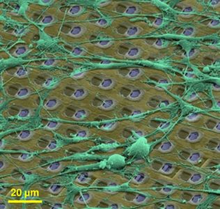
title = {Growing cells atop microelectronic chips: Interfacing electrogenic cells in vitro with CMOS-based microelectrode arrays},
author = {Andreas Hierlemann and Urs Frey and Sadik Hafizovic and Flavio Heer},
url = {http://ieeexplore.ieee.org/document/5594982/},
doi = {10.1109/JPROC.2010.2066532},
issn = {00189219},
year = {2011},
date = {2011-02-01},
journal = {Proceedings of the IEEE},
volume = {99},
number = {2},
pages = {252-284},
abstract = {Complementary semiconductor-metal-oxide (CMOS) technology is a very powerful technology that can be more or less directly interfaced to electrogenic cells, like heart or brain cells in vitro. To this end, the cells are cultured directly atop the CMOS chips, which usually undergo dedicated postprocessing to obtain a reliable bidirectional interface via noble-metal microelectrodes or high-k dielectrics. The big advantages of using CMOS integrated circuits (ICs) include connectivity, the possibility to address a large number of microelectrodes on a tiny chip, and signal quality, the possibility to condition small signals right at the spot of their generation. CMOS will be demonstrated to constitute an enabling technology that opens a route to high-spatio-temporal-resolution and low-noise electrophysiological recordings from a variety of biological preparations, such as brain slices, or cultured cardiac and brain cells. The recording technique is extracellular and noninvasive, and the CMOS chips do not leak out any toxic compounds, so that the cells remain viable for extended times. In turn, the CMOS chips have been demonstrated to survive several months of culturing while being fully immersed in saline solution and being exposed to cellular metabolic products. The latter requires dedicated passivation and packaging techniques as will be shown. Fully integrated, monolithic microelectrode systems, which feature large numbers of tightly spaced microelectrodes and the associated circuitry units for bidirectional interaction (stimulation and recording), will be in the focus of this review. The respective dense microelectrode arrays (MEAs) with small pixels enable subcellular-resolution investigation of regions of interest in, e.g., neurobiological preparations, and, at the same time, the large number of electrodes allows for studying the activity of entire neuronal networks . Application areas include neuroscience, as the devices enable fundamental neurophysiological insights at the cellular and circuit level, as well as medical diagnostics and pharmacology.},
keywords = {ETH-CMOS-MEA, MEA Technology, Review},
pubstate = {published},
tppubtype = {article}
}
2010
title = {Compact voltage and current stimulation buffer for high-density microelectrode arrays},
author = {Paolo Livi and Flavio Heer and Urs Frey and Douglas J Bakkum and Andreas Hierlemann},
url = {http://ieeexplore.ieee.org/document/5617318/},
doi = {10.1109/TBCAS.2010.2080676},
issn = {19324545},
year = {2010},
date = {2010-11-01},
journal = {IEEE Transactions on Biomedical Circuits and Systems},
volume = {4},
number = {6},
pages = {372-378},
abstract = {We report on a compact (0.02 mm2 ) buffer for both voltage and current stimulation of electrogenic cells on a complementary metal-oxide semiconductor microelectrode array. In voltage mode, the circuit is a high-current class-AB voltage follower, based on a local common-mode feedback (LCMFB) amplifier. In current mode, the circuit is a current conveyor of type II, using the same LCMFB amplifier with cascode stages to increase the gain. The circuit shows good linearity in the 0.5-3.5 V input range and has extensively been used for stimulation of neuronal cultures.},
keywords = {ETH-CMOS-MEA, MEA Technology, Stimulation},
pubstate = {published},
tppubtype = {article}
}
title = {Switch-matrix-based high-density microelectrode array in CMOS technology},
author = {Urs Frey and Jan Sedivy and Flavio Heer and Rene Pedron and Marco Ballini and Jan Müller and Douglas J Bakkum and Sadik Hafizovic and Francesca D Faraci and Frauke Greve and Kay Uwe Kirstein and Andreas Hierlemann},
url = {http://ieeexplore.ieee.org/document/5405139/},
doi = {10.1109/JSSC.2009.2035196},
issn = {00189200},
year = {2010},
date = {2010-02-02},
journal = {IEEE Journal of Solid-State Circuits},
volume = {45},
number = {2},
pages = {467-482},
abstract = {We report on a CMOS-based microelectrode array (MEA) featuring 11, 011 metal electrodes and 126 channels, each of which comprises recording and stimulation electronics, for extracellular bidirectional communication with electrogenic cells, such as neurons or cardiomyocytes. The important features include: (i) high spatial resolution at (sub)cellular level with 3150 electrodes per mm2 (electrode diameter 7 um, electrode pitch 18 um); (ii) a reconflgurable routing of the recording sites to the 126 channels; and (iii) low noise levels.},
keywords = {ETH-CMOS-MEA, MEA Technology},
pubstate = {published},
tppubtype = {article}
}
2007

title = {A perforated CMOS microchip platform for immobilization and activity monitoring of electrogenic cells},
author = {Frauke Greve and Jan Lichtenberg and Kay Uwe Kirstein and Urs Frey and Jean Claude Perriard and Andreas Hierlemann},
url = {http://iopscience.iop.org/article/10.1088/0960-1317/17/3/007/},
doi = {10.1088/0960-1317/17/3/007},
issn = {0960-1317},
year = {2007},
date = {2007-01-30},
journal = {Journal of Micromechanics and Microengineering},
volume = {17},
number = {3},
pages = {462-471},
abstract = {CMOS-based microelectrode systems offer decisive advantages over conventional micro-electrode arrays, which include the possibility to perform on-chip signal conditioning or to efficiently use larger numbers of electrodes to obtain statistically relevant data, e.g., in pharmacological drug screening. A larger number of electrodes can only be realized with the help of on-chip multiplexing and readout schemes, which require integrated electronics. Another fundamental issue in performing high-fidelity recordings from electrogenic cells is a good electrical coupling between the cells and the microelectrodes, in particular, since the recorded extracellular signals are in the range of only 10–1000 µV. In this paper we present the first CMOS microelectrode system with integrated micromechanical cell-placement features fabricated in a commercial CMOS process with subsequent post-CMOS bulk micromachining. This new microdevice aims at enabling the precise placement of single cells in the center of the electrodes to ensure an efficient use of the available electrodes, even for low-density cell cultures. Small through-chip holes have been generated at the metal-electrode sites by using a combination of bulk micromachining and reactive-ion etching. These holes act as orifices so that cell immobilization can be achieved by means of pneumatic anchoring. The chip additionally hosts integrated circuitry, i.e., multiplexers to select the respective readout electrodes, an amplifier with selectable gain (2×, 10×, 100×), and a high-pass filter (100 Hz cut-off). In this paper we show that electrical signals from most of the electrodes can be recorded, even in low-density cultures of neonatal rat cardiomyocytes, by using perforated metal electrodes and by applying a small underpressure from the backside of the chip. The measurements evidenced that, in most cases, about 90% of the electrodes were covered with single cells, approximately 4% were covered with more than one cell due to clustering and approximately 6% were not covered with any cell, mostly as a consequence of orifice clogging. After 4 days of culturing, the cells were still in place on the electrodes so that the cell electrical activity could be measured using the on-chip circuitry. Measured signal amplitudes were in the range of 500–700 µV, while the input-referred noise of the readout was below 15 µVrms (100 Hz–4 kHz bandwidth). We report on the development and fabrication of this new cell-biological tool and present first results collected during the characterization and evaluation of the chip. The recordings of electrical potentials of neonatal rat cardiomyocytes after several days in vitro, which, on the one hand, were conventionally cultured (no pneumatic anchoring) and, on the other hand, were anchored and immobilized, will be detailed.},
keywords = {2D Neuronal Culture, Cardiomyocytes, ETH-CMOS-MEA, MEA Technology},
pubstate = {published},
tppubtype = {article}
}
2006
title = {CMOS microelectrode array for bidirectional interaction with neuronal networks},
author = {Flavio Heer and Sadik Hafizovic and Wendy Franks and Axel Blau and Christiane Ziegler and Andreas Hierlemann},
url = {http://ieeexplore.ieee.org/document/1644873/},
doi = {10.1109/JSSC.2006.873677},
issn = {0018-9200},
year = {2006},
date = {2006-07-07},
journal = {IEEE Journal of Solid-State Circuits},
volume = {41},
number = {7},
pages = {1620-1629},
abstract = {A CMOS metal-electrode-based micro system for bidirectional communication (stimulation and recording) with neuronal cells in vitro is presented. The chip overcomes the interconnect challenge that limits today's bidirectional microelectrode arrays. The microsystem has been fabricated in an industrial CMOS technology with several post-CMOS processing steps to realize 128 biocompatible electrodes and to ensure chip stability in physiological saline. The system comprises all necessary control circuitry and on-chip A/D and D/A conversion. A modular design has been implemented, where individual stimulation- and signal-conditioning circuitry units are associated with each electrode. Stimulation signals with a resolution of 8 bits can be sent to any subset of electrodes at a rate of 60 kHz, while all electrodes of the chip are continuously sampled at a rate of 20 kHz. The circuitry at each electrode can be individually reset to its operating point in order to suppress artifacts evoked by the stimulation pulses. Biological measurements from cultured neuronal networks originating from dissociated cortical tissue of fertilized chicken eggs with amplitudes of up to 500 muVpp are presented.},
keywords = {2D Neuronal Culture, ETH-CMOS-MEA, MEA Technology, Neuronal Networks, Stimulation},
pubstate = {published},
tppubtype = {article}
}
2005
title = {Impedance characterization and modeling of electrodes for biomedical applications},
author = {Wendy Franks and Iwan Schenker and Patrik Schmutz and Andreas Hierlemann},
url = {http://ieeexplore.ieee.org/document/1440608/},
doi = {10.1109/TBME.2005.847523},
issn = {00189294},
year = {2005},
date = {2005-06-13},
journal = {IEEE Transactions on Biomedical Engineering},
volume = {52},
number = {7},
pages = {1295-1302},
abstract = {A low electrode-electrolyte impedance interface is critical in the design of electrodes for biomedical applications. To design low-impedance interfaces a complete understanding of the physical processes contributing to the impedance is required. In this work a model describing these physical processes is validated and extended to quantify the effect of organic coatings and incubation time. Electrochemical impedance spectroscopy has been used to electrically characterize the interface for various electrode materials: platinum, platinum black, and titanium nitride; and varying electrode sizes: 1 cm2, and 900 mu m2. An equivalent circuit model comprising an interface capacitance, shunted by a charge transfer resistance, in series with the solution resistance has been fitted to the experimental results. Theoretical equations have been used to calculate the interface capacitance impedance and the solution resistance, yielding results that correspond well with the fitted parameter values, thereby confirming the validity of the equations. The effect of incubation time, and two organic cell-adhesion promoting coatings, poly-L-lysine and laminin, on the interface impedance has been quantified using the model. This demonstrates the benefits of using this model in developing better understanding of the physical processes occurring at the interface in more complex, biomedically relevant situations.},
keywords = {MEA Technology},
pubstate = {published},
tppubtype = {article}
}
2004
title = {Cell-based CMOS sensor and actuator arrays},
author = {Martin Jenkner and Marco Tartagni and Andreas Hierlemann and Roland Thewes},
url = {http://ieeexplore.ieee.org/document/1362853/},
doi = {10.1109/JSSC.2004.837082},
issn = {00189200},
year = {2004},
date = {2004-11-30},
journal = {IEEE Journal of Solid-State Circuits},
volume = {39},
number = {12},
pages = {2431-2437},
abstract = {In recent years, increasing knowledge about in vitro cell handling and culturing has encouraged a variety of CMOS-based approaches to stimulate and detect electrical activity of biological cells. This paper outlines in a topical review the scope of cell-based biosensors and actuators for in vitro applications ranging from single-cell detection to multisite probing of complex neural tissue. Recent examples are selected to demonstrate how standard CMOS processes have been used to engineer arrays with different functionality.},
keywords = {MEA Technology, Review},
pubstate = {published},
tppubtype = {article}
}
title = {CMOS microelectrode array for the monitoring of electrogenic cells},
author = {Flavio Heer and Wendy Franks and Axel Blau and S Taschini and Christiane Ziegler and Andreas Hierlemann and Henry Baltes},
url = {http://www.sciencedirect.com/science/article/pii/S0956566304000806?via%3Dihub},
doi = {10.1016/j.bios.2004.02.006},
issn = {0956-5663},
year = {2004},
date = {2004-03-19},
journal = {Biosensors & Bioelectronics},
volume = {20},
number = {2},
pages = {358-366},
abstract = {Signal degradation and an array size dictated by the number of available interconnects are the two main limitations inherent to standalone microelectrode arrays (MEAs). A new biochip consisting of an array of microelectrodes with fully-integrated analog and digital circuitry realized in an industrial CMOS process addresses these issues. The device is capable of on-chip signal filtering for improved signal-to-noise ratio (SNR), on-chip analog and digital conversion, and multiplexing, thereby facilitating simultaneous stimulation and recording of electrogenic cell activity. The designed electrode pitch of 250 mu m significantly limits the space available for circuitry: a repeated unit of circuitry associated with each electrode comprises a stimulation buffer and a bandpass filter for readout. The bandpass filter has corner frequencies of 100 Hz and 50 kHz, and a gain of 1000. Stimulation voltages are generated from an 8-bit digital signal and converted to an analog signal at a frequency of 120 kHz. Functionality of the read-out circuitry is demonstrated by the measurement of cardiomyocyte activity. The microelectrode is realized in a shifted design for flexibility and biocompatibility. Several microelectrode materials (platinum, platinum black and titanium nitride) have been electrically characterized. An equivalent circuit model, where each parameter represents a macroscopic physical quantity contributing to the interface impedance, has been successfully fitted to experimental results.},
keywords = {ETH-CMOS-MEA, MEA Technology},
pubstate = {published},
tppubtype = {article}
}