 日本語
日本語 繁體中文
繁體中文 简体中文
简体中文 English
Publications
Selected Publications

High-resolution CMOS MEA platform to study neurons at subcellular, cellular, and network levels

Presenting measurements of neuronal preparations with a novel CMOS-based microelectrode array at high-spatiotemporal-resolution on subcellular, cellular, and network level.
J. Müller, M. Ballini, P. Livi, Y. Chen, M. Radivojevic, A. Shadmani, V. Viswam, I. L. Jones, M. Fiscella, R. Diggelmann, A. Stettler, U. Frey, D. J. Bakkum, and A. Hierlemann, “High-resolution CMOS MEA platform to study neurons at subcellular, cellular, and network levels,” Lab Chip, vol. 15, no. 13, pp. 2767–2780, May 2015.
Revealing Neuronal Function through Microelectrode Array Recordings

Reviewing the current understanding of microelectrode signals and the techniques for analyzing them, with focus on the ongoing advancements in microelectrode technology (in vivo and in vitro) and recent advanced microelectrode array measurement methods that facilitate the understanding of single neurons and network function.
M. E. J. Obien, K. Deligkaris, T. Bullmann, D. J. Bakkum, and U. Frey, “Revealing Neuronal Function through Microelectrode Array Recordings,” Front. Neurosci., 8:423, Jan 2015.

A 1024-Channel CMOS Microelectrode Array With 26,400 Electrodes for Recording and Stimulation of Electrogenic Cells In Vitro
A high-resolution CMOS-based microelectrode array featuring 1,024 low-noise readout channels, 26,400 electrodes at a density of 3,265 electrodes per mm2, including on-chip 10bit ADCs and consuming only 75 mW.
M. Ballini, J. Muller, P. Livi, Y. Chen, U. Frey, A. Stettler, A. Shadmani, V. Viswam, I. L. Jones, D. Jackel, M. Radivojevic, M. K. Lewandowska, W. Gong, M. Fiscella, D. J. Bakkum, F. Heer, and A. Hierlemann, “A 1024-Channel CMOS Microelectrode Array With 26,400 Electrodes for Recording and Stimulation of Electrogenic Cells In Vitro,” IEEE Journal of Solid-State Circuits, vol. 49, no. 11, pp. 2705-2719, 2014.

Tracking axonal action potential propagation on a high-density microelectrode array across hundreds of sites
Demonstrating a method to electrically visualize action potential propagation on axons and revealing
large variations in velocity.
D. J. Bakkum, U. Frey, M. Radivojevic, T. L. Russell, J. Muller, M. Fiscella, H. Takahashi, and A. Hierlemann, “Tracking axonal action potential propagation on a high-density microelectrode array across hundreds of sites,” Nature Communications, 4:2181, Jul 2013.

Microelectronic System for High-Resolution Mapping of Extracellular Electric Fields Applied to Brain Slices
Recording and modeling extracellular action potentials of Purkinje cells at subcellular resolution.
U. Frey, U. Egert, F. Heer, S. Hafizovic, and A. Hierlemann, “Microelectronic System for High-Resolution Mapping of Extracellular Electric Fields Applied to Brain Slices,” Biosensors and Bioelectronics, vol. 24, no. 7, pp. 2191-2198, 2009.

Modulation of Cardiomyocyte Electrical Properties Using Regulated Bone Morphogenetic Protein-2 Expression
Controlling BMP-2 expression to modulate the electrophysiological properties of cardiomyocytes using an HD-MEA for detailed monitoring.
C. D. Sanchez-Bustamante, U. Frey, J. M. Kelm, A. Hierlemann, and M. Fussenegger,
“Modulation of Cardiomyocyte Electrical Properties Using Regulated Bone Morphogenetic Protein-2 Expression,” Tissue Engineering Part A, vol. 14, no. 12, pp. 1969-1988, 2008.
All Publications
| 21. | Bartram, Julian; Franke, Felix; Kumar, Sreedhar Saseendran; Buccino, Alessio Paolo; Xue, Xiaohan; Gänswein, Tobias; Schröter, Manuel; Kim, Taehoon; Kasuba, Krishna Chaitanya; Hierlemann, Andreas: Parallel reconstruction of the excitatory and inhibitory inputs received by single neurons reveals the synaptic basis of recurrent spiking. In: eLife, 2023. (Type: Journal Article | Abstract | Links | BibTeX) @article{Bartram2023b, title = {Parallel reconstruction of the excitatory and inhibitory inputs received by single neurons reveals the synaptic basis of recurrent spiking}, author = {Julian Bartram and Felix Franke and Sreedhar Saseendran Kumar and Alessio Paolo Buccino and Xiaohan Xue and Tobias Gänswein and Manuel Schröter and Taehoon Kim and Krishna Chaitanya Kasuba and Andreas Hierlemann}, url = {https://elifesciences.org/reviewed-preprints/86820}, doi = {10.7554/eLife.86820}, year = {2023}, date = {2023-05-17}, journal = {eLife}, abstract = {Self-sustained recurrent activity in cortical networks is thought to be important for multiple crucial processes, including circuit development and homeostasis. Yet, the precise relationship between the synaptic input patterns and the spiking output of individual neurons remains largely unresolved. Here, we developed, validated and applied a novel in vitro experimental platform and analytical procedures that provide – for individual neurons – simultaneous excitatory and inhibitory synaptic activity estimates during recurrent network activity. Our approach combines whole-network high-density microelectrode array (HD-MEA) recordings from rat neuronal cultures with patch clamping and enables a comprehensive mapping and characterization of active incoming connections to single postsynaptic neurons. We found that, during network states with excitation(E)-inhibition(I) balance, postsynaptic spiking coincided precisely with the maxima of fast fluctuations in the input E/I ratio. These spike-associated E/I ratio escalations were largely due to a rapid bidirectional change in synaptic inhibition that was modulated by the network-activity level. Our approach also uncovered the underlying circuit architecture and we show that individual neurons received a few key inhibitory connections – often from special hub neurons – that were instrumental in controlling postsynaptic spiking. Balanced network theory predicts dynamical regimes governed by small and rapid input fluctuation and featuring a fast neuronal responsiveness. Our findings – obtained in self-organized neuronal cultures – suggest that the emergence of these favorable regimes and associated network architectures is an inherent property of cortical networks in general.}, keywords = {}, pubstate = {published}, tppubtype = {article} } Self-sustained recurrent activity in cortical networks is thought to be important for multiple crucial processes, including circuit development and homeostasis. Yet, the precise relationship between the synaptic input patterns and the spiking output of individual neurons remains largely unresolved. Here, we developed, validated and applied a novel in vitro experimental platform and analytical procedures that provide – for individual neurons – simultaneous excitatory and inhibitory synaptic activity estimates during recurrent network activity. Our approach combines whole-network high-density microelectrode array (HD-MEA) recordings from rat neuronal cultures with patch clamping and enables a comprehensive mapping and characterization of active incoming connections to single postsynaptic neurons. We found that, during network states with excitation(E)-inhibition(I) balance, postsynaptic spiking coincided precisely with the maxima of fast fluctuations in the input E/I ratio. These spike-associated E/I ratio escalations were largely due to a rapid bidirectional change in synaptic inhibition that was modulated by the network-activity level. Our approach also uncovered the underlying circuit architecture and we show that individual neurons received a few key inhibitory connections – often from special hub neurons – that were instrumental in controlling postsynaptic spiking. Balanced network theory predicts dynamical regimes governed by small and rapid input fluctuation and featuring a fast neuronal responsiveness. Our findings – obtained in self-organized neuronal cultures – suggest that the emergence of these favorable regimes and associated network architectures is an inherent property of cortical networks in general. |
| 22. | Duru, Jens; Maurer, Benedikt; Doran, Ciara Giles; Jelitto, Robert; Küchler, Joël; Ihle, Stephan J; Ruff, Tobias; John, Robert; Genocchi, Barbara; Vörös, János: Investigation of the input-output relationship of engineered neural networks using high-density microelectrode arrays. In: SSRN, 2023. (Type: Journal Article | Abstract | Links | BibTeX) @article{Duru2023, title = {Investigation of the input-output relationship of engineered neural networks using high-density microelectrode arrays}, author = {Jens Duru and Benedikt Maurer and Ciara Giles Doran and Robert Jelitto and Joël Küchler and Stephan J. Ihle and Tobias Ruff and Robert John and Barbara Genocchi and János Vörös}, url = {https://www.ssrn.com/abstract=4427959}, doi = {DOI: 10.2139/ssrn.4427959}, year = {2023}, date = {2023-04-24}, journal = {SSRN}, abstract = {Bottom-up neuroscience utilizes small, engineered biological neural networks to study neuronal activity in systems of reduced complexity. We present a platform that establishes up to six independent networks formed by primary rat neurons on planar complementary metal–oxide–semiconductor (CMOS) microelectrode arrays (MEAs). We introduce an approach that allows repetitive stimulation and recording of network activity at any of the over 700 electrodes underlying a network. We demonstrate that the continuous application of a repetitive super-threshold stimulus yields a reproducible network answer within a 15 ms post-stimulus window. This response can be tracked with high spatiotemporal resolution across the whole extent of the network. Moreover, we show that the location of the stimulation plays a significant role in the networks’ early response to the stimulus. By applying a stimulation pattern to all network-underlying electrodes in sequence, the sensitivity of the whole network to the stimulus can be visualized. We demonstrate that microchannels reduce the voltage stimulation threshold and induce the strongest network response. By varying the stimulation amplitude and frequency we reveal discrete network transition points. Finally, we introduce vector fields to follow stimulation-induced spike propagation pathways within the network. Overall we show that our defined neural networks on CMOS MEAs enable us to elicit highly reproducible activity patterns that can be precisely modulated by stimulation amplitude, stimulation frequency and the site of stimulation.}, keywords = {}, pubstate = {published}, tppubtype = {article} } Bottom-up neuroscience utilizes small, engineered biological neural networks to study neuronal activity in systems of reduced complexity. We present a platform that establishes up to six independent networks formed by primary rat neurons on planar complementary metal–oxide–semiconductor (CMOS) microelectrode arrays (MEAs). We introduce an approach that allows repetitive stimulation and recording of network activity at any of the over 700 electrodes underlying a network. We demonstrate that the continuous application of a repetitive super-threshold stimulus yields a reproducible network answer within a 15 ms post-stimulus window. This response can be tracked with high spatiotemporal resolution across the whole extent of the network. Moreover, we show that the location of the stimulation plays a significant role in the networks’ early response to the stimulus. By applying a stimulation pattern to all network-underlying electrodes in sequence, the sensitivity of the whole network to the stimulus can be visualized. We demonstrate that microchannels reduce the voltage stimulation threshold and induce the strongest network response. By varying the stimulation amplitude and frequency we reveal discrete network transition points. Finally, we introduce vector fields to follow stimulation-induced spike propagation pathways within the network. Overall we show that our defined neural networks on CMOS MEAs enable us to elicit highly reproducible activity patterns that can be precisely modulated by stimulation amplitude, stimulation frequency and the site of stimulation. |
| 23. | Xu, He Jax; Yao, Yao; Yao, Fenyong; Chen, Jiehui; Li, Meishi; Yang, Xianfa; Li, Sheng; Lu, Fangru; Hu, Ping; He, Shuijin; Peng, Guangdun; Jing, Naihe: Generation of functional posterior spinal motor neurons from hPSCs-derived human spinal cord neural progenitor cells. In: Cell Regeneration, 2023. (Type: Journal Article | Abstract | Links | BibTeX) @article{Xu2023, title = {Generation of functional posterior spinal motor neurons from hPSCs-derived human spinal cord neural progenitor cells}, author = {He Jax Xu and Yao Yao and Fenyong Yao and Jiehui Chen and Meishi Li and Xianfa Yang and Sheng Li and Fangru Lu and Ping Hu and Shuijin He and Guangdun Peng and Naihe Jing}, url = {https://cellregeneration.springeropen.com/articles/10.1186/s13619-023-00159-6}, doi = {10.1186/s13619-023-00159-6}, year = {2023}, date = {2023-03-23}, journal = {Cell Regeneration}, abstract = {Spinal motor neurons deficiency results in a series of devastating disorders such as amyotrophic lateral sclerosis (ALS), spinal muscular atrophy (SMA) and spinal cord injury (SCI). These disorders are currently incurable, while human pluripotent stem cells (hPSCs)-derived spinal motor neurons are promising but suffered from inappropriate regional identity and functional immaturity for the study and treatment of posterior spinal cord related injuries. In this study, we have established human spinal cord neural progenitor cells (hSCNPCs) via hPSCs differentiated neuromesodermal progenitors (NMPs) and demonstrated the hSCNPCs can be continuously expanded up to 40 passages. hSCNPCs can be rapidly differentiated into posterior spinal motor neurons with high efficiency. The functional maturity has been examined in detail. Moreover, a co-culture scheme which is compatible for both neural and muscular differentiation is developed to mimic the neuromuscular junction (NMJ) formation in vitro. Together, these studies highlight the potential avenues for generating clinically relevant spinal motor neurons and modeling neuromuscular diseases through our defined hSCNPCs.}, keywords = {}, pubstate = {published}, tppubtype = {article} } Spinal motor neurons deficiency results in a series of devastating disorders such as amyotrophic lateral sclerosis (ALS), spinal muscular atrophy (SMA) and spinal cord injury (SCI). These disorders are currently incurable, while human pluripotent stem cells (hPSCs)-derived spinal motor neurons are promising but suffered from inappropriate regional identity and functional immaturity for the study and treatment of posterior spinal cord related injuries. In this study, we have established human spinal cord neural progenitor cells (hSCNPCs) via hPSCs differentiated neuromesodermal progenitors (NMPs) and demonstrated the hSCNPCs can be continuously expanded up to 40 passages. hSCNPCs can be rapidly differentiated into posterior spinal motor neurons with high efficiency. The functional maturity has been examined in detail. Moreover, a co-culture scheme which is compatible for both neural and muscular differentiation is developed to mimic the neuromuscular junction (NMJ) formation in vitro. Together, these studies highlight the potential avenues for generating clinically relevant spinal motor neurons and modeling neuromuscular diseases through our defined hSCNPCs. |
| 24. | Radivojevic, Milos; Punga, Anna Rostedt: Functional imaging of conduction dynamics in cortical and spinal axons. In: BioRxiv, 2023. (Type: Journal Article | Abstract | Links | BibTeX) @article{Radivojevic2023, title = {Functional imaging of conduction dynamics in cortical and spinal axons}, author = {Milos Radivojevic and Anna Rostedt Punga}, url = {https://www.biorxiv.org/content/10.1101/2023.02.28.530461v1}, doi = {https://doi.org/10.1101/2023.02.28.530461}, year = {2023}, date = {2023-03-01}, journal = {BioRxiv}, abstract = {Mammalian axons are specialized for transmitting action potentials to targets within the central and peripheral nervous system. A growing body of evidence suggests that, besides signal conduction, axons play essential roles in neural information processing, and their malfunctions are common hallmarks of neurodegenerative diseases. The technologies available to study axonal function and structure integrally limit the comprehension of axon neurobiology. High-density microelectrode arrays (HD-MEAs) allow for accessing axonal action potentials at high spatiotemporal resolution, but provide no insights on axonal morphology. Here we demonstrate a method for electrical visualization of axonal morphologies based on extracellular action potentials recorded from cortical and motor neurons using HD-MEAs. The method enabled us to reconstruct up to 5-centimeter-long axonal arbors and directly monitor axonal conduction across thousands of recording sites. We reconstructed 1.86 meters of cortical and spinal axons in total and found specific features in their structure and function.}, keywords = {}, pubstate = {published}, tppubtype = {article} } Mammalian axons are specialized for transmitting action potentials to targets within the central and peripheral nervous system. A growing body of evidence suggests that, besides signal conduction, axons play essential roles in neural information processing, and their malfunctions are common hallmarks of neurodegenerative diseases. The technologies available to study axonal function and structure integrally limit the comprehension of axon neurobiology. High-density microelectrode arrays (HD-MEAs) allow for accessing axonal action potentials at high spatiotemporal resolution, but provide no insights on axonal morphology. Here we demonstrate a method for electrical visualization of axonal morphologies based on extracellular action potentials recorded from cortical and motor neurons using HD-MEAs. The method enabled us to reconstruct up to 5-centimeter-long axonal arbors and directly monitor axonal conduction across thousands of recording sites. We reconstructed 1.86 meters of cortical and spinal axons in total and found specific features in their structure and function. |
| 25. | Cai, Hongwei; Ao, Zheng; Tian, Chunhui; Wu, Zhuhao; Liu, Hongcheng; Tchieu, Jason; Gu, Mingxia; Mackie, Ken; and Guo, Feng: Brain Organoid Computing for Artificial Intelligence. In: bioRxiv, 2023. (Type: Journal Article | Abstract | Links | BibTeX) @article{Cai2023, title = {Brain Organoid Computing for Artificial Intelligence}, author = {Hongwei Cai and Zheng Ao and Chunhui Tian and Zhuhao Wu and Hongcheng Liu and Jason Tchieu and Mingxia Gu and Ken Mackie and and Feng Guo}, url = {https://www.biorxiv.org/content/10.1101/2023.02.28.530502v1}, doi = {10.1101/2023.02.28.530502}, year = {2023}, date = {2023-03-01}, journal = {bioRxiv}, abstract = {Brain-inspired hardware emulates the structure and working principles of a biological brain and may address the hardware bottleneck for fast-growing artificial intelligence (AI). Current brain-inspired silicon chips are promising but still limit their power to fully mimic brain function for AI computing. Here, we develop Brainoware, living AI hardware that harnesses the computation power of 3D biological neural networks in a brain organoid. Brain-like 3D in vitro cultures compute by receiving and sending information via a multielectrode array. Applying spatiotemporal electrical stimulation, this approach not only exhibits nonlinear dynamics and fading memory properties but also learns from training data. Further experiments demonstrate real-world applications in solving non-linear equations. This approach may provide new insights into AI hardware. }, keywords = {}, pubstate = {published}, tppubtype = {article} } Brain-inspired hardware emulates the structure and working principles of a biological brain and may address the hardware bottleneck for fast-growing artificial intelligence (AI). Current brain-inspired silicon chips are promising but still limit their power to fully mimic brain function for AI computing. Here, we develop Brainoware, living AI hardware that harnesses the computation power of 3D biological neural networks in a brain organoid. Brain-like 3D in vitro cultures compute by receiving and sending information via a multielectrode array. Applying spatiotemporal electrical stimulation, this approach not only exhibits nonlinear dynamics and fading memory properties but also learns from training data. Further experiments demonstrate real-world applications in solving non-linear equations. This approach may provide new insights into AI hardware. |
| 26. | Kim, Eunhee; Jeon, Sungwoong; Yang, Yoon-Sil; Jin, Chaewon; Kim, Jin-young; Oh, Yong- Seok; Rah, Jong-Cheol; and Choi, Hongsoo: A Neurospheroid-Based Microrobot for Targeted Neural Connections in a Hippocampal Slice. In: Advanced Materials, 2023. (Type: Journal Article | Abstract | Links | BibTeX) @article{EunheeKim2023, title = {A Neurospheroid-Based Microrobot for Targeted Neural Connections in a Hippocampal Slice}, author = {Eunhee Kim and Sungwoong Jeon and Yoon-Sil Yang and Chaewon Jin and Jin-young Kim and Yong- Seok Oh and Jong-Cheol Rah and and Hongsoo Choi}, url = {https://onlinelibrary.wiley.com/doi/10.1002/adma.202208747?af=R}, doi = {https://doi.org/10.1002/adma.202208747}, year = {2023}, date = {2023-01-14}, journal = {Advanced Materials}, abstract = {Functional restoration by the re-establishment of cellular or neural connections remains a major challenge in targeted cell therapy and regenerative medicine. Recent advances in magnetically powered microrobots have shown potential for use in controlled and targeted cell therapy. In this study, a magnetic neurospheroid (Mag-Neurobot) that could form both structural and functional connections with an organotypic hippocampal slice (OHS) was assessed using an ex vivo model as a bridge toward in vivo application. The Mag-Neurobot consists of hippocampal neurons and superparamagnetic nanoparticles (SPIONs); it is precisely and skillfully manipulated by an external magnetic field. Furthermore, the results of patch-clamp recordings of hippocampal neurons indicated that neither the neuronal excitabilities nor the synaptic functions of SPION-loaded cells were significantly affected. Analysis of neural activity propagation using high-density multi-electrode arrays showed that the delivered Mag-Neurobot was functionally connected with the OHS. The applications of this study include functional verification for targeted cell delivery through the characterization of novel synaptic connections and the functionalities of transported and transplanted cells. The success of the Mag-Neurobot opens up new avenues of research and application; it offers a test platform for functional neural connections and neural regenerative processes through cell transplantation.}, keywords = {}, pubstate = {published}, tppubtype = {article} } Functional restoration by the re-establishment of cellular or neural connections remains a major challenge in targeted cell therapy and regenerative medicine. Recent advances in magnetically powered microrobots have shown potential for use in controlled and targeted cell therapy. In this study, a magnetic neurospheroid (Mag-Neurobot) that could form both structural and functional connections with an organotypic hippocampal slice (OHS) was assessed using an ex vivo model as a bridge toward in vivo application. The Mag-Neurobot consists of hippocampal neurons and superparamagnetic nanoparticles (SPIONs); it is precisely and skillfully manipulated by an external magnetic field. Furthermore, the results of patch-clamp recordings of hippocampal neurons indicated that neither the neuronal excitabilities nor the synaptic functions of SPION-loaded cells were significantly affected. Analysis of neural activity propagation using high-density multi-electrode arrays showed that the delivered Mag-Neurobot was functionally connected with the OHS. The applications of this study include functional verification for targeted cell delivery through the characterization of novel synaptic connections and the functionalities of transported and transplanted cells. The success of the Mag-Neurobot opens up new avenues of research and application; it offers a test platform for functional neural connections and neural regenerative processes through cell transplantation. |
| 27. | Sato, Yuya; Yamamoto, Hideaki; Kato, Hideyuki; Tanii, Takashi; Sato, Shigeo; Hirano-Iwata, Ayumi: Microfluidic cell engineering on high-density microelectrode arrays for assessing structure-function relationships in living neuronal networks. In: Frontiers in Neuroscience, 2023. (Type: Journal Article | Abstract | Links | BibTeX) @article{Sato2023, title = {Microfluidic cell engineering on high-density microelectrode arrays for assessing structure-function relationships in living neuronal networks}, author = {Yuya Sato and Hideaki Yamamoto and Hideyuki Kato and Takashi Tanii and Shigeo Sato and Ayumi Hirano-Iwata}, url = {https://www.frontiersin.org/articles/10.3389/fnins.2022.943310/full}, doi = {doi: 10.3389/fnins.2022.943310}, year = {2023}, date = {2023-01-09}, journal = {Frontiers in Neuroscience}, abstract = {Neuronal networks in dissociated culture combined with cell engineering technology offer a pivotal platform to constructively explore the relationship between structure and function in living neuronal networks. Here, we fabricated defined neuronal networks possessing a modular architecture on high-density microelectrode arrays (HD-MEAs), a state-of-the-art electrophysiological tool for recording neural activity with high spatial and temporal resolutions. We first established a surface coating protocol using a cell-permissive hydrogel to stably attach a polydimethylsiloxane microfluidic film on the HD-MEA. We then recorded the spontaneous neural activity of the engineered neuronal network, which revealed an important portrait of the engineered neuronal network–modular architecture enhances functional complexity by reducing the excessive neural correlation between spatially segregated modules. The results of this study highlight the impact of HD- MEA recordings combined with cell engineering technologies as a novel tool in neuroscience to constructively assess the structure-function relationships in neuronal networks.}, keywords = {}, pubstate = {published}, tppubtype = {article} } Neuronal networks in dissociated culture combined with cell engineering technology offer a pivotal platform to constructively explore the relationship between structure and function in living neuronal networks. Here, we fabricated defined neuronal networks possessing a modular architecture on high-density microelectrode arrays (HD-MEAs), a state-of-the-art electrophysiological tool for recording neural activity with high spatial and temporal resolutions. We first established a surface coating protocol using a cell-permissive hydrogel to stably attach a polydimethylsiloxane microfluidic film on the HD-MEA. We then recorded the spontaneous neural activity of the engineered neuronal network, which revealed an important portrait of the engineered neuronal network–modular architecture enhances functional complexity by reducing the excessive neural correlation between spatially segregated modules. The results of this study highlight the impact of HD- MEA recordings combined with cell engineering technologies as a novel tool in neuroscience to constructively assess the structure-function relationships in neuronal networks. |
| 28. | Han, Xiaobo; Matsuda, Naoki; Ishibashi, Yuto; Odawara, Aoi; Takahashi, Sayuri; Tooi, Norie; Kinoshita, Koshi; Suzuki, Ikuro: A functional neuron maturation device provides convenient application on microelectrode array for neural network measurement. In: Biomaterials Research, 2022. (Type: Journal Article | Abstract | Links | BibTeX) @article{Han2022, title = {A functional neuron maturation device provides convenient application on microelectrode array for neural network measurement}, author = {Xiaobo Han and Naoki Matsuda and Yuto Ishibashi and Aoi Odawara and Sayuri Takahashi and Norie Tooi and Koshi Kinoshita and Ikuro Suzuki }, url = {https://biomaterialsres.biomedcentral.com/articles/10.1186/s40824-022-00324-z}, doi = {https://doi.org/10.1186/s40824-022-00324-z}, year = {2022}, date = {2022-12-20}, journal = {Biomaterials Research}, abstract = {Background Microelectrode array (MEA) systems are valuable for in vitro assessment of neurotoxicity and drug efficiency. However, several difficulties such as protracted functional maturation and high experimental costs hinder the use of MEA analysis requiring human induced pluripotent stem cells (hiPSCs). Neural network functional parameters are also needed for in vitro to in vivo extrapolation. Methods In the present study, we produced a cost effective nanofiber culture platform, the SCAD device, for long-term culture of hiPSC-derived neurons and primary peripheral neurons. The notable advantage of SCAD device is convenient application on multiple MEA systems for neuron functional analysis. Results We showed that the SCAD device could promote functional maturation of cultured hiPSC-derived neurons, and neurons responded appropriately to convulsant agents. Furthermore, we successfully analyzed parameters for in vitro to in vivo extrapolation, i.e., low-frequency components and synaptic propagation velocity of the signal, potentially reflecting neural network functions from neurons cultured on SCAD device. Finally, we measured the axonal conduction velocity of peripheral neurons. Conclusions: Neurons cultured on SCAD devices might constitute a reliable in vitro platform to investigate neuron functions, drug efficacy and toxicity, and neuropathological mechanisms by MEA.}, keywords = {}, pubstate = {published}, tppubtype = {article} } Background Microelectrode array (MEA) systems are valuable for in vitro assessment of neurotoxicity and drug efficiency. However, several difficulties such as protracted functional maturation and high experimental costs hinder the use of MEA analysis requiring human induced pluripotent stem cells (hiPSCs). Neural network functional parameters are also needed for in vitro to in vivo extrapolation. Methods In the present study, we produced a cost effective nanofiber culture platform, the SCAD device, for long-term culture of hiPSC-derived neurons and primary peripheral neurons. The notable advantage of SCAD device is convenient application on multiple MEA systems for neuron functional analysis. Results We showed that the SCAD device could promote functional maturation of cultured hiPSC-derived neurons, and neurons responded appropriately to convulsant agents. Furthermore, we successfully analyzed parameters for in vitro to in vivo extrapolation, i.e., low-frequency components and synaptic propagation velocity of the signal, potentially reflecting neural network functions from neurons cultured on SCAD device. Finally, we measured the axonal conduction velocity of peripheral neurons. Conclusions: Neurons cultured on SCAD devices might constitute a reliable in vitro platform to investigate neuron functions, drug efficacy and toxicity, and neuropathological mechanisms by MEA. |
| 29. | Tran, Hoang-Dai; Shin, Min-Kyoung; Denman, Charlotte; Han, Run-Run; Kuhn, Bernd; Arbuthnott, Gordon; Jo, Junghyun: Generation of Human Striatal-Midbrain Assembloids From Human Pluripotent Stem Cells to Model Alpha-Synuclein Propagation. In: Sneak Peek - Cell Press, 2022. (Type: Journal Article | Abstract | Links | BibTeX) @article{Tran2022, title = {Generation of Human Striatal-Midbrain Assembloids From Human Pluripotent Stem Cells to Model Alpha-Synuclein Propagation}, author = {Hoang-Dai Tran and Min-Kyoung Shin and Charlotte Denman and Run-Run Han and Bernd Kuhn and Gordon Arbuthnott and Junghyun Jo}, url = {https://papers.ssrn.com/sol3/papers.cfm?abstract_id=4288935}, doi = {http://dx.doi.org/10.2139/ssrn.4288935}, year = {2022}, date = {2022-12-05}, journal = {Sneak Peek - Cell Press}, abstract = {Animal models of the pathology of Parkinson’s disease (PD) have provided most of the treatments to date, but the disease is restricted to human patients. In vitro models using human pluripotent stem cell-derived neural organoids have provided improved access to study PD etiology. Here, we generated human striatal and midbrain organoids and assembled both regionalized neural organoids to form human striatal-midbrain assembloids (hSMAs), mimicking a part of basal ganglia. Both the nigrostriatal and striatonigral pathways are present and electrophysiologically active in the hSMAs. hSMA development in the presence of increased alpha-synuclein (α-syn) from SNCA overexpression, induced nigrostriatal system damage, which is typical of the disease. Using the α-syn-mKO2 reporter and bimolecular fluorescence complementation system, we demonstrated that fluorescent α-syn is transported from the striatal area tothe dopaminergic (DA) neurons of the midbrain area. Furthermore, insoluble α-syn aggregated over time in DA neurons similar to Lewy bodies in human patients. Such assembloids are a compelling new platform to develop novel PD therapeutic strategies.}, keywords = {}, pubstate = {published}, tppubtype = {article} } Animal models of the pathology of Parkinson’s disease (PD) have provided most of the treatments to date, but the disease is restricted to human patients. In vitro models using human pluripotent stem cell-derived neural organoids have provided improved access to study PD etiology. Here, we generated human striatal and midbrain organoids and assembled both regionalized neural organoids to form human striatal-midbrain assembloids (hSMAs), mimicking a part of basal ganglia. Both the nigrostriatal and striatonigral pathways are present and electrophysiologically active in the hSMAs. hSMA development in the presence of increased alpha-synuclein (α-syn) from SNCA overexpression, induced nigrostriatal system damage, which is typical of the disease. Using the α-syn-mKO2 reporter and bimolecular fluorescence complementation system, we demonstrated that fluorescent α-syn is transported from the striatal area tothe dopaminergic (DA) neurons of the midbrain area. Furthermore, insoluble α-syn aggregated over time in DA neurons similar to Lewy bodies in human patients. Such assembloids are a compelling new platform to develop novel PD therapeutic strategies. |
| 30. | Akarca, Danyal; Dunn, Alexander W E; Hornauer, Philipp J; Ronchi, Silvia; Fiscella, Michele; Wang, Congwei; Terrigno, Marco; Jagasia, Ravi; Vértes, Petra E; Mierau, Susanna B; Paulsen, Ole; Eglen, Stephen J; Hierlemann, Andreas; Astle, Duncan E; Schröter, Manuel: Homophilic wiring principles underpin neuronal network topology in vitro. In: BioRxiv, 2022. (Type: Journal Article | Abstract | Links | BibTeX) @article{Akarca2022, title = {Homophilic wiring principles underpin neuronal network topology in vitro}, author = {Danyal Akarca and Alexander W. E. Dunn and Philipp J. Hornauer and Silvia Ronchi and Michele Fiscella and Congwei Wang and Marco Terrigno and Ravi Jagasia and Petra E. Vértes and Susanna B. Mierau and Ole Paulsen and Stephen J. Eglen and Andreas Hierlemann and Duncan E. Astle and Manuel Schröter}, url = {https://www.biorxiv.org/content/10.1101/2022.03.09.483605v2.abstract}, doi = {https://doi.org/10.1101/2022.03.09.483605}, year = {2022}, date = {2022-12-01}, journal = {BioRxiv}, abstract = {Economic efficiency has been a popular explanation for how networks self-organize within the developing nervous system. However, the precise nature of the economic negotiations governing this putative organizational principle remains unclear. Here, we address this question further by combining large-scale electrophysiological recordings, to characterize the functional connectivity of developing neuronal networks in vitro, with a generative modeling approach capable of simulating network formation. We find that the best fitting model uses a homophilic generative wiring principle in which neurons form connections to other neurons which are spatially proximal and have similar connectivity patterns to themselves. Homophilic generative models outperform more canonical models in which neurons wire depending upon their spatial proximity either alone or in combination with the extent of their local connectivity. This homophily-based mechanism for neuronal network emergence accounts for a wide range of observations that are described, but not sufficiently explained, by traditional analyses of network topology. Using rodent and human monolayer and organoid cultures, we show that homophilic generative mechanisms can accurately recapitulate the topology of emerging cellular functional connectivity, representing an important wiring principle and determining factor of neuronal network formation in vitro.}, keywords = {}, pubstate = {published}, tppubtype = {article} } Economic efficiency has been a popular explanation for how networks self-organize within the developing nervous system. However, the precise nature of the economic negotiations governing this putative organizational principle remains unclear. Here, we address this question further by combining large-scale electrophysiological recordings, to characterize the functional connectivity of developing neuronal networks in vitro, with a generative modeling approach capable of simulating network formation. We find that the best fitting model uses a homophilic generative wiring principle in which neurons form connections to other neurons which are spatially proximal and have similar connectivity patterns to themselves. Homophilic generative models outperform more canonical models in which neurons wire depending upon their spatial proximity either alone or in combination with the extent of their local connectivity. This homophily-based mechanism for neuronal network emergence accounts for a wide range of observations that are described, but not sufficiently explained, by traditional analyses of network topology. Using rodent and human monolayer and organoid cultures, we show that homophilic generative mechanisms can accurately recapitulate the topology of emerging cellular functional connectivity, representing an important wiring principle and determining factor of neuronal network formation in vitro. |
| 31. | Habibollahi, Forough; Khajehnejad, Moein; Gaurav, Amitesh; Kagan, Brett Joseph: Biological Neurons vs Deep Reinforcement Learning: Sample efficiency in a simulated game-world . 2022. (Type: Conference | Abstract | Links | BibTeX) @conference{Habibollahi2022, title = {Biological Neurons vs Deep Reinforcement Learning: Sample efficiency in a simulated game-world }, author = {Forough Habibollahi and Moein Khajehnejad and Amitesh Gaurav and Brett Joseph Kagan}, url = {https://openreview.net/forum?id=N5qLXpc7HQy}, year = {2022}, date = {2022-11-28}, journal = {OpenReview.net}, abstract = {How do synthetic biological systems and artificial neural networks compete in their performance in a game environment? Reinforcement learning has undergone significant advances, however remains behind biological neural intelligence in terms of sample efficiency. Yet most biological systems are significantly more complicated than most algorithms. Here we compare the inherent intelligence of in vitro biological neuronal networks to state-of-the-art deep reinforcement learning algorithms in the arcade game 'pong'. We employed DishBrain, a system that embodies in vitro neural networks with in silico computation using a high-density multielectrode array. We compared the learning curve and the performance of these biological systems against time-matched learning from DQN, A2C, and PPO algorithms. Agents were implemented in a reward-based environment of the `Pong' game. Key learning characteristics of the deep reinforcement learning agents were tested with those of the biological neuronal cultures in the same game environment. We find that even these very simple biological cultures typically outperform deep reinforcement learning systems in terms of various game performance characteristics, such as the average rally length implying a higher sample efficiency. Furthermore, the human cell cultures proved to have the overall highest relative improvement in the average number of hits in a rally when comparing the initial 5 minutes and the last 15 minutes of each designed gameplay session. }, keywords = {}, pubstate = {published}, tppubtype = {conference} } How do synthetic biological systems and artificial neural networks compete in their performance in a game environment? Reinforcement learning has undergone significant advances, however remains behind biological neural intelligence in terms of sample efficiency. Yet most biological systems are significantly more complicated than most algorithms. Here we compare the inherent intelligence of in vitro biological neuronal networks to state-of-the-art deep reinforcement learning algorithms in the arcade game 'pong'. We employed DishBrain, a system that embodies in vitro neural networks with in silico computation using a high-density multielectrode array. We compared the learning curve and the performance of these biological systems against time-matched learning from DQN, A2C, and PPO algorithms. Agents were implemented in a reward-based environment of the `Pong' game. Key learning characteristics of the deep reinforcement learning agents were tested with those of the biological neuronal cultures in the same game environment. We find that even these very simple biological cultures typically outperform deep reinforcement learning systems in terms of various game performance characteristics, such as the average rally length implying a higher sample efficiency. Furthermore, the human cell cultures proved to have the overall highest relative improvement in the average number of hits in a rally when comparing the initial 5 minutes and the last 15 minutes of each designed gameplay session. |
| 32. | McSweeney, Danny; Gabriel, Rafael; Jin, Kang; Pang, Zhiping P; Aronow, Bruce; and Pak, ChangHui: CASK loss of function differentially regulates neuronal maturation and synaptic function in human induced cortical excitatory neurons. In: iScience, 2022. (Type: Journal Article | Abstract | Links | BibTeX) @article{McSweeney2022b, title = {CASK loss of function differentially regulates neuronal maturation and synaptic function in human induced cortical excitatory neurons}, author = {Danny McSweeney and Rafael Gabriel and Kang Jin and Zhiping P. Pang and Bruce Aronow and and ChangHui Pak}, url = {https://www.cell.com/iscience/fulltext/S2589-0042(22)01459-6?_returnURL=https%3A%2F%2Flinkinghub.elsevier.com%2Fretrieve%2Fpii%2FS2589004222014596%3Fshowall%3Dtrue}, doi = {https://doi.org/10.1016/j.isci.2022.105187}, year = {2022}, date = {2022-10-21}, journal = {iScience}, abstract = {Loss-of-function (LOF) mutations in CASK cause severe developmental pheno- types, including microcephaly with pontine and cerebellar hypoplasia, X-linked in- tellectual disability, and autism. Unraveling the pathological mechanisms of CASK-related disorders has been challenging owing to limited human cellular models to study the dynamic roles of this molecule during neuronal maturation and synapse development. Here, we investigate cell-autonomous functions of CASK in cortical excitatory induced neurons (iNs) generated from CASK knockout (KO) isogenic human embryonic stem cells (hESCs) using gene expression, mor- phometrics, and electrophysiology. While immature CASK KO iNs show robust neuronal outgrowth, mature CASK KO iNs display severe defects in syn- aptic transmission and synchronized network activity without compromising neuronal morphology and synapse numbers. In the developing human cortical excitatory neurons, CASK functions to promote both structural integrity and establishment of cortical excitatory neuronal networks. These results lay the foundation for future studies identifying suppressors of such phenotypes rele- vant to human patients.}, keywords = {}, pubstate = {published}, tppubtype = {article} } Loss-of-function (LOF) mutations in CASK cause severe developmental pheno- types, including microcephaly with pontine and cerebellar hypoplasia, X-linked in- tellectual disability, and autism. Unraveling the pathological mechanisms of CASK-related disorders has been challenging owing to limited human cellular models to study the dynamic roles of this molecule during neuronal maturation and synapse development. Here, we investigate cell-autonomous functions of CASK in cortical excitatory induced neurons (iNs) generated from CASK knockout (KO) isogenic human embryonic stem cells (hESCs) using gene expression, mor- phometrics, and electrophysiology. While immature CASK KO iNs show robust neuronal outgrowth, mature CASK KO iNs display severe defects in syn- aptic transmission and synchronized network activity without compromising neuronal morphology and synapse numbers. In the developing human cortical excitatory neurons, CASK functions to promote both structural integrity and establishment of cortical excitatory neuronal networks. These results lay the foundation for future studies identifying suppressors of such phenotypes rele- vant to human patients. |
| 33. | Kagan, Brett J; Kitchen, Andy C; Tran, Nhi T; Habibollahi, Forough; Khajehnejad, Moein; Parker, Bradyn J; Bhat, Anjali; Rollo, Ben; Razi, Adeel; Friston, Karl J: In vitro neurons learn and exhibit sentience when embodied in a simulated game-world. In: Neuron, 2022. (Type: Journal Article | Abstract | Links | BibTeX) @article{Kagan2022, title = {In vitro neurons learn and exhibit sentience when embodied in a simulated game-world}, author = {Brett J. Kagan and Andy C. Kitchen and Nhi T. Tran and Forough Habibollahi and Moein Khajehnejad and Bradyn J. Parker and Anjali Bhat and Ben Rollo and Adeel Razi and Karl J. Friston}, url = {https://www.cell.com/neuron/fulltext/S0896-6273(22)00806-6?_returnURL=https%3A%2F%2Flinkinghub.elsevier.com%2Fretrieve%2Fpii%2FS0896627322008066%3Fshowall%3Dtrue#articleInformation}, doi = {https://doi.org/10.1016/j.neuron.2022.09.001}, year = {2022}, date = {2022-10-12}, journal = {Neuron}, abstract = {Integrating neurons into digital systems may enable performance infeasible with silicon alone. Here, we develop DishBrain, a system that harnesses the inherent adaptive computation of neurons in a structured environment. In vitro neural networks from human or rodent origins are integrated with in silico computing via a high-density multielectrode array. Through electrophysiological stimulation and recording, cultures are embedded in a simulated game-world, mimicking the arcade game ‘‘Pong.’’ Applying implications from the theory of active inference via the free energy principle, we find apparent learning within five minutes of real-time gameplay not observed in control conditions. Further experiments demonstrate the importance of closed-loop structured feedback in eliciting learning over time. Cultures display the ability to self-organize activity in a goal-directed manner in response to sparse sensory information about the consequences of their actions, which we term synthetic biological intelligence. Future applications may provide further insights into the cellular correlates of intelligence.}, keywords = {}, pubstate = {published}, tppubtype = {article} } Integrating neurons into digital systems may enable performance infeasible with silicon alone. Here, we develop DishBrain, a system that harnesses the inherent adaptive computation of neurons in a structured environment. In vitro neural networks from human or rodent origins are integrated with in silico computing via a high-density multielectrode array. Through electrophysiological stimulation and recording, cultures are embedded in a simulated game-world, mimicking the arcade game ‘‘Pong.’’ Applying implications from the theory of active inference via the free energy principle, we find apparent learning within five minutes of real-time gameplay not observed in control conditions. Further experiments demonstrate the importance of closed-loop structured feedback in eliciting learning over time. Cultures display the ability to self-organize activity in a goal-directed manner in response to sparse sensory information about the consequences of their actions, which we term synthetic biological intelligence. Future applications may provide further insights into the cellular correlates of intelligence. |
| 34. | Habibey, Rouhollah; Striebel, Johannes; Schmieder, Felix; Czarske, Jürgen; Busskamp, Volker: Long-term morphological and functional dynamics of human stem cell-derived neuronal networks on high-density micro-electrode arrays. In: Frontiers in Neuroscience, 2022. (Type: Journal Article | Abstract | Links | BibTeX) @article{Habibey2022, title = {Long-term morphological and functional dynamics of human stem cell-derived neuronal networks on high-density micro-electrode arrays}, author = {Rouhollah Habibey and Johannes Striebel and Felix Schmieder and Jürgen Czarske and Volker Busskamp}, url = {https://www.frontiersin.org/articles/10.3389/fnins.2022.951964/full}, doi = {10.3389/fnins.2022.951964}, year = {2022}, date = {2022-10-04}, journal = {Frontiers in Neuroscience}, abstract = {Comprehensive electrophysiological characterizations of human induced pluripotent stem cell (hiPSC)-derived neuronal networks are essential to determine to what extent these in vitro models recapitulate the functional features of in vivo neuronal circuits. High-density micro-electrode arrays (HD-MEAs) offer non-invasive recording with the best spatial and temporal resolution possible to date. For 3 months, we tracked the morphology and activity features of developing networks derived from a transgenic hiPSC line in which neurogenesis is inducible by neurogenic transcription factor overexpression. Our morphological data revealed large-scale structural changes from homogeneously distributed neurons in the first month to the formation of neuronal clusters over time. This led to a constant shift in position of neuronal cells and clusters on HD-MEAs and corresponding changes in spatial distribution of the network activity maps. Network activity appeared as scarce action potentials (APs), evolved as local bursts with longer duration and changed to network-wide synchronized bursts with higher frequencies but shorter duration over time, resembling the emerging burst features found in the developing human brain. Instantaneous firing rate data indicated that the fraction of fast spiking neurons (150–600 Hz) increases sharply after 63 days post induction (dpi). Inhibition of glutamatergic synapses erased burst features from network activity profiles and confirmed the presence of mature excitatory neurotransmission. The application of GABAergic receptor antagonists profoundly changed the bursting profile of the network at 120 dpi. This indicated a GABAergic switch from excitatory to inhibitory neurotransmission during circuit development and maturation. Our results suggested that an emerging GABAergic system at older culture ages is involved in regulating spontaneous network bursts. In conclusion, our data showed that long-term and continuous microscopy and electrophysiology readouts are crucial for a meaningful characterization of morphological and functional maturation in stem cell-derived human networks. Most importantly, assessing the level and duration of functional maturation is key to subject these human neuronal circuits on HD-MEAs for basic and biomedical applications.}, keywords = {}, pubstate = {published}, tppubtype = {article} } Comprehensive electrophysiological characterizations of human induced pluripotent stem cell (hiPSC)-derived neuronal networks are essential to determine to what extent these in vitro models recapitulate the functional features of in vivo neuronal circuits. High-density micro-electrode arrays (HD-MEAs) offer non-invasive recording with the best spatial and temporal resolution possible to date. For 3 months, we tracked the morphology and activity features of developing networks derived from a transgenic hiPSC line in which neurogenesis is inducible by neurogenic transcription factor overexpression. Our morphological data revealed large-scale structural changes from homogeneously distributed neurons in the first month to the formation of neuronal clusters over time. This led to a constant shift in position of neuronal cells and clusters on HD-MEAs and corresponding changes in spatial distribution of the network activity maps. Network activity appeared as scarce action potentials (APs), evolved as local bursts with longer duration and changed to network-wide synchronized bursts with higher frequencies but shorter duration over time, resembling the emerging burst features found in the developing human brain. Instantaneous firing rate data indicated that the fraction of fast spiking neurons (150–600 Hz) increases sharply after 63 days post induction (dpi). Inhibition of glutamatergic synapses erased burst features from network activity profiles and confirmed the presence of mature excitatory neurotransmission. The application of GABAergic receptor antagonists profoundly changed the bursting profile of the network at 120 dpi. This indicated a GABAergic switch from excitatory to inhibitory neurotransmission during circuit development and maturation. Our results suggested that an emerging GABAergic system at older culture ages is involved in regulating spontaneous network bursts. In conclusion, our data showed that long-term and continuous microscopy and electrophysiology readouts are crucial for a meaningful characterization of morphological and functional maturation in stem cell-derived human networks. Most importantly, assessing the level and duration of functional maturation is key to subject these human neuronal circuits on HD-MEAs for basic and biomedical applications. |
| 35. | Lee, Jihyun; Gänswein, Tobias; Ulusan, Hasan; Emmenegger, Vishalini; Saguner, Ardan M; Duru, Firat; and Hierlemann, Andreas: Repeated and On-Demand Intracellular Recordings of Cardiomyocytes Derived from Human Induced Pluripotent Stem Cells. In: ACS Sensors, 2022. (Type: Journal Article | Abstract | Links | BibTeX) @article{Lee2022, title = {Repeated and On-Demand Intracellular Recordings of Cardiomyocytes Derived from Human Induced Pluripotent Stem Cells}, author = {Jihyun Lee and Tobias Gänswein and Hasan Ulusan and Vishalini Emmenegger and Ardan M. Saguner and Firat Duru and and Andreas Hierlemann}, url = {https://pubs.acs.org/doi/10.1021/acssensors.2c01678}, doi = {https://doi.org/10.1021/acssensors.2c01678}, year = {2022}, date = {2022-09-27}, journal = {ACS Sensors}, abstract = {Pharmaceutical compounds may have cardiotoxic properties, triggering potentially life-threatening arrhythmi- as. To investigate proarrhythmic effects of drugs, the patch clamp technique has been used as the gold standard for charac- terizing the electrophysiology of cardiomyocytes in vitro. However, the applicability of this technology for drug screening is limited, as it is complex to use and features low throughput. Recent studies have demonstrated that 3D-nanostructured electrodes enable to obtain intracellular signals from many cardiomyocytes in parallel; however, the tedious electrode fab- rication and limited measurement duration still remain major issues for cardiotoxicity testing. Here, we demonstrate how porous Pt-black electrodes, arranged in high-density microelectrode arrays, can be used to record intracellular-like signals of cardiomyocytes at large-scale repeatedly over an extended period of time. The developed technique, which yields highly parallelized electroporations by using stimulation voltages around 1 Volt peak-to-peak amplitude, enabled intracellular-like recordings at high success rates without causing significant alteration in key electrophysiological features. In a proof of concept study, we investigated electrophysiological modulations induced by two clinically applied drugs, nifedipine and quinidine. As the obtained results were in good agreement with previously published data, we are confident that the devel- oped technique has the potential to be routinely used in in vitro platforms for cardiotoxicity screening.}, keywords = {}, pubstate = {published}, tppubtype = {article} } Pharmaceutical compounds may have cardiotoxic properties, triggering potentially life-threatening arrhythmi- as. To investigate proarrhythmic effects of drugs, the patch clamp technique has been used as the gold standard for charac- terizing the electrophysiology of cardiomyocytes in vitro. However, the applicability of this technology for drug screening is limited, as it is complex to use and features low throughput. Recent studies have demonstrated that 3D-nanostructured electrodes enable to obtain intracellular signals from many cardiomyocytes in parallel; however, the tedious electrode fab- rication and limited measurement duration still remain major issues for cardiotoxicity testing. Here, we demonstrate how porous Pt-black electrodes, arranged in high-density microelectrode arrays, can be used to record intracellular-like signals of cardiomyocytes at large-scale repeatedly over an extended period of time. The developed technique, which yields highly parallelized electroporations by using stimulation voltages around 1 Volt peak-to-peak amplitude, enabled intracellular-like recordings at high success rates without causing significant alteration in key electrophysiological features. In a proof of concept study, we investigated electrophysiological modulations induced by two clinically applied drugs, nifedipine and quinidine. As the obtained results were in good agreement with previously published data, we are confident that the devel- oped technique has the potential to be routinely used in in vitro platforms for cardiotoxicity screening. |
| 36. | Al-Absi, Abdel-Rahman; Thambiappaa, Sakeerthi Kethees; Khanc, Ahmad Raza; Glerup, Simon; Sanchez, Connie; Landau, Anne M; Nyengaard, Jens R: Df(h22q11)/+ mouse model exhibits reduced binding levels of GABAA receptors and structural and functional dysregulation in the inhibitory and excitatory networks of hippocampus. In: Molecular and Cellular Neuroscience, 2022. (Type: Journal Article | Abstract | Links | BibTeX) @article{Al-Absi2022, title = {Df(h22q11)/+ mouse model exhibits reduced binding levels of GABAA receptors and structural and functional dysregulation in the inhibitory and excitatory networks of hippocampus}, author = {Abdel-Rahman Al-Absi and Sakeerthi Kethees Thambiappaa and Ahmad Raza Khanc and Simon Glerup and Connie Sanchez and Anne M. Landau and Jens R. Nyengaard}, url = {https://www.sciencedirect.com/science/article/pii/S1044743122000756?via%3Dihub}, doi = {https://doi.org/10.1016/j.mcn.2022.103769}, year = {2022}, date = {2022-08-18}, journal = {Molecular and Cellular Neuroscience}, abstract = {The 22q11.2 hemizygous deletion confers high risk for multiple neurodevelopmental disorders. Inhibitory signaling, largely regulated through GABAA receptors, is suggested to serve a multitude of brain functions that are disrupted in the 22q11.2 deletion syndrome. We investigated the putative deficit of GABAA receptors and the potential substrates contributing to the inhibitory and excitatory dysregulations in hippocampal networks of the Df(h22q11)/+ mouse model of the 22q11.2 hemizygous deletion. The Df(h22q11)/+ mice exhibited impairments in several hippocampus-related functional domains, represented by impaired spatial memory and sensory gating functions. Autoradiography using the [3H]muscimol tracer revealed a significant reduction in GABAA receptor binding in the CA1 and CA3 subregions, together with a loss of GAD67+ interneurons in CA1 of Df(h22q11)/+ mice. Furthermore, electro- physiology recordings exhibited significantly higher neuronal activity in CA3, in response to the GABAA receptor antagonist, bicuculline, as compared with wild type mice. Density and volume of dendritic spines in pyramidal neurons were reduced and Sholl analysis also showed a reduction in the complexity of basal dendritic tree in CA1 and CA3 subregions of Df(h22q11)/+ mice. Overall, our findings demonstrate that hemizygous deletion in the 22q11.2 locus leads to dysregulations in the inhibitory circuits, involving reduced binding levels of GABAA receptors, in addition to functional and structural modulations of the excitatory networks of hippocampus.}, keywords = {}, pubstate = {published}, tppubtype = {article} } The 22q11.2 hemizygous deletion confers high risk for multiple neurodevelopmental disorders. Inhibitory signaling, largely regulated through GABAA receptors, is suggested to serve a multitude of brain functions that are disrupted in the 22q11.2 deletion syndrome. We investigated the putative deficit of GABAA receptors and the potential substrates contributing to the inhibitory and excitatory dysregulations in hippocampal networks of the Df(h22q11)/+ mouse model of the 22q11.2 hemizygous deletion. The Df(h22q11)/+ mice exhibited impairments in several hippocampus-related functional domains, represented by impaired spatial memory and sensory gating functions. Autoradiography using the [3H]muscimol tracer revealed a significant reduction in GABAA receptor binding in the CA1 and CA3 subregions, together with a loss of GAD67+ interneurons in CA1 of Df(h22q11)/+ mice. Furthermore, electro- physiology recordings exhibited significantly higher neuronal activity in CA3, in response to the GABAA receptor antagonist, bicuculline, as compared with wild type mice. Density and volume of dendritic spines in pyramidal neurons were reduced and Sholl analysis also showed a reduction in the complexity of basal dendritic tree in CA1 and CA3 subregions of Df(h22q11)/+ mice. Overall, our findings demonstrate that hemizygous deletion in the 22q11.2 locus leads to dysregulations in the inhibitory circuits, involving reduced binding levels of GABAA receptors, in addition to functional and structural modulations of the excitatory networks of hippocampus. |
| 37. | Buccino Alessio Paolo; Damart, Tanguy; Bartram Julian; Mandge Darshan; Xue Xiaohan; Zbili Mickael; Gänswein Tobias; Jaquier Aurélien; Emmenegger Vishalini; Markram Henry; Hierlemann Andreas; Van Geit Werner. : A multi-modal fitting approach to construct single-neuron models with patch clamp and high-density microelectrode arrays. In: bioRxiv, 2022. (Type: Journal Article | Abstract | Links | BibTeX) @article{Buccino2022, title = {A multi-modal fitting approach to construct single-neuron models with patch clamp and high-density microelectrode arrays}, author = {Buccino, Alessio Paolo; Damart, Tanguy; Bartram, Julian; Mandge, Darshan; Xue, Xiaohan; Zbili, Mickael; Gänswein, Tobias; Jaquier, Aurélien; Emmenegger, Vishalini; Markram, Henry; Hierlemann, Andreas; Van Geit, Werner.}, doi = {https://doi.org/10.1101/2022.08.03.502468}, year = {2022}, date = {2022-08-11}, journal = {bioRxiv}, abstract = {In computational neuroscience, multicompartment models are among the most biophysically realistic representations of single neurons. Constructing such models usually involves the use of the patch-clamp technique to record somatic voltage signals under different experimental conditions. The experimental data are then used to fit the many parameters of the model. While patching of the soma is currently the gold-standard approach to build multicompartment models, several studies have also evidenced a richness of dynamics in dendritic and axonal sections. Recording from the soma alone makes it hard to observe and correctly parameterize the activity of non-somatic compartments. In order to provide a richer set of data as input to multicompartment models, we here investigate the combination of somatic patch-clamp recordings with recordings of high-density micro-electrode arrays (HD-MEAs). HD-MEAs enable the observation of extracellular potentials and neural activity of neuronal compartments at sub-cellular resolution. In this work, we introduce a novel framework to combine patch-clamp and HD-MEA data to construct multicompartment models. We first validate our method on a ground-truth model with known parameters and show that the use of features extracted from extracellular signals, in addition to intracellular ones, yields models enabling better fits than using intracellular features alone. We also demonstrate our procedure using experimental data by constructing cell models from in vitro cell cultures. The proposed multi-modal fitting procedure has the potential to augment the modeling efforts of the computational neuroscience community and to provide the field with neuronal models that are more realistic and can be better validated. Author Summary Multicompartment models are one of the most biophysically detailed representations of single neurons. The vast majority of these models are built using experimental data from somatic recordings. However, neurons are much more than just their soma and one needs recordings from distal neurites to build an accurate model. In this article, we combine the patch-clamp technique with extracellular high-density microelectrode arrays (HD-MEAs) to compensate this shortcoming. In fact, HD-MEAs readouts allow one to record the neuronal signal in the entire axonal arbor. We show that the proposed multi-modal strategy is superior to the use of patch clamp alone using an existing model as ground-truth. Finally, we show an application of this strategy on experimental data from cultured neurons.}, keywords = {}, pubstate = {published}, tppubtype = {article} } In computational neuroscience, multicompartment models are among the most biophysically realistic representations of single neurons. Constructing such models usually involves the use of the patch-clamp technique to record somatic voltage signals under different experimental conditions. The experimental data are then used to fit the many parameters of the model. While patching of the soma is currently the gold-standard approach to build multicompartment models, several studies have also evidenced a richness of dynamics in dendritic and axonal sections. Recording from the soma alone makes it hard to observe and correctly parameterize the activity of non-somatic compartments. In order to provide a richer set of data as input to multicompartment models, we here investigate the combination of somatic patch-clamp recordings with recordings of high-density micro-electrode arrays (HD-MEAs). HD-MEAs enable the observation of extracellular potentials and neural activity of neuronal compartments at sub-cellular resolution. In this work, we introduce a novel framework to combine patch-clamp and HD-MEA data to construct multicompartment models. We first validate our method on a ground-truth model with known parameters and show that the use of features extracted from extracellular signals, in addition to intracellular ones, yields models enabling better fits than using intracellular features alone. We also demonstrate our procedure using experimental data by constructing cell models from in vitro cell cultures. The proposed multi-modal fitting procedure has the potential to augment the modeling efforts of the computational neuroscience community and to provide the field with neuronal models that are more realistic and can be better validated. Author Summary Multicompartment models are one of the most biophysically detailed representations of single neurons. The vast majority of these models are built using experimental data from somatic recordings. However, neurons are much more than just their soma and one needs recordings from distal neurites to build an accurate model. In this article, we combine the patch-clamp technique with extracellular high-density microelectrode arrays (HD-MEAs) to compensate this shortcoming. In fact, HD-MEAs readouts allow one to record the neuronal signal in the entire axonal arbor. We show that the proposed multi-modal strategy is superior to the use of patch clamp alone using an existing model as ground-truth. Finally, we show an application of this strategy on experimental data from cultured neurons. |
| 38. | Xue, Xiaohan; Buccino, Alessio Paolo; Kumar, Sreedhar Saseendran; Hierlemann, Andreas; Bartram, Julian: Inferring monosynaptic connections from paired dendritic spine Ca2+ imaging and large-scale recording of extracellular spiking. In: Journal of Neural Engineering, 2022. (Type: Journal Article | Abstract | Links | BibTeX) @article{Xue2022b, title = {Inferring monosynaptic connections from paired dendritic spine Ca2+ imaging and large-scale recording of extracellular spiking}, author = {Xiaohan Xue and Alessio Paolo Buccino and Sreedhar Saseendran Kumar and Andreas Hierlemann and Julian Bartram}, doi = {https://doi.org/10.1088/1741-2552/ac8765}, year = {2022}, date = {2022-08-11}, journal = {Journal of Neural Engineering}, abstract = {Techniques to identify monosynaptic connections between neurons have been vital for neuroscience research, facilitating important advancements concerning network topology, synaptic plasticity, and synaptic integration, among others. Here, we introduce a novel approach to identify and monitor monosynaptic connections using high-resolution dendritic spine Ca2+ imaging combined with simultaneous large-scale recording of extracellular electrical activity by means of high-density microelectrode arrays (HD-MEAs). We introduce an easily adoptable analysis pipeline that associates the imaged spine with its presynaptic unit and test it on in vitro recordings. The method is further validated and optimized by simulating synaptically-evoked spine Ca2+ transients based on measured spike trains in order to obtain simulated ground-truth connections. The proposed approach offers unique advantages as i) it can be used to identify monosynaptic connections with an accurate localization of the synapse within the dendritic tree, ii) it provides precise information of presynaptic spiking, and iii) postsynaptic spine Ca2+ signals and, finally, iv) the non-invasive nature of the proposed method allows for long-term measurements. The analysis toolkit together with the rich data sets that were acquired are made publicly available for further exploration by the research community.}, keywords = {}, pubstate = {published}, tppubtype = {article} } Techniques to identify monosynaptic connections between neurons have been vital for neuroscience research, facilitating important advancements concerning network topology, synaptic plasticity, and synaptic integration, among others. Here, we introduce a novel approach to identify and monitor monosynaptic connections using high-resolution dendritic spine Ca2+ imaging combined with simultaneous large-scale recording of extracellular electrical activity by means of high-density microelectrode arrays (HD-MEAs). We introduce an easily adoptable analysis pipeline that associates the imaged spine with its presynaptic unit and test it on in vitro recordings. The method is further validated and optimized by simulating synaptically-evoked spine Ca2+ transients based on measured spike trains in order to obtain simulated ground-truth connections. The proposed approach offers unique advantages as i) it can be used to identify monosynaptic connections with an accurate localization of the synapse within the dendritic tree, ii) it provides precise information of presynaptic spiking, and iii) postsynaptic spine Ca2+ signals and, finally, iv) the non-invasive nature of the proposed method allows for long-term measurements. The analysis toolkit together with the rich data sets that were acquired are made publicly available for further exploration by the research community. |
| 39. | Sharf Tal; Molen, Tjitse; Glasauer Stella; Guzman Elmer; Buccino Alessio; Luna Gabriel; Cheng Zhuowei; Audouard Morgane; Ranasinghe Kamalini; Kudo Kiwamu; Nagarajan Srikantan; Tovar Kenneth; Petzold Linda; Hierlemann Andreas; Hansma Paul; ; Kosik, Kenneth; : Functional neuronal circuitry and oscillatory dynamics in human brain organoids. In: Nature Communications, 2022. (Type: Journal Article | Abstract | Links | BibTeX) @article{Sharf2022, title = {Functional neuronal circuitry and oscillatory dynamics in human brain organoids}, author = {Sharf, Tal; Molen, Tjitse; Glasauer, Stella; Guzman, Elmer; Buccino, Alessio; Luna, Gabriel; Cheng, Zhuowei; Audouard, Morgane; Ranasinghe, Kamalini; Kudo, Kiwamu; Nagarajan, Srikantan; Tovar, Kenneth; Petzold, Linda; Hierlemann, Andreas; Hansma, Paul; and Kosik, Kenneth; }, doi = {https://doi.org/10.1038/s41467-022-32115-4}, year = {2022}, date = {2022-07-29}, journal = {Nature Communications}, abstract = {Human brain organoids replicate much of the cellular diversity and developmental anatomy of the human brain. However, the physiology of neuronal circuits within organoids remains under-explored. With high-density CMOS microelectrode arrays and shank electrodes, we captured spontaneous extracellular activity from brain organoids derived from human induced pluripotent stem cells. We inferred functional connectivity from spike timing, revealing a large number of weak connections within a skeleton of significantly fewer strong connections. A benzodiazepine increased the uniformity of firing patterns and decreased the relative fraction of weakly connected edges. Our analysis of the local field potential demonstrate that brain organoids contain neuronal assemblies of sufficient size and functional connectivity to co-activate and generate field potentials from their collective transmembrane currents that phase-lock to spiking activity. These results point to the potential of brain organoids for the study of neuropsychiatric diseases, drug action, and the effects of external stimuli upon neuronal networks.}, keywords = {}, pubstate = {published}, tppubtype = {article} } Human brain organoids replicate much of the cellular diversity and developmental anatomy of the human brain. However, the physiology of neuronal circuits within organoids remains under-explored. With high-density CMOS microelectrode arrays and shank electrodes, we captured spontaneous extracellular activity from brain organoids derived from human induced pluripotent stem cells. We inferred functional connectivity from spike timing, revealing a large number of weak connections within a skeleton of significantly fewer strong connections. A benzodiazepine increased the uniformity of firing patterns and decreased the relative fraction of weakly connected edges. Our analysis of the local field potential demonstrate that brain organoids contain neuronal assemblies of sufficient size and functional connectivity to co-activate and generate field potentials from their collective transmembrane currents that phase-lock to spiking activity. These results point to the potential of brain organoids for the study of neuropsychiatric diseases, drug action, and the effects of external stimuli upon neuronal networks. |
| 40. | Idrees, Saad; Baumann, Matthias-Philipp; Korympidou, Maria M; Schubert, Timm; Kling, Alexandra; Franke, Katrin; Hafed, Ziad M; Franke, Felix; Münch, Thomas A: Suppression without inhibition: how retinal computation contributes to saccadic suppression. In: Communications Biology, 2022. (Type: Journal Article | Abstract | Links | BibTeX) @article{Idrees2022, title = {Suppression without inhibition: how retinal computation contributes to saccadic suppression}, author = {Saad Idrees and Matthias-Philipp Baumann and Maria M. Korympidou and Timm Schubert and Alexandra Kling and Katrin Franke and Ziad M. Hafed and Felix Franke and Thomas A. Münch }, url = {https://www.nature.com/articles/s42003-022-03526-2}, year = {2022}, date = {2022-07-12}, journal = {Communications Biology}, abstract = {Visual perception remains stable across saccadic eye movements, despite the concurrent strongly disruptive visual flow. This stability is partially associated with a reduction in visual sensitivity, known as saccadic suppression, which already starts in the retina with reduced ganglion cell sensitivity. However, the retinal circuit mechanisms giving rise to such sup- pression remain unknown. Here, we describe these mechanisms using electrophysiology in mouse, pig, and macaque retina, 2-photon calcium imaging, computational modeling, and human psychophysics. We find that sequential stimuli, like those that naturally occur during saccades, trigger three independent suppressive mechanisms in the retina. The main mechanism is triggered by contrast-reversing sequential stimuli and originates within the receptive field center of ganglion cells. It does not involve inhibition or other known sup- pressive mechanisms like saturation or adaptation. Instead, it relies on temporal filtering of the inherently slow response of cone photoreceptors coupled with downstream non- linearities. Two further mechanisms of suppression are present predominantly in ON ganglion cells and originate in the receptive field surround, highlighting another disparity between ON and OFF ganglion cells. The mechanisms uncovered here likely play a role in shaping the retinal output following eye movements and other natural viewing conditions where sequential stimulation is ubiquitous.}, keywords = {}, pubstate = {published}, tppubtype = {article} } Visual perception remains stable across saccadic eye movements, despite the concurrent strongly disruptive visual flow. This stability is partially associated with a reduction in visual sensitivity, known as saccadic suppression, which already starts in the retina with reduced ganglion cell sensitivity. However, the retinal circuit mechanisms giving rise to such sup- pression remain unknown. Here, we describe these mechanisms using electrophysiology in mouse, pig, and macaque retina, 2-photon calcium imaging, computational modeling, and human psychophysics. We find that sequential stimuli, like those that naturally occur during saccades, trigger three independent suppressive mechanisms in the retina. The main mechanism is triggered by contrast-reversing sequential stimuli and originates within the receptive field center of ganglion cells. It does not involve inhibition or other known sup- pressive mechanisms like saturation or adaptation. Instead, it relies on temporal filtering of the inherently slow response of cone photoreceptors coupled with downstream non- linearities. Two further mechanisms of suppression are present predominantly in ON ganglion cells and originate in the receptive field surround, highlighting another disparity between ON and OFF ganglion cells. The mechanisms uncovered here likely play a role in shaping the retinal output following eye movements and other natural viewing conditions where sequential stimulation is ubiquitous. |
McSweeney, Danny; Gabriel, Rafael; Jin, Kang; Pang, Zhiping P; Aronow, Bruce; Pak, ChangHui Loss of Neurodevelopmental Gene CASK Disrupts Neural Connectivity in Human Cortical Excitatory Neurons Journal Article BioRxiv, 2022. Abstract | Links | BibTeX | タグ: IPSC, MaxOne, Neuronal Networks Paulsen, Bruna; Velasco, Silvia; Kedaigle, Amanda J; Pigoni, Martina; Quadrato, Giorgia; Deo, Anthony J; Adiconis, Xian; Uzquiano, Ana; Sartore, Rafaela; Yang, Sung Min; Simmons, Sean K; Symvoulidis, Panagiotis; Kim, Kwanho; Tsafou, Kalliopi; Podury, Archana; Abbate, Catherine; Tucewicz, Ashley; Smith, Samantha N; Albanese, Alexandre; Barrett, Lindy; Sanjana, Neville E; Shi, Xi; Chung, Kwanghun; Lage, Kasper; Boyden, Edward S; andJoshua Levin, Aviv Regev Z; Arlotta, Paola Autism genes converge on asynchronous development of shared neuron classes Journal Article Nature, 602 , pp. 268–273, 2022. Abstract | Links | BibTeX | タグ: HD-MEA, MaxOne, Organoids Kagan, Brett J; Kitchen, Andy C; Tran, Nhi T; Parker, Bradyn J; Bhat, Anjali; Rollo, Ben; Razi, Adeel; Friston, Karl J In vitro neurons learn and exhibit sentience when embodied in a simulated game-world Journal Article BioRxiv, 2021. Abstract | Links | BibTeX | タグ: ETH-CMOS-MEA, In-Vitro, MaxOne Kubota, Tomoyuki; Takahashi, Hirokazu; and Nakajima, Kohei Unifying framework for information processing in stochastically driven dynamical systems Journal Article Physical Review Research, 2021. Abstract | Links | BibTeX | タグ: 2D Neuronal Culture, HD-MEA, MaxOne, Primary Neuronal Cell Culture, Stimulation Schenke, Maren; Prause, Hélène-Christine; Bergforth, Wiebke; Przykopanski, Adina Human-Relevant Sensitivity of iPSC-Derived Human Motor Neurons to BoNT/A1 and B1 Journal Article toxins, 2021. Abstract | Links | BibTeX | タグ: MaxOne Sundberg, Maria; Pinson, Hannah; Smith, Richard S; Winden, Kellen D; Venugopal, Pooja; Tai, Derek J C; Gusella, James F; Talkowski, Michael E; Walsh, Christopher A; Tegmark, Max; Sahin, Mustafa Nature Communications, 12 (2897 ), 2021. Abstract | Links | BibTeX | タグ: ETH-CMOS-MEA, IPSC, MaxOne Kajiwara Motoki; Nomura, Ritsuki; Goetze Felix; Kawabata Masanori; Isomura Yoshikazu; Akutsu Tatsuya; Shimono Masanori; Inhibitory neurons exhibit high controlling ability in the cortical microconnectome Journal Article PLOS Computational Biology, 2021. Abstract | Links | BibTeX | タグ: Brain Slice, MaxOne Idrees, Saad; Münch, Thomas A Different contrast encoding in ON and OFF visual pathways Journal Article BioRxiv, 2020. Abstract | Links | BibTeX | タグ: MaxOne, Retina Ricci, Chiara; Frey, Urs; Obien, Marie Engelene J IEEE, 2020. Abstract | Links | BibTeX | タグ: Activity Scan Assay, HD-MEA, MaxOne, MEA Technology Al-Absi, Abdel-Rahman; Qvist, Per; Okujeni, Samora; Khan, Ahmad Raza; Glerup, Simon; Sanchez, Connie; Nyengaard, Jens R Molecular Neurobiology, 2020. Abstract | Links | BibTeX | タグ: MaxOne, Slices Idrees, Saad; Baumann, Matthias P; Franke, Felix; Münch, Thomas A; Hafed, Ziad M Perceptual saccadic suppression starts in the retina Journal Article Nature Communications, 11 (1977), 2020. Links | BibTeX | タグ: ETH-CMOS-MEA, MaxOne, Retina Kubota, Tomoyuki; Nakajima, Kohei; Takahashi, Hirokazu Echo State Property of Neuronal Cell Cultures Book Chapter 11731 , Springer, 2019, ISBN: 978-3-030-30493-5. Abstract | Links | BibTeX | タグ: Action Potential, MaxOne, Neuronal cell culture Obien, Marie Engelene J; Zorzi, Giulio; Fiscella, Michele; Leary, Noelle; Hierlemann, Andreas Comparison of axonal-conduction velocity in developing primary cells and human iPSC-derived neurons Conference 11th International Meeting on Substrate Integrated Microelectrode Arrays (MEA Meeting) Reutlingen, Germany, 2018. Abstract | Links | BibTeX | タグ: ETH-CMOS-MEA, MaxOne Zorzi, Giulio; Obien, Marie Engelene J; Fiscella, Michele; Leary, Noelle; Hierlemann, Andreas Automatic extraction of axonal arbor morphology applied to h-iPSC-derived neurons Conference 11th International Meeting on Substrate Integrated Microelectrode Arrays (MEA Meeting) Reutlingen, Germany, 2018. Abstract | Links | BibTeX | タグ: ETH-CMOS-MEA, MaxOne Shein-Idelson, Mark; Pammer, Lorenz; Hemberger, Mike; Laurent, Gilles Large-scale mapping of cortical synaptic projections with extracellular electrode arrays Journal Article Nature Methods, 14 (9), pp. 882–889, 2017, ISSN: 1548-7091. Abstract | Links | BibTeX | タグ: Brain Slice, MaxOne, Neuronal Networks Hillier, Daniel; Fiscella, Michele; Drinnenberg, Antonia; Trenholm, Stuart; Rompani, Santiago B; Raics, Zoltan; Katona, Gergely; Jüttner, Josephine; Hierlemann, Andreas; Rozsa, Balazs; Roska, Botond Causal evidence for retina-dependent and -independent visual motion computations in mouse cortex Journal Article Nature Neuroscience, 20 (7), pp. 960–968, 2017, ISSN: 1097-6256. Abstract | Links | BibTeX | タグ: MaxOne, Retina Lewandowska, Marta K; Radivojevic, Milos; Jäckel, David; Müller, Jan; Hierlemann, Andreas Cortical axons, isolated in channels, display activity-dependent signal modulation as a result of targeted stimulation Journal Article Frontiers in Neuroscience, 10 , pp. 83, 2016, ISSN: 1662453X. Abstract | Links | BibTeX | タグ: MaxOne, Neuronal Networks, u-Tunnels Müller, Jan; Ballini, Marco; Livi, Paolo; Chen, Yihui; Radivojevic, Milos; Shadmani, Amir; Viswam, Vijay; Jones, Ian L; Fiscella, Michele; Diggelmann, Roland; Stettler, Alexander; Frey, Urs; Bakkum, Douglas J; Hierlemann, Andreas High-resolution CMOS MEA platform to study neurons at subcellular, cellular, and network levels Journal Article Lab Chip, 15 (13), pp. 2767-2780, 2015, ISSN: 1473-0197. Abstract | Links | BibTeX | タグ: MaxOne, Neuronal Networks Krol, Jacek; Krol, Ilona; Alvarez, Claudia Patricia Patino; Fiscella, Michele; Hierlemann, Andreas; Roska, Botond; Filipowicz, Witold A network comprising short and long noncoding RNAs and RNA helicase controls mouse retina architecture. Journal Article Nature Communications, 6 , pp. 7305, 2015, ISSN: 2041-1723. Abstract | Links | BibTeX | タグ: MaxOne, Retina Ballini, Marco; Müller, Jan; Livi, Paolo; Chen, Yihui; Frey, Urs; Stettler, Alexander; Shadmani, Amir; Viswam, Vijay; Jones, Ian L; Jäckel, David; Radivojevic, Milos; Lewandowska, Marta K; Gong, Wei; Fiscella, Michele; Bakkum, Douglas J; Heer, Flavio; Hierlemann, Andreas A 1024-channel CMOS microelectrode array with 26,400 electrodes for recording and stimulation of electrogenic cells in vitro Journal Article IEEE Journal of Solid-State Circuits, 49 (11), pp. 2705-2719, 2014, ISSN: 00189200. Abstract | Links | BibTeX | タグ: MaxOne, MEA Technology
2022
![]()
title = {Loss of Neurodevelopmental Gene CASK Disrupts Neural Connectivity in Human Cortical Excitatory Neurons},
author = {Danny McSweeney and Rafael Gabriel and Kang Jin and Zhiping P. Pang and Bruce Aronow and ChangHui Pak},
url = {https://doi.org/10.1101/2022.02.14.480404},
doi = {10.1101/2022.02.14.480404},
year = {2022},
date = {2022-02-15},
journal = {BioRxiv},
abstract = {Loss-of-function (LOF) mutations in CASK cause severe developmental phenotypes, including microcephaly with pontine and cerebellar hypoplasia, X-linked intellectual disability, and autism. Unraveling the pathogenesis of CASK-related disorders has been challenging due to limited human cellular models to study the dynamic roles of this molecule during neuronal and synapse development. Here, we generated CASK knockout (KO) isogenic cell lines from human embryonic stem cells (hESCs) using CRISPR/Cas9 and examined gene expression, morphometrics and synaptic function of induced neuronal cells during development. While young (immature) CASK KO neurons show robust neuronal outgrowth, mature CASK KO neurons displayed severe defects in synaptic transmission and synchronized burst activity without compromising neuronal morphology and synapse numbers. In developing human cortical neurons, CASK functions to promote both structural integrity and establishment of cortical excitatory neuronal networks. These results lay the foundation for future studies identifying suppressors of such phenotypes relevant to human patients.},
keywords = {IPSC, MaxOne, Neuronal Networks},
pubstate = {published},
tppubtype = {article}
}

title = {Autism genes converge on asynchronous development of shared neuron classes},
author = {Bruna Paulsen and Silvia Velasco and Amanda J. Kedaigle and Martina Pigoni and Giorgia Quadrato and Anthony J. Deo and Xian Adiconis and Ana Uzquiano and Rafaela Sartore and Sung Min Yang and Sean K. Simmons and Panagiotis Symvoulidis and Kwanho Kim and Kalliopi Tsafou and Archana Podury and Catherine Abbate and Ashley Tucewicz and Samantha N. Smith and Alexandre Albanese and Lindy Barrett and Neville E. Sanjana and Xi Shi and Kwanghun Chung and Kasper Lage and Edward S. Boyden and Aviv Regev andJoshua Z. Levin and Paola Arlotta },
url = {https://www.nature.com/articles/s41586-021-04358-6},
doi = {10.1038/s41586-021-04358-6},
year = {2022},
date = {2022-02-02},
journal = {Nature},
volume = {602},
pages = {268–273},
abstract = {Genetic risk for autism spectrum disorder (ASD) is associated with hundreds of genes spanning a wide range of biological functions1,2,3,4,5,6. The alterations in the human brain resulting from mutations in these genes remain unclear. Furthermore, their phenotypic manifestation varies across individuals7,8. Here we used organoid models of the human cerebral cortex to identify cell-type-specific developmental abnormalities that result from haploinsufficiency in three ASD risk genes—SUV420H1 (also known as KMT5B), ARID1B and CHD8—in multiple cell lines from different donors, using single-cell RNA-sequencing (scRNA-seq) analysis of more than 745,000 cells and proteomic analysis of individual organoids, to identify phenotypic convergence. Each of the three mutations confers asynchronous development of two main cortical neuronal lineages—γ-aminobutyric-acid-releasing (GABAergic) neurons and deep-layer excitatory projection neurons—but acts through largely distinct molecular pathways. Although these phenotypes are consistent across cell lines, their expressivity is influenced by the individual genomic context, in a manner that is dependent on both the risk gene and the developmental defect. Calcium imaging in intact organoids shows that these early-stage developmental changes are followed by abnormal circuit activity. This research uncovers cell-type-specific neurodevelopmental abnormalities that are shared across ASD risk genes and are finely modulated by human genomic context, finding convergence in the neurobiological basis of how different risk genes contribute to ASD pathology.},
keywords = {HD-MEA, MaxOne, Organoids},
pubstate = {published},
tppubtype = {article}
}
2021
![]()
title = {In vitro neurons learn and exhibit sentience when embodied in a simulated game-world},
author = {Brett J. Kagan and Andy C. Kitchen and Nhi T. Tran and Bradyn J. Parker and Anjali Bhat and Ben Rollo and Adeel Razi and Karl J. Friston},
url = {https://www.biorxiv.org/content/10.1101/2021.12.02.471005v1 },
doi = {10.1101/2021.12.02.471005},
year = {2021},
date = {2021-12-03},
journal = {BioRxiv},
abstract = {Human brain organoids replicate much of the cellular diversity and developmental anatomy of the human brain. However, the physiological behavior of neuronal circuits within organoids remains relatively under-explored. With high-density CMOS microelectrode arrays (26,400 electrodes) and shank electrodes (960 electrodes), we probed broadband and three-dimensional extracellular field recordings generated by spontaneous activity of human brain organoids. These recordings simultaneously captured local field potentials (LFPs) and single-unit activity extracted through spike sorting. From spiking activity, we estimated a directed functional connectivity graph of synchronous neural network activity, which showed a large number of weak functional connections enmeshed within a network skeleton of significantly fewer strong connections. Treatment of the organoid with a benzodiazepine induced a reproducible signature response that shortened the inter-burst intervals, increased the uniformity of the firing pattern within each burst and decreased the population of weakly connected edges. Simultaneously examining the spontaneous LFPs and their phase alignment to spiking showed that spike bursts were coherent with theta oscillations in the LFPs. Our results demonstrate that human brain organoids have self-organized neuronal assemblies of sufficient size, cellular orientation, and functional connectivity to co-activate and generate field potentials from their collective transmembrane currents that phase-lock to spiking activity. These results point to the potential of brain organoids for the study of neuropsychiatric diseases, drug mechanisms, and the effects of external stimuli upon neuronal networks.},
keywords = {ETH-CMOS-MEA, In-Vitro, MaxOne},
pubstate = {published},
tppubtype = {article}
}

title = {Unifying framework for information processing in stochastically driven dynamical systems},
author = {Tomoyuki Kubota and Hirokazu Takahashi and and Kohei Nakajima},
url = {https://journals.aps.org/prresearch/abstract/10.1103/PhysRevResearch.3.043135},
doi = {https://doi.org/10.1103/PhysRevResearch.3.043135},
year = {2021},
date = {2021-11-23},
journal = {Physical Review Research},
abstract = {A dynamical system is an information processing apparatus that encodes input streams from the external environment to its state and processes them through state transitions. The information processing capacity (IPC) is an excellent tool that comprehensively evaluates these processed inputs, providing details of unknown information processing in black box systems; however, this measure can be applied only to time-invariant systems. This paper extends the applicable range to time-variant systems and further reveals that the IPC is equivalent to coefficients of polynomial chaos (PC) expansion in more general dynamical systems. To achieve this objective, we tackle three issues. First, we establish a connection between the IPC for time-invariant systems and PC expansion, which is a type of polynomial expansion using orthogonal functions of input history as bases. We prove that the IPC corresponds to the squared norm of the coefficient vector of the basis in the PC expansion. Second, we show that an input following an arbitrary distribution can be used for the IPC, removing previous restrictions to specific input distributions. Third, we extend the conventional orthogonal bases to functions of both time and input history and propose the IPC for time-variant systems. To show the significance of our approach, we demonstrate that our measure can reveal information representations in not only machine learning networks but also a real, cultured neural network. Our generalized measure paves the way for unveiling the information processing capabilities of a wide variety of physical dynamics which have been left behind in nature.},
keywords = {2D Neuronal Culture, HD-MEA, MaxOne, Primary Neuronal Cell Culture, Stimulation},
pubstate = {published},
tppubtype = {article}
}
![]()
title = {Human-Relevant Sensitivity of iPSC-Derived Human Motor Neurons to BoNT/A1 and B1},
author = {Maren Schenke and Hélène-Christine Prause and Wiebke Bergforth and Adina Przykopanski},
url = {https://www.mdpi.com/2072-6651/13/8/585},
doi = {https://doi.org/10.3390/toxins13080585},
year = {2021},
date = {2021-08-22},
journal = {toxins},
abstract = {The application of botulinum neurotoxins (BoNTs) for medical treatments necessitates a potency quantification of these lethal bacterial toxins, resulting in the use of a large number of test animals. Available alternative methods are limited in their relevance, as they are based on rodent cells or neuroblastoma cell lines or applicable for single toxin serotypes only. Here, human motor neurons (MNs), which are the physiological target of BoNTs, were generated from induced pluripotent stem cells (iPSCs) and compared to the neuroblastoma cell line SiMa, which is often used in cell-based assays for BoNT potency determination. In comparison with the mouse bioassay, human MNs exhibit a superior sensitivity to the BoNT serotypes A1 and B1 at levels that are reflective of human sensitivity. SiMa cells were able to detect BoNT/A1, but with much lower sensitivity than human MNs and appear unsuitable to detect any BoNT/B1 activity. The MNs used for these experiments were generated according to three differentiation protocols, which resulted in distinct sensitivity levels. Molecular parameters such as receptor protein concentration and electrical activity of the MNs were analyzed, but are not predictive for BoNT sensitivity. These results show that human MNs from several sources should be considered in BoNT testing and that human MNs are a physiologically relevant model, which could be used to optimize current BoNT potency testing.},
keywords = {MaxOne},
pubstate = {published},
tppubtype = {article}
}

title = {16p11.2 deletion is associated with hyperactivation of human iPSC-derived dopaminergic neuron networks and is rescued by RHOA inhibition in vitro},
author = {Maria Sundberg and Hannah Pinson and Richard S. Smith and Kellen D. Winden and Pooja Venugopal and Derek J. C. Tai and James F. Gusella and Michael E. Talkowski and Christopher A. Walsh and Max Tegmark and Mustafa Sahin },
url = {https://www.nature.com/articles/s41467-021-23113-z},
doi = {10.1038/s41467-021-23113-z},
year = {2021},
date = {2021-05-18},
journal = {Nature Communications},
volume = {12},
number = {2897 },
abstract = {Reciprocal copy number variations (CNVs) of 16p11.2 are associated with a wide spectrum of neuropsychiatric and neurodevelopmental disorders. Here, we use human induced pluripotent stem cells (iPSCs)-derived dopaminergic (DA) neurons carrying CNVs of 16p11.2 duplication (16pdup) and 16p11.2 deletion (16pdel), engineered using CRISPR-Cas9. We show that 16pdel iPSC-derived DA neurons have increased soma size and synaptic marker expression compared to isogenic control lines, while 16pdup iPSC-derived DA neurons show deficits in neuronal differentiation and reduced synaptic marker expression. The 16pdel iPSC-derived DA neurons have impaired neurophysiological properties. The 16pdel iPSC-derived DA neuronal networks are hyperactive and have increased bursting in culture compared to controls. We also show that the expression of RHOA is increased in the 16pdel iPSC-derived DA neurons and that treatment with a specific RHOA-inhibitor, Rhosin, rescues the network activity of the 16pdel iPSC-derived DA neurons. Our data suggest that 16p11.2 deletion-associated iPSC-derived DA neuron hyperactivation can be rescued by RHOA inhibition.},
keywords = {ETH-CMOS-MEA, IPSC, MaxOne},
pubstate = {published},
tppubtype = {article}
}
![]()
title = {Inhibitory neurons exhibit high controlling ability in the cortical microconnectome},
author = {Kajiwara, Motoki; Nomura, Ritsuki; Goetze, Felix; Kawabata, Masanori; Isomura, Yoshikazu; Akutsu, Tatsuya; Shimono, Masanori; },
url = {https://journals.plos.org/ploscompbiol/article?id=10.1371/journal.pcbi.1008846},
year = {2021},
date = {2021-04-08},
journal = {PLOS Computational Biology},
abstract = {The brain is a network system in which excitatory and inhibitory neurons keep activity bal- anced in the highly non-random connectivity pattern of the microconnectome. It is well known that the relative percentage of inhibitory neurons is much smaller than excitatory neu- rons in the cortex. So, in general, how inhibitory neurons can keep the balance with the sur- rounding excitatory neurons is an important question. There is much accumulated knowledge about this fundamental question. This study quantitatively evaluated the rela- tively higher functional contribution of inhibitory neurons in terms of not only properties of individual neurons, such as firing rate, but also in terms of topological mechanisms and con- trolling ability on other excitatory neurons. We combined simultaneous electrical recording (~2.5 hours) of ~1000 neurons in vitro, and quantitative evaluation of neuronal interactions including excitatory-inhibitory categorization. This study accurately defined recording brain anatomical targets, such as brain regions and cortical layers, by inter-referring MRI and immunostaining recordings. The interaction networks enabled us to quantify topological influence of individual neurons, in terms of controlling ability to other neurons. Especially, the result indicated that highly influential inhibitory neurons show higher controlling ability of other neurons than excitatory neurons, and are relatively often distributed in deeper layers of the cortex. Furthermore, the neurons having high controlling ability are more effectively limited in number than central nodes of k-cores, and these neurons also participate in more clustered motifs. In summary, this study suggested that the high controlling ability of inhibi- tory neurons is a key mechanism to keep balance with a large number of other excitatory neurons beyond simple higher firing rate. Application of the selection method of limited important neurons would be also applicable for the ability to effectively and selectively stimu- late E/I imbalanced disease states.},
keywords = {Brain Slice, MaxOne},
pubstate = {published},
tppubtype = {article}
}
2020

title = {Different contrast encoding in ON and OFF visual pathways},
author = {Saad Idrees and Thomas A. Münch },
url = {https://www.biorxiv.org/content/10.1101/2020.11.25.398230v1},
doi = {10.1101/2020.11.25.398230},
year = {2020},
date = {2020-11-26},
journal = {BioRxiv},
abstract = {Subjective visual experience builds on sensory encoding of light reflected by different objects in our environment. Most retinal ganglion cells encode changes in light intensity, quantified as contrast, rather than the absolute intensity. Mathematically, contrast is often defined as a relative change in light intensity. Activity in the visual system and perceptual responses are usually explained with such definitions of contrast. Here, for the first time, we explicitly explored how contrast is actually represented in the visual system. Using mouse retina electrophysiology, we show that response strength of OFF retinal ganglion cells does not represent relative, but absolute changes in light intensity. ON RGC response strength is governed by a combination of absolute and relative change in light intensity. This is true for a wide range of ambient light levels, at least from scotopic to high mesopic regimes. Consequently, light decrements and increments are represented asymmetrically in the retina, which may explain the asymmetries in responses to negative and positive contrast observed throughout the visual system. These findings may help to more thoroughly design and interpret vision science studies where responses are driven by contrast of the visual stimuli.},
keywords = {MaxOne, Retina},
pubstate = {published},
tppubtype = {article}
}

title = {MAPSYNE: Miniaturized micropipette system combined with high-density microelectrode arrays for automated manipulation of neuronal networks in-vitro},
author = {Chiara Ricci and Urs Frey and Marie Engelene J. Obien},
url = {https://ieeexplore.ieee.org/document/9175797/},
doi = {10.1109/EMBC44109.2020.9175797},
year = {2020},
date = {2020-10-08},
journal = {IEEE},
abstract = {We present MAPSYNE, a miniaturized and automated system combining a high-density microelectrode array (HD-MEA) and a movable micropipette for studying, monitoring, and perturbing neurons in vitro. The system involves an all-electrical approach to automatically move a glass micropipette towards a target location on the HD-MEA surface, without the need for a microscope. Two methods of performing blind navigation are employed, (i) stop-measure-go approach wherein the pipette moves for a predefined distance before measuring its location then the process is repeated until the pipette reaches its destination, and (ii) predictive approach wherein the pipette is continuously tracked and moved. This automated system can be applied for unsupervised single-cell manipulation of neurons in a network, such as electroporation and local delivery of compounds.},
keywords = {Activity Scan Assay, HD-MEA, MaxOne, MEA Technology},
pubstate = {published},
tppubtype = {article}
}

title = {Layers II/III of Prefrontal Cortex in Df(h22q11)/+ Mouse Model of the 22q11.2 Deletion Display Loss of Parvalbumin Interneurons and Modulation of Neuronal Morphology and Excitability},
author = {Abdel-Rahman Al-Absi and Per Qvist and Samora Okujeni and Ahmad Raza Khan and Simon Glerup and Connie Sanchez and Jens R. Nyengaard },
url = {https://link.springer.com/article/10.1007/s12035-020-02067-1},
doi = {https://doi.org/10.1007/s12035-020-02067-1},
year = {2020},
date = {2020-08-20},
journal = {Molecular Neurobiology},
abstract = {The 22q11.2 deletion has been identified as a risk factor for multiple neurodevelopmental disorders. Behavioral and cognitive impairments are common among carriers of the 22q11.2 deletion. Parvalbumin expressing (PV+) interneurons provide perisomatic inhibition of excitatory neuronal circuits through GABAA receptors, and a deficit of PV+ inhibitory circuits may underlie a multitude of the behavioral and functional deficits in the 22q11.2 deletion syndrome. We investigated putative deficits of PV+ inhibitory circuits and the associated molecular, morphological, and functional alterations in the prefrontal cortex (PFC) of the Df(h22q11)/+ mouse model of the 22q11.2 hemizygous deletion. We detected a significant decrease in the number of PV+ interneurons in layers II/III of PFC in Df(h22q11)/+ mice together with a reduction in the mRNA and protein levels of GABAA (α3), a PV+ putative postsynaptic receptor subunit. Pyramidal neurons from the same layers further experienced morphological reorganizations of spines and dendrites. Accordingly, a decrease in the levels of the postsynaptic density protein 95 (PSD95) and a higher neuronal activity in response to the GABAA antagonist bicuculline were measured in these layers in PFC of Df(h22q11)/+ mice compared with their wild-type littermates. Our study shows that a hemizygotic deletion of the 22q11.2 locus leads to deficit in the GABAergic control of network activity and involves molecular and morphological changes in both the inhibitory and excitatory synapses of parvalbumin interneurons and pyramidal neurons specifically in layers II/III PFC.},
keywords = {MaxOne, Slices},
pubstate = {published},
tppubtype = {article}
}

title = {Perceptual saccadic suppression starts in the retina},
author = {Saad Idrees and Matthias P. Baumann and Felix Franke and Thomas A. Münch and Ziad M. Hafed},
url = {https://www.nature.com/articles/s41467-020-15890-w},
doi = {10.1038/s41467-020-15890-w},
year = {2020},
date = {2020-04-24},
journal = {Nature Communications},
volume = {11},
number = {1977},
keywords = {ETH-CMOS-MEA, MaxOne, Retina},
pubstate = {published},
tppubtype = {article}
}
2019

title = {Echo State Property of Neuronal Cell Cultures},
author = {Tomoyuki Kubota and Kohei Nakajima and Hirokazu Takahashi },
url = {https://link.springer.com/chapter/10.1007%2F978-3-030-30493-5_13},
doi = {10.1007/978-3-030-30493-5_13},
isbn = {978-3-030-30493-5},
year = {2019},
date = {2019-09-09},
volume = {11731},
publisher = {Springer},
abstract = {Physical reservoir computing (PRC) utilizes the nonlinear dynamics of physical systems, which is called a reservoir, as a computational resource. The prerequisite for physical dynamics to be a successful reservoir is to have the echo state property (ESP), asymptotic properties of transient trajectory to driving signals, with some memory held in the system. In this study, the prerequisites in dissociate cultures of cortical neuronal cells are estimated. With a state-of-the-art measuring system of high-dense CMOS array, our experiments demonstrated that each neuron exhibited reproducible spike trains in response to identical driving stimulus. Additionally, the memory function was estimated, which found that input information in the dynamics of neuronal activities in the culture up to at least 20 ms was retrieved. These results supported the notion that the cultures had ESP and could thereby serve as PRC.},
keywords = {Action Potential, MaxOne, Neuronal cell culture},
pubstate = {published},
tppubtype = {inbook}
}
2018

title = {Comparison of axonal-conduction velocity in developing primary cells and human iPSC-derived neurons},
author = {Marie Engelene J. Obien and Giulio Zorzi and Michele Fiscella and Noelle Leary and Andreas Hierlemann},
url = {https://www.frontiersin.org/10.3389/conf.fncel.2018.38.00095/event_abstract},
doi = {10.3389/conf.fncel.2018.38.00095},
year = {2018},
date = {2018-07-04},
address = {Reutlingen, Germany},
organization = {11th International Meeting on Substrate Integrated Microelectrode Arrays (MEA Meeting)},
abstract = {Neurons communicate through action potentials propagating along axons. In developing cell cultures, axonal arbor outgrowth indicates the formation of synaptic connections between neurons, which form networks. As axons regulate the transfer of information, we hypothesize that axonal conduction characteristics, e.g., axonal action potential amplitude and propagation velocity, may be indicative of the maturation state of cells and the strength of interneuronal connections.},
keywords = {ETH-CMOS-MEA, MaxOne},
pubstate = {published},
tppubtype = {conference}
}
title = {Automatic extraction of axonal arbor morphology applied to h-iPSC-derived neurons},
author = {Giulio Zorzi and Marie Engelene J. Obien and Michele Fiscella and Noelle Leary and Andreas Hierlemann},
url = {https://www.frontiersin.org/10.3389/conf.fncel.2018.38.00049/event_abstract},
doi = {10.3389/conf.fncel.2018.38.00049},
year = {2018},
date = {2018-07-04},
address = {Reutlingen, Germany},
organization = {11th International Meeting on Substrate Integrated Microelectrode Arrays (MEA Meeting)},
abstract = {Neurons derived from human induced pluripotent stem cells (h-iPSCs) offer tremendous opportunities to investigate the mechanisms involved in brain function and to model neurodegenerative diseases. Analyzing the behavior of h-iPSC-derived neurons that represent the phenotypes of human neurological disorders paves the way for the development of physiologically-relevant models and assays for drug discovery. In this framework, we utilize a CMOS-based high-density microelectrode array (HD-MEA, MaxWell Biosystems) to investigate h-iPSC neurons at sub-cellular resolution. Recording extracellular action potentials (EAPs or spikes) of cultured neurons through microelectrode arrays (MEAs) is a well-established technique for extracting valuable features of neuronal function and network connectivity (Obien et al., Frontiers in Neuroscience, 2015). },
keywords = {ETH-CMOS-MEA, MaxOne},
pubstate = {published},
tppubtype = {conference}
}
2017

title = {Large-scale mapping of cortical synaptic projections with extracellular electrode arrays},
author = {Mark Shein-Idelson and Lorenz Pammer and Mike Hemberger and Gilles Laurent},
url = {http://www.nature.com/doifinder/10.1038/nmeth.4393},
doi = {10.1038/nmeth.4393},
issn = {1548-7091},
year = {2017},
date = {2017-08-14},
journal = {Nature Methods},
volume = {14},
number = {9},
pages = {882--889},
abstract = {Understanding circuit computation in the nervous system requires sampling activity over large neural populations and maximizing the number of features that can be extracted. By combining planar arrays of extracellular electrodes with the three-layered cortex of turtles, we show that synaptic signals induced along individual axons as well as action potentials can be easily captured. Two types of information can be extracted from these signals, the neuronal subtype (inhibitory or excitatory)—whose identification is more reliable than with traditional measures such as action potential width—and a (partial) spatial map of functional axonal projections from individual neurons. Because our approach is algorithmic, it can be carried out in parallel on hundreds of simultaneously recorded neurons. Combining our approach with soma triangulation, we reveal an axonal projection bias among a population of pyramidal neurons in turtle cortex and confirm this bias through anatomical reconstructions.},
keywords = {Brain Slice, MaxOne, Neuronal Networks},
pubstate = {published},
tppubtype = {article}
}

title = {Causal evidence for retina-dependent and -independent visual motion computations in mouse cortex},
author = {Daniel Hillier and Michele Fiscella and Antonia Drinnenberg and Stuart Trenholm and Santiago B Rompani and Zoltan Raics and Gergely Katona and Josephine Jüttner and Andreas Hierlemann and Balazs Rozsa and Botond Roska},
url = {http://www.nature.com/doifinder/10.1038/nn.4566},
doi = {10.1038/nn.4566},
issn = {1097-6256},
year = {2017},
date = {2017-05-22},
journal = {Nature Neuroscience},
volume = {20},
number = {7},
pages = {960--968},
abstract = {How neuronal computations in the sensory periphery contribute to computations in the cortex is not well understood. We examined this question in the context of visual-motion processing in the retina and primary visual cortex (V1) of mice. We disrupted retinal direction selectivity, either exclusively along the horizontal axis using FRMD7 mutants or along all directions by ablating starburst amacrine cells, and monitored neuronal activity in layer 2/3 of V1 during stimulation with visual motion. In control mice, we found an over-representation of cortical cells preferring posterior visual motion, the dominant motion direction an animal experiences when it moves forward. In mice with disrupted retinal direction selectivity, the over-representation of posterior-motion-preferring cortical cells disappeared, and their responses at higher stimulus speeds were reduced. This work reveals the existence of two functionally distinct, sensory-periphery-dependent and -independent computations of visual motion in the cortex.},
keywords = {MaxOne, Retina},
pubstate = {published},
tppubtype = {article}
}
2016

title = {Cortical axons, isolated in channels, display activity-dependent signal modulation as a result of targeted stimulation},
author = {Marta K Lewandowska and Milos Radivojevic and David Jäckel and Jan Müller and Andreas Hierlemann},
url = {https://www.frontiersin.org/articles/10.3389/fnins.2016.00083/full},
doi = {10.3389/fnins.2016.00083},
issn = {1662453X},
year = {2016},
date = {2016-03-07},
journal = {Frontiers in Neuroscience},
volume = {10},
pages = {83},
abstract = {Mammalian cortical axons are extremely thin processes that are difficult to study as a result of their small diameter: they are too narrow to patch while intact, and super-resolution microscopy is needed to resolve single axons. We present a method for studying axonal physiology by pairing a high-density microelectrode array with a microfluidic axonal isolation device, and use it to study activity-dependent modulation of axonal signal propagation evoked by stimulation near the soma. Up to three axonal branches from a single neuron, isolated in different channels, were recorded from simultaneously using 10-20 electrodes per channel. The axonal channels amplified spikes such that propagations of individual signals along tens of electrodes could easily be discerned with high signal to noise. Stimulation from 10 up to 160 Hz demonstrated similar qualitative results from all of the cells studied: extracellular action potential characteristics changed drastically in response to stimulation. Spike height decreased, spike width increased, and latency increased, as a result of reduced propagation velocity, as the number of stimulations and the stimulation frequencies increased. Quantitatively, the strength of these changes manifested itself differently in cells at different frequencies of stimulation. Some cells' signal fidelity fell to 80% already at 10 Hz, while others maintained 80% signal fidelity at 80 Hz. Differences in modulation by axonal branches of the same cell were also seen for different stimulation frequencies, starting at 10 Hz. Potassium ion concentration changes altered the behavior of the cells causing propagation failures at lower concentrations and improving signal fidelity at higher concentrations.},
keywords = {MaxOne, Neuronal Networks, u-Tunnels},
pubstate = {published},
tppubtype = {article}
}
2015
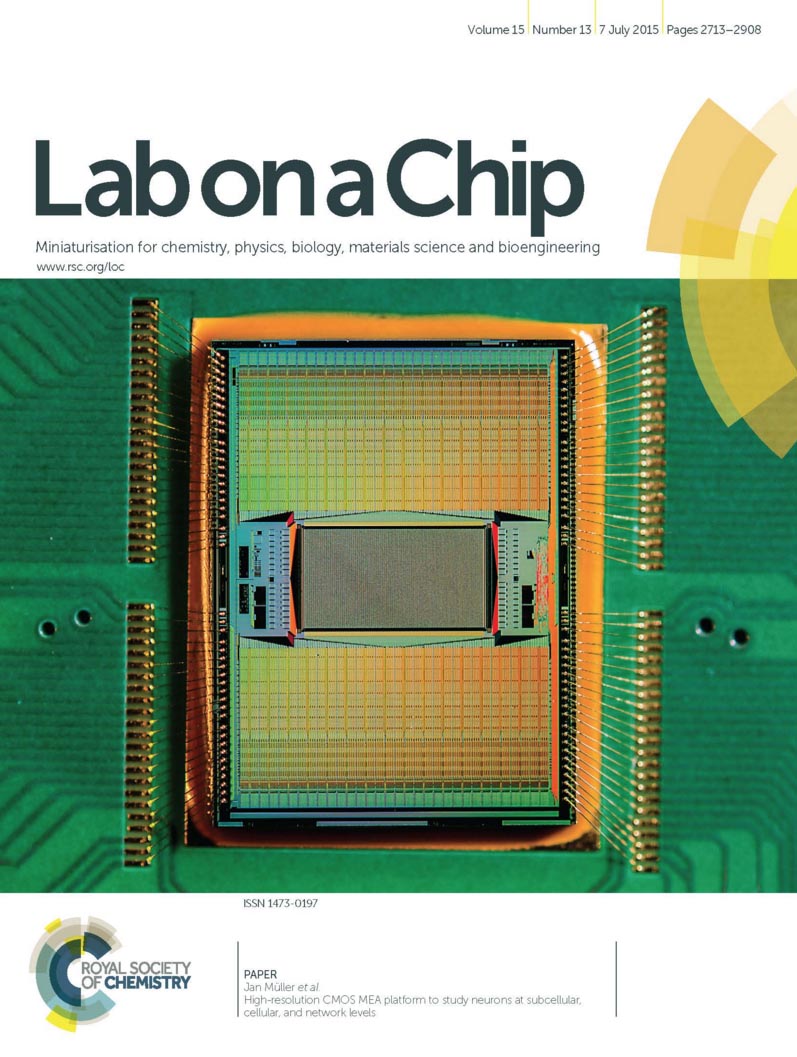
title = {High-resolution CMOS MEA platform to study neurons at subcellular, cellular, and network levels},
author = {Jan Müller and Marco Ballini and Paolo Livi and Yihui Chen and Milos Radivojevic and Amir Shadmani and Vijay Viswam and Ian L Jones and Michele Fiscella and Roland Diggelmann and Alexander Stettler and Urs Frey and Douglas J Bakkum and Andreas Hierlemann},
url = {http://pubs.rsc.org/en/Content/ArticleLanding/2015/LC/C5LC00133A#!divAbstract},
doi = {10.1039/C5LC00133A},
issn = {1473-0197},
year = {2015},
date = {2015-07-07},
journal = {Lab Chip},
volume = {15},
number = {13},
pages = {2767-2780},
publisher = {Royal Society of Chemistry},
abstract = {Studies on information processing and learning properties of neuronal networks would benefit from simultaneous and parallel access to the activity of a large fraction of all neurons in such networks. Here, we present a CMOS-based device, capable of simultaneously recording the electrical activity of over a thousand cells in in vitro neuronal networks. The device provides sufficiently high spatiotemporal resolution to enable, at the same time, access to neuronal preparations on subcellular, cellular, and network level. The key feature is a rapidly reconfigurable array of 26 400 microelectrodes arranged at low pitch (17.5 um) within a large overall sensing area (3.85 × 2.10 mm2). An arbitrary subset of the electrodes can be simultaneously connected to 1024 low-noise readout channels as well as 32 stimulation units. Each electrode or electrode subset can be used to electrically stimulate or record the signals of virtually any neuron on the array. We demonstrate the applicability and potential of this device for various different experimental paradigms: large-scale recordings from whole networks of neurons as well as investigations of axonal properties of individual neurons.},
keywords = {MaxOne, Neuronal Networks},
pubstate = {published},
tppubtype = {article}
}

title = {A network comprising short and long noncoding RNAs and RNA helicase controls mouse retina architecture.},
author = {Jacek Krol and Ilona Krol and Claudia Patricia Patino Alvarez and Michele Fiscella and Andreas Hierlemann and Botond Roska and Witold Filipowicz},
url = {https://www.nature.com/articles/ncomms8305},
doi = {10.1038/ncomms8305},
issn = {2041-1723},
year = {2015},
date = {2015-06-04},
journal = {Nature Communications},
volume = {6},
pages = {7305},
publisher = {Nature Publishing Group},
abstract = {Brain regions, such as the cortex and retina, are composed of layers of uniform thickness. The molecular mechanism that controls this uniformity is not well understood. Here we show that during mouse postnatal development the timed expression of Rncr4, a retina-specific long noncoding RNA, regulates the similarly timed processing of pri-miR-183/96/182, which is repressed at an earlier developmental stage by RNA helicase Ddx3x. Shifting the timing of mature miR-183/96/182 accumulation or interfering with Ddx3x expression leads to the disorganization of retinal architecture, with the photoreceptor layer being most affected. We identify Crb1, a component of the adhesion belt between glial and photoreceptor cells, as a link between Rncr4-regulated miRNA metabolism and uniform retina layering. Our results suggest that the precise timing of glia-neuron interaction controlled by noncoding RNAs and Ddx3x is important for the even distribution of cells across layers.},
keywords = {MaxOne, Retina},
pubstate = {published},
tppubtype = {article}
}
2014
title = {A 1024-channel CMOS microelectrode array with 26,400 electrodes for recording and stimulation of electrogenic cells in vitro},
author = {Marco Ballini and Jan Müller and Paolo Livi and Yihui Chen and Urs Frey and Alexander Stettler and Amir Shadmani and Vijay Viswam and Ian L Jones and David Jäckel and Milos Radivojevic and Marta K Lewandowska and Wei Gong and Michele Fiscella and Douglas J Bakkum and Flavio Heer and Andreas Hierlemann},
url = {http://ieeexplore.ieee.org/document/6923484/},
doi = {10.1109/JSSC.2014.2359219},
issn = {00189200},
year = {2014},
date = {2014-10-14},
journal = {IEEE Journal of Solid-State Circuits},
volume = {49},
number = {11},
pages = {2705-2719},
abstract = {To advance our understanding of the functioning of neuronal ensembles, systems are needed to enable simultaneous recording from a large number of individual neurons at high spa-tiotemporal resolution and good signal-to-noise ratio. Moreover, stimulation capability is highly desirable for investigating, for example, plasticity and learning processes. Here, we present a microelectrode array (MEA) system on a single CMOS die for in vitro recording and stimulation. The system incorporates 26,400 platinum electrodes, fabricated by in-house post-processing, over a large sensing area (3.85 2.10 mm) with sub-cellular spatial resolution (pitch of 17.5 µm). Owing to an area and power efficient implementation, we were able to integrate 1024 readout channels on chip to record extracellular signals from a user-specified selection of electrodes. These channels feature noise values of 2.4 µV in the action-potential band (300 Hz–10 kHz) and 5.4 µV in the local-field-potential band (1 Hz–300 Hz), and provide programmable gain (up to 78 dB) to accommodate various biological preparations. Amplified and filtered signals are digitized by 10 bit parallel single-slope ADCs at 20 kSamples/s. The system also includes 32 stimulation units, which can elicit neural spikes through either current or voltage pulses. The chip consumes only 75 mW in total, which obviates the need of active cooling even for sensitive cell cultures.},
keywords = {MaxOne, MEA Technology},
pubstate = {published},
tppubtype = {article}
}