Brain Slices
Every Cell has a Story to Tell.
Let’s Discover Yours
Brain slices offer a powerful ex vivo model to study defined brain regions and circuits with both physiological relevance and experimental control. Traditional techniques such as imaging and patch-clamp provide invaluable mechanistic insights but are often limited in scale, speed, or resolution.
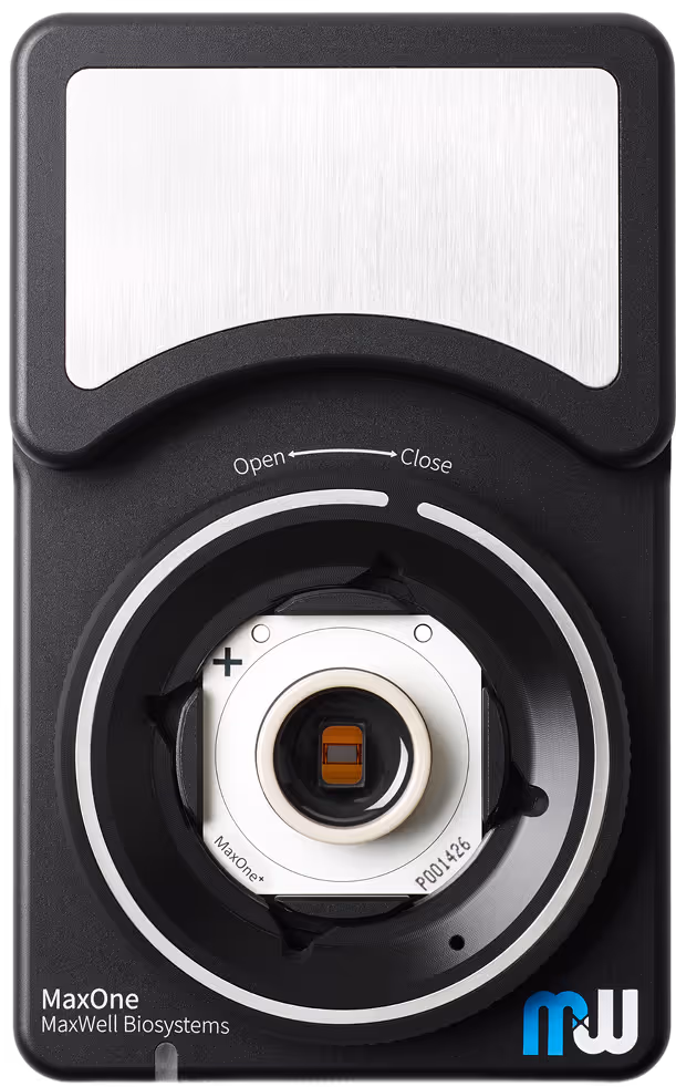
Our High-Density Microelectrode Array (HD-MEA) technology delivers precise spatial localization for recording and stimulation, enabling detailed dissection of neuronal circuits across distinct compartments, thanks to its high electrode density. Capture rich, reproducible data at subcellular, single-cell, and network levels across the full spectrum of brain oscillatory activity, allowing detection of subtle functional differences other systems may miss.
Uncover functional insight across all scales
Capture circuit dynamics across the entire brain slice, from single-neuron spikes to the full range of brain oscillations, leveraging the signal quality and high spatial and temporal resolution of our HD-MEA technology.
Always on target, never miss an event
Dissect activity within and across brain regions using a streamlined setup; without the need for complex rigs. With 26’400 electrodes, precisely target specific areas to capture key events and subtle functional differences, recording exactly where it matters every time.
Scale up your electrophysiology workflow
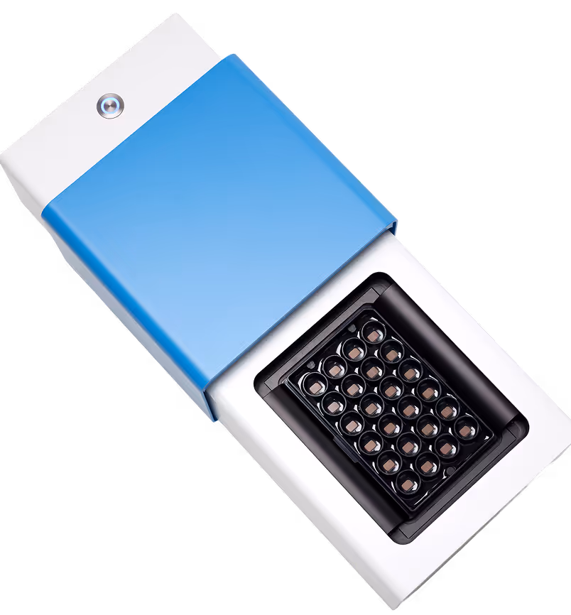
Run high-throughput slice experiments with our platform featuring the first-ever perfusion system optimized for the MaxTwo Multi-Well HD-MEA System, maintaining slice health and signal quality and fidelity across wells.
Probe target brain regions with precise stimulations
Activate defined circuits or test plasticity mechanisms using flexible stimulation paradigms, from simple pulses to complex, customizable protocols, delivered with precise spatial and temporal control.
Optimized light stimulation for your studies
Perform high-resolution optogenetics experiments with our HD-MEA technology, optimized to reduce optical artifacts while preserving signal quality.
.avif)
Case studies

Recording of extracellular action potentials in cerebellum slices
Identify functionally active regions in cerebellar slices by mapping spike amplitudes across the tissue with high spatial resolution. This enables rapid localization of high-activity regions and quantitative comparison of spike amplitudes across cerebellar layers.
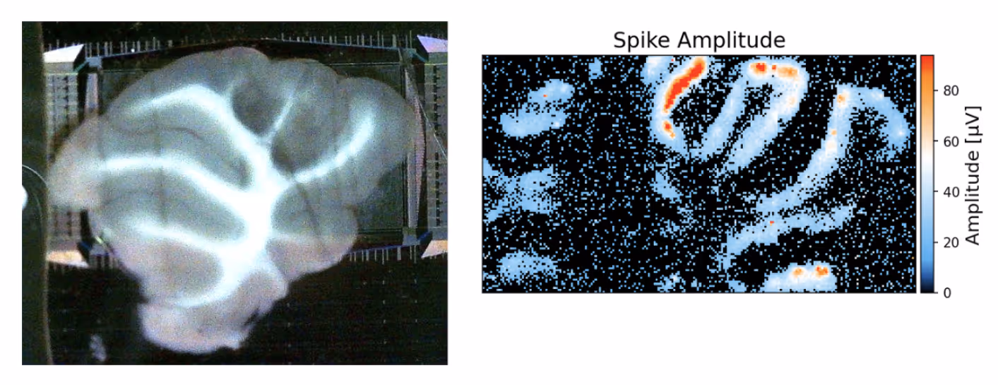

Acute extracellular recordings from cerebellum slices.
Left: Brightfield image of an acute cerebellar slice positioned on the MaxOne HD-MEA chip.
Right: Spike amplitude map from the ActivityScan Assay, derived from extracellular recordings, highlighting spatially localized high-amplitude activity within distinct cerebellar lobules.
Chemically induced epileptiform activity across multiple brain regions
Take full advantage of the large electrode array on the MaxOne Single-Well and MaxTwo Multi-Well HD-MEA System to monitor chemically induced epileptiform activity simultaneously in hippocampus and cortex. In the panel below, electrical events resembling epileptic states were observed in the hippocampus and cortex of a freshly prepared brain slice treated with 4-aminopyridine.
Epileptiform activity in acute brain slice treated with 4-aminopyridine.
Top: Acute hippocampal brain slice positioned on a MaxOne HD-MEA Chip.
Bottom: Spatially distributed epileptiform activity with local field potential traces illustrating abnormal events in hippocampus and cortex following 4-aminopyridine treatment.
Courtesy of Prof. Ed Mann, University of Oxford.
Selected Resources
Chronic silencing of Drd1a-Cre+ neurons impairs dopaminergic-driven cortical activation

An orexin-sensitive subpopulation of layer 6 neurons regulates cortical excitability and anxiety behaviour
Multimodal evaluation of network activity and optogenetic interventions in human hippocampal slices

Mutual generation in neuronal activity across the brain via deep neural approach, and its network interpretation

MaxOne Acute Brain Slice Protocol
Capture high-density electrophysiology recordings from acute brain slices with the MaxOne Tissue Holder and Perfusion System for stable, reproducible results.
MaxOne Acute Brain Slice Protocol - Cerebellum
Capture high-density electrophysiology recordings from acute cerebellar brain slices with the MaxOne Tissue Holder and Perfusion System for stable, reproducible results.
MaxOne Digital Input Extension Board Instructions
User guide for setting up the Digital Input Extension Board (DigiPins) to synchronize external events with MaxOne recordings using Python scripts.
Brain Slice Application Brochure
MaxTwo Perfusion System Brochure
Introducing the first fully integrated multiwell MEA perfusion system, MaxTwo Perfusion system ensures continuous medium flow and high-resolution data for advanced cell research.
Relevant
Applications
Relevant Biological Models
.avif)
Dr. Kateryna Voitiuk
“The MaxOne HD-MEA Chips allowed us to align neural activity to histology at comparable resolution. Also, it enabled us to study network activity and the structured spatial dynamics across the sub-regions of the hippocampus during seizure-like activity and optogenetic silencing.”