Organoids
Every Cell has a Story to Tell.
Let’s Discover Yours
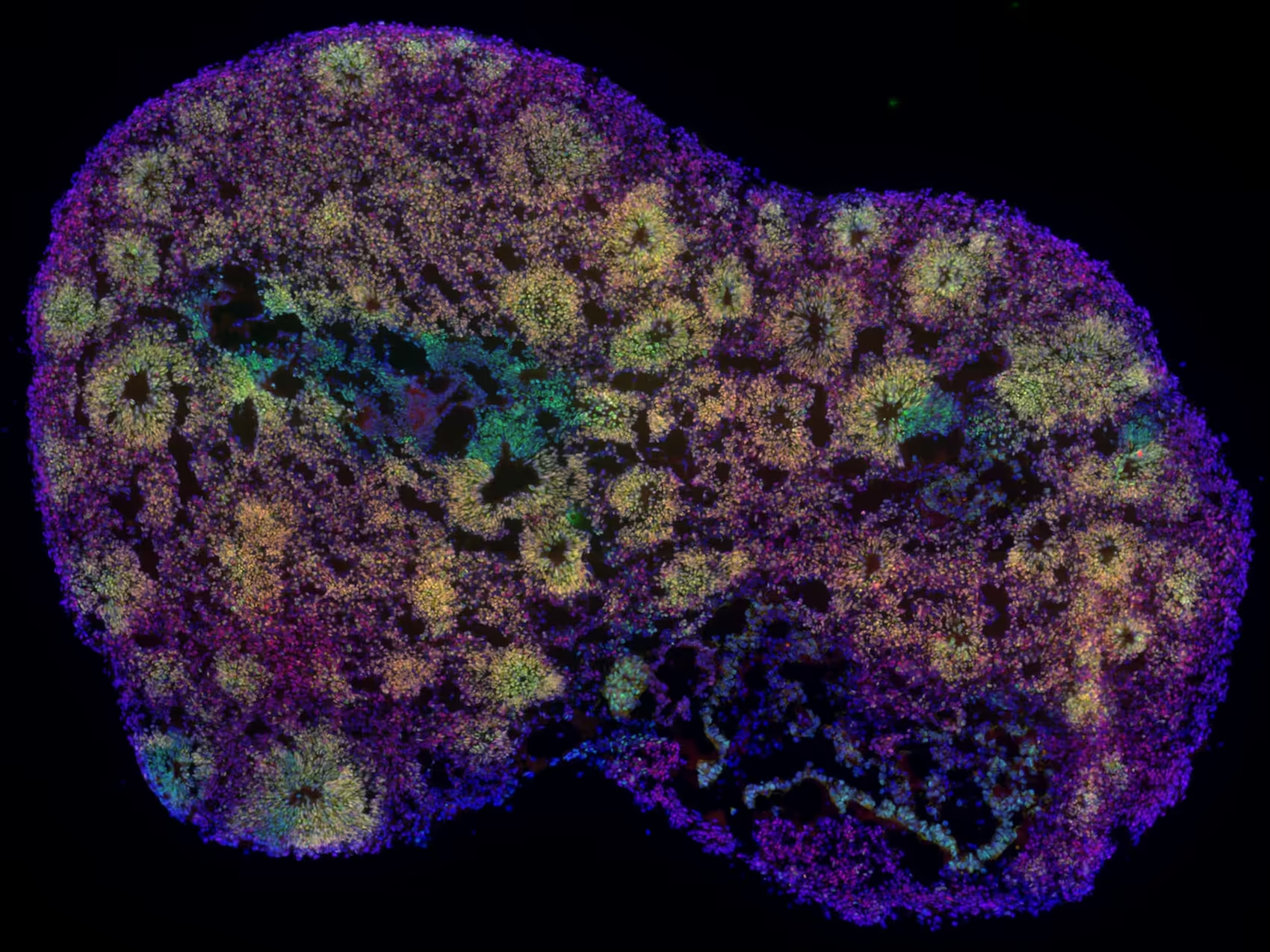
Organoids are precious. They require expertise, months of effort, and significant resources to develop, making every sample valuable. Often patient-derived and biologically complex, each organoid provides a physiologically relevant model of human neurodevelopment or neurological disease. Their full value can only be realized with the right tools.
Our High-Density Microelectrode Array (HD-MEA) technology is designed to make every organoid count, enabling detailed functional characterization at single-cell and network levels, even at early stages of development, with unmatched spatial and temporal resolution, in both acute and longitudinal recording paradigms.
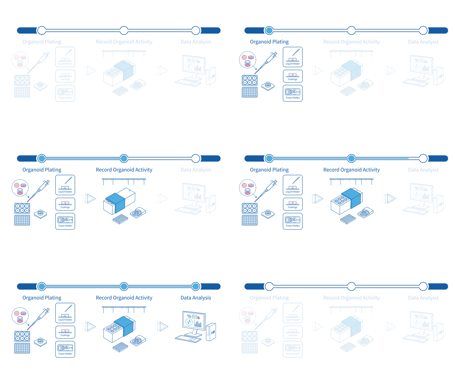
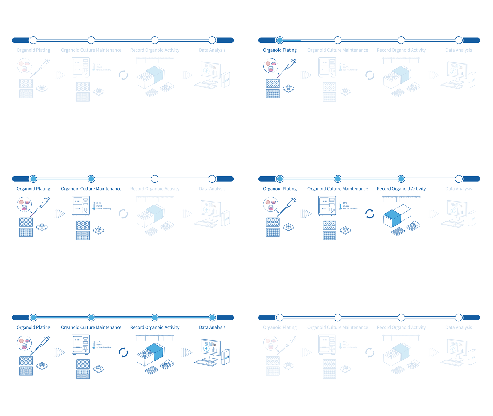
Experimental Workflows
Uncover the full functional profile of every organoid
Your organoids are precious, make them count and unlock insights that optical imaging alone cannot provide. Reveal how organoids fire, connect, and develop, even at early stages, with functional insights captured at subcellular, single-cell, and network level by our HD-MEA technology.
Greater reproducibility with fewer samples
Minimize biological variability with reproducible, high-quality functional data across wells and timepoints. Our high electrode count ensures reliable, statistically robust results, especially critical when organoid samples are limited.
Always at the right spot
Optimize recording quality with our flexible, high-density electrode configuration, ensuring you capture signals directly beneath active cells, including the center of the organoid. Obtain high-quality data across your organoids without requiring perfect placement every time.
Biologically relevant and non-invasive functional readouts
Capture the true activity of your organoids without compromising viability. Our system records from connected networks on the organoid surface, without invasive probes or dyes, providing biologically relevant signals that reflect how your model actually behaves.
Fit for a wide range of organoids
Organoids vary in protocol, structure, and handling, but our technology adapts. Whether you are performing acute or longitudinal recordings, our platform has been proven across a broad range of organoid use cases by researchers worldwide.
Relevant applications:


Case studies

Functional profiling of different neural organoid models
For every neural organoid model, extract a comprehensive functional signature using our HD-MEA technology. The representative recordings below highlight the high-quality characterization of a wide range of organoids, extending down to sub-cellular resolution.
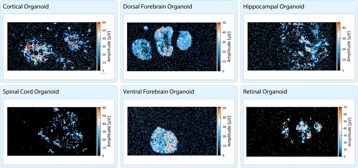

Whole sample functional characterization of neural organoids.
ActivityScan Assay heatmaps depicting the activity patterns from representative neural organoids of various types including cortical, dorsal-forebrain, hippocampal, spinal cord, ventral-forebrain and retinal organoids.
Data courtesy of multiple MaxWell Biosystems’ users.
Acquire instant readouts from your organoids
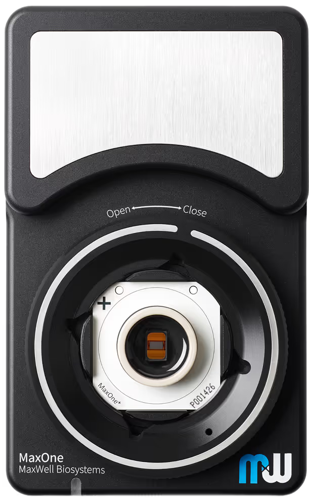
Extract rich functional data acutely from organoids with ease using our HD-MEA technology. While multiple approaches exist to stabilize organoids on the MaxOne Chips and MaxTwo Multi-Well Plates, an innovative, user-driven patented approach involves a Liquid Holder known as perfluorodecalin (PFD) (Duenki et al., Advanced Healthcare Materials, 2025). This biocompatible liquid gently anchors organoids onto the electrode array, ensuring optimal contact without compromising tissue integrity. Perform reliable acute recordings of your organoids with ease.
Acute recording of a cortical organoid
Top: Zoom into the MaxOne Chip electrode array (left) showing a light microscopy image of the organoid on the chip (middle), with the corresponding recording overlaid, where each red triangle represents an action potential. To the right, the electrical footprint of a single neuron is mapped across multiple electrodes.
Bottom: Example functional readouts from MaxLab Live, including an ActivityScan Assay heatmap depicting the organoid-wide activity patterns (left) and Network Assay raster and network activity plots (middle, right) highlighting synchronous network bursts across numerous cells.
Data collected in collaboration with the Biomolecular and Cellular Engineering Lab headed by Professor Yoshiho Ikeuchi at the Institute of Industrial Science, University of Tokyo.
Multiscale profiling of hiPSC-derived midbrain organoid maturation and axonal propagation
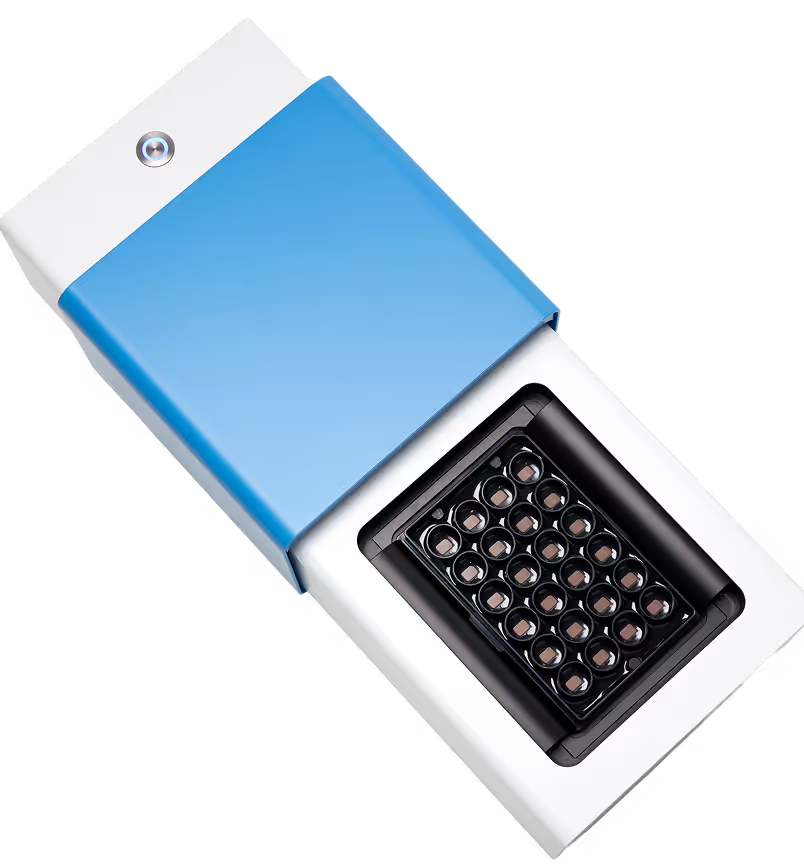
Track the functional evolution of STEMCELL Technologies hiPSC-derived midbrain organoids over time with scalable, label-free, non-invasive recordings on the MaxTwo HD-MEA 24-Well Plate. The workflow enables repeated measurements of the same organoid over time, while preserving its native 3D architecture, allowing downstream analyses without labelling or repositioning.
From electrical footprints and spontaneous activity to population-level network dynamics, MaxWell Biosystems’ HD-MEA technology captures the emergence of coordinated network activity and high-frequency bursting that are characteristic of maturing midbrain circuitry.
The unique AxonTracking Assay further extends functional profiling to the subcellular level by resolving action potential propagation within intact organoids, enabling quantitative investigation of axonal dysfunction in 3D disease models.
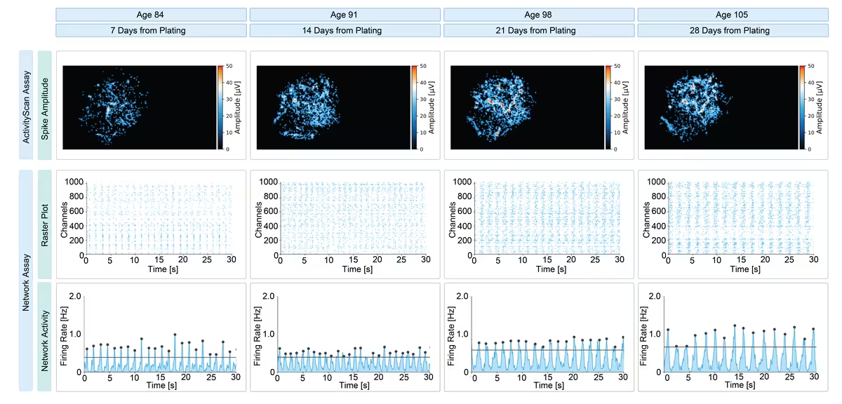
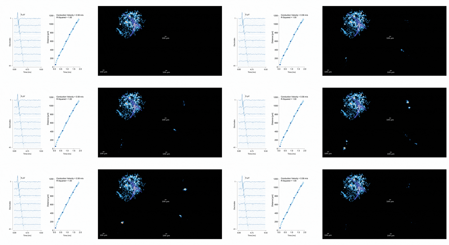
Multiscale longitudinal profiling of hiPSC-derived midbrain organoids on the MaxTwo 24-Well Plate.
Top: ActivityScan and Network Assay readouts reveal longitudinal maturation of the organoid electrical footprint and population dynamic with raster and network activity plots showing the emergence of high-frequency bursting characteristic of maturing midbrain circuitry.
Bottom: The unique AxonTracking Assay resolves subcellular action potential propagation at the organoid–electrode interface, visualizing three axonal branches extending through the 3D organoid footprint.
Data collected in-house using STEMCELL Technologies human midbrain organoids differentiated from human induced pluripotent (hiPSC) stem cell line SCTi003-A.
Selected Resources
Preconfigured neuronal firing sequences in human brain organoids

A human striatal-midbrain assembloid model of alpha-synuclein propagation
Human neural organoid microphysiological systems show the building blocks necessary for basic learning and memory

FGF8-mediated gene regulation affects regional identity in human cerebral organoids

Functional neuronal circuitry and oscillatory dynamics in human brain organoids

Functional imaging of brain organoids using high-density microelectrode arrays
Acute Brain Organoid Plating Protocol with Liquid Holder
Acquire functional HD-MEA recordings from your brain organoids in no time with this unique and easy-to-use protocol.
Brain Organoid Supplementary Guide
Discover helpful tips for optimizing your organoid and spheroid cultures and their recordings on the MaxOne and MaxTwo systems.
MaxOne+ and MaxOne Brain Organoid Plating Protocol
Use this brain organoid plating protocol for MaxOne+& MaxOne to achieve optimal attachment and enable longitudinal electrophysiology recordings of your neural organoids.
MaxTwo Brain Organoid Plating Protocol
Use this Brain Organoid Plating Protocol for MaxTwo to achieve high-throughput, longitudinal electrophysiology recordings of your neural organoids.
Sliced Brain Organoid Protocol
This protocol describes the preparation, slicing, and recovery of human brain organoid sections optimized for high-density electrophysiological recordings on MaxWell Biosystems platforms
Organoids Application Brochure
MxW - ACROBiosystems Application Note
Acute recordings of spinal cord organoids generated via a commercially available kit from ACROBiosystems, performed on MaxTwo 6-Well Plate with Liquid Holder.
MxW - InSphero Application Note
Application note with InSphero: pharmacological profiling of 3D human neural microtissues on the MaxTwo HD-MEA for scalable, label-free CNS drug screening.
STEMCELL Technologies Midbrain Organoid Poster
Longitudinal profiling of midbrain organoid maturation, pharmacological response, and axon dysfunction–relevant functional readout
Breathe-Easy Foil MaxTwo 6-Well Plate Video Instruction
See how easy it is to apply the Breathe-Easy® sealing membranes on our MaxTwo Multiwell Plates, minimizing evaporation and ensuring optimal conditions for your sample.

MaxOne Organoid Plating Video Instruction
Plating organoids on MaxOne Chips has never been so easy, thanks to our detailed step-by-step instructions, with practical tips at key stages.
MaxTwo Brain Organoid Plating Protocol Video Instructions
Plating organoids on MaxTwo Multiwell Plates has never been so easy, thanks to our detailed step-by-step instructions, with practical tips at key stages.

Prof. Yoshiho Ikeuchi
“Today, our MaxOne System plays a central role in our work. In fact, it has become a defining element of our research. It allows us to grow organoid-derived neural networks directly on the array surface and record their activity over long periods. This enables us to observe temporal-spatial dynamics, correlations, and sequential patterns of activity, all within a single, integrated system. Given the complexity of the networks we’re studying, the fact that the technology is also easy to use makes it even more valuable.”

Prof. Kenneth Kosik
“MaxOne opened so many doors in regards to questions of brain wiring, genetics, drug discovery, access to circuitry through organoids, which could not be addressed in any other way.”