Neuronal Cell Cultures
Every Cell has a Story to Tell.
Let’s Discover Yours
Neuronal cell cultures are powerful in vitro models for investigating development, connectivity, and dysfunction of the nervous system. But to truly understand how neurons behave, you need more than structure; you need their detailed function.
Our High-Density Microelectrode Array (HD-MEA) technology captures rich, reproducible functional data at subcellular, single-cell, and network levels, providing you with scalable workflows to reveal subtle phenotypes, track maturation, and confidently screen compounds with ease and precision.
Complete the picture of your neuronal activity
Get more meaningful data on how your neurons fire, connect, and mature, going beyond standard assays with rich functional readouts at subcellular, single-cell, and network levels thanks to the 26’400 electrode per well.
Reveal hidden action potential insights
Acquire reliable action potentials, from tiniest to strongest, to reconstruct the subcellular axonal behavior of your cells. Uncover novel functional mechanisms and phenotypes others cannot see.
Sensitivity to subtle and unseen phenotypes
Biologically relevant changes are not always obvious. Detect nuanced functional differences other systems might miss, even in sparse, co-cultured or early-stage neuronal models, thanks to our high-resolution and low-noise signal acquisition.
Always at the right spot, never miss a neuron
Record high-quality signals with thousands of electrodes per well, precisely positioned beneath your cells of interest, and capture activity with unmatched resolution.
Reproducibility at the core
Neuronal cell cultures can be biologically variable, but your recordings do not have to be. See functional differences that you can trust: engineered for long-term, stable experiments and consistent signal acquisition across sessions.
Case studies

Comprehensive characterization across neuronal culture types
For every neuronal culture model, our HD-MEA systems are designed to reveal their full functional profile. A broad range of cultures can be characterized at multiple levels, from activity across the whole sample at single-cell resolution with the ActivityScan Assay, population network dynamics with the Network Assay, to electrically reconstructed axonal branches with the AxonTracking Assay, a unique capability of our HD-MEA technology.
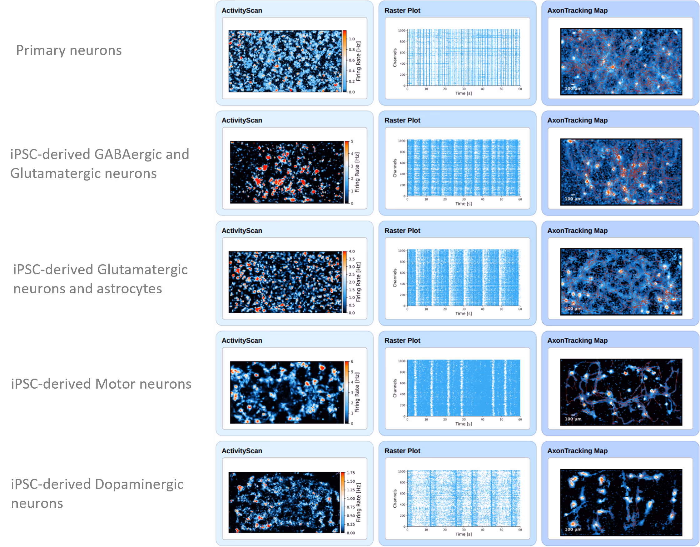

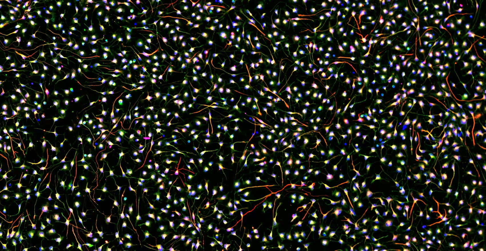
Example dataset illustrating the multi-scale functional characterization of neuronal cell cultures recorded with MaxWell’s HD-MEAs and analyzed using MaxLab Live.
From left to right: ActivityScan Assay heatmaps display spatial patterns of spiking activity across the culture; Network Assay raster plots capture temporal dynamics of population bursts; and AxonTracking Assay axonal footprint reconstructions reveal subcellular signal propagation paths in representative neuronal cultures.
Data courtesy of multiple MaxWell Biosystems’ users.
Reproducible functional insights across neuronal models
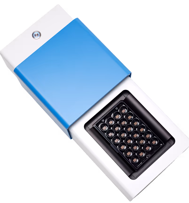
Capture robust, reproducible neuronal phenotypes across both healthy and disease model cultures using the MaxTwo HD-MEA System. The panel below highlights spontaneous network bursting features recorded from FUJIFILM Cellular Dynamics, Inc. (FCDI, Madison, Wisconsin) human iPSC-derived control and ALS-associated mutant motor neuron cultures, co-cultured with iCell Astrocytes 2.0 (FCDI) and recorded on a MaxTwo 24-Well Plate.
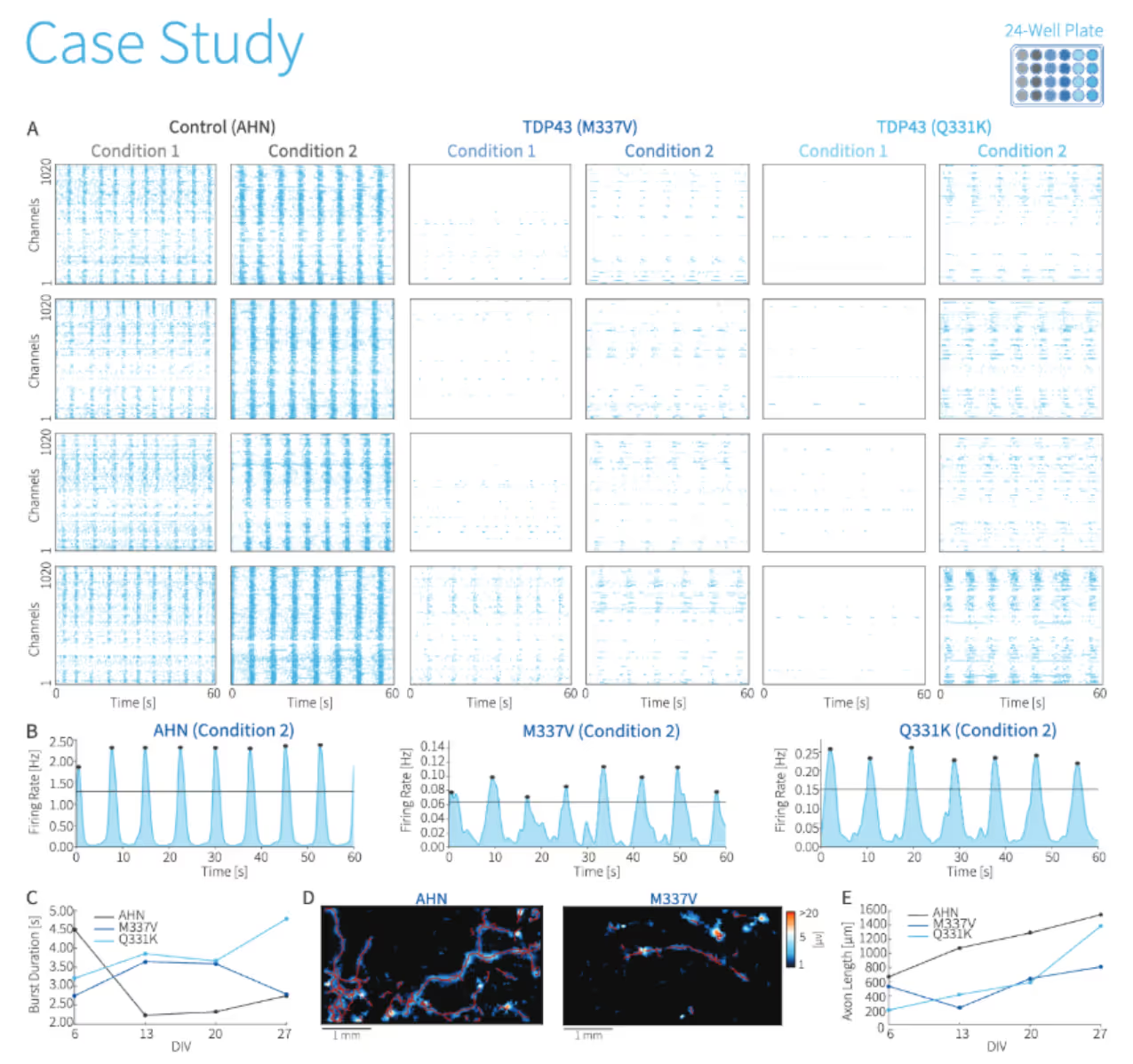
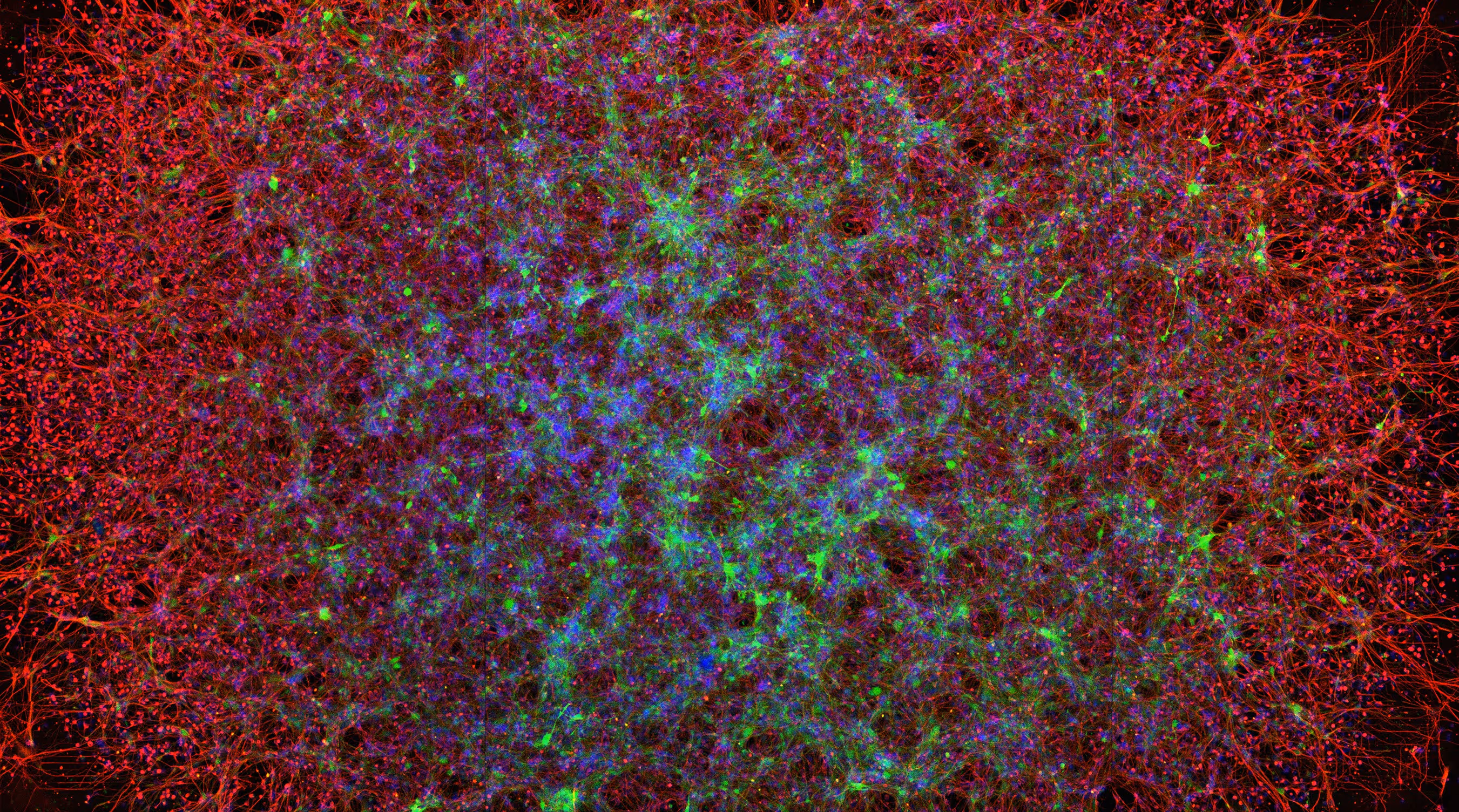
Functional characterization of ALS-associated TDP-43 mutant neurons reveals robust and reproducible phenotypes.
Human iPSC-derived motor neuron lines carrying ALS-associated TARBDP mutation (M337V and Q331K; FUJIFILM Cellular Dynamics, Inc.) were co-cultured with iCell Astrocytes 2.0 and compared to a control iPSC-derived motor neuron line derived from apparently healthy neurons (AHN). All recordings were performed on a single MaxTwo 24-Well Plate. (A) Raster plots illustrate spontaneous network bursting activity, where each dot represents an action potential, and vertical groupings indicate synchronized firing (network bursts). (B) Both mutant lines exhibited consistent decreases in network synchrony, demonstrating the sensitivity and reproducibility of HD-MEA recordings across wells and genotypes. (C) Quantification of burst duration over days in vitro (DIV) reveals diverging temporal evolution of network synchrony among AHN and mutant cultures. (D) Axonal footprint reconstructions illustrate differences in axonal morphology and signal propagation between AHN and M337V neuronal cultures. (E) Axon length measurements over time demonstrate distinct growth trajectories for control versus mutant motor neuron cultures.
Data courtesy of Fujifilm Cellular Dynamics, Inc.
Selected Resources
Human neuron subtype programming via single-cell transcriptome-coupled patterning screens

A model of human neural networks reveals NPTX2 pathology in ALS and FTLD

Multiplex epigenome editing of MECP2 to rescue Rett syndrome neurons
16p11.2 deletion is associated with hyperactivation of human iPSC-derived dopaminergic neuron networks and is rescued by RHOA inhibition in vitro

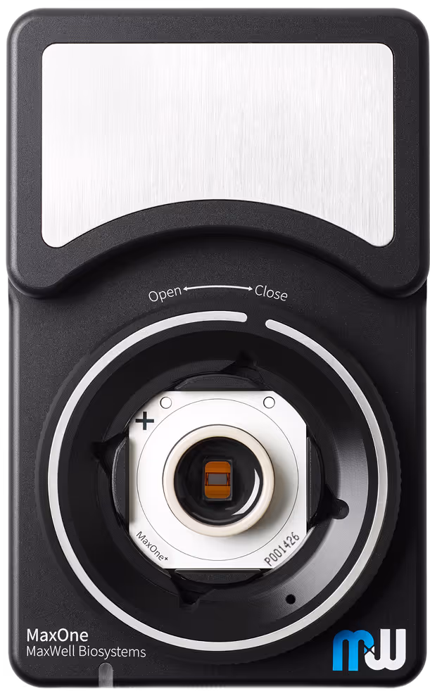
MaxOne+ and MaxOne Neuronal Cell Plating Protocol
Follow this easy-to-use neuronal cell plating protocol for MaxOne,ensuring optimal cell growth and attachment with clear step-by-step instructions.
MxW - bit.bio Application Protocol
Co-culturing ioMotor Neurons and rat cortical astrocytes for HD-MEA
MxW - Elixirgen Application Protocol
Culturing Human iPSC-derived Mixed Neurons on Multielectrode Arrays: MaxTwo Multiwell MEA
MxW - Neucyte Application Protocol
SynFire®iN plating protocol and chemical long-term potentiation on MaxWell Biosystems Multi-Well High-Density Microelectrode Array (HD-MEA)
Neuronal Cell Cultures Application Brochure
Application Note with CRL and bit.bio
Developing next-generation in-vitro phenotypic assays for Huntington's disease by combining precision reprogrammed hiPSC-derived disease models with high-density microelectrode arrays.
Elixirgen Scientific - MxW Application Note
Long-term Characterization of Quick-NeuronTMExciatory - Human iPSC-derived Neurons with High-Density Microelectrode Arrays
MxW - FCDI Application Note
Longitudinal Functional Profiling of HumaniPSC-derived Frontotemporal Dementia Neuronson HD-MEAs
MxW - NeuCyte Application Note
Long-term functional characterization of SynFire®iNs and astrocytes co-cultured on MaxTwo multi-well high-density microelectrode arrays
Nikon Application Note
Episcopic brightfield imaging of neuronal cells on high-densitymicroelectrode arrays enables prediction of cell region through AI learning
Breathe-Easy Foil MaxTwo 6-Well Plate Video Instruction
See how easy it is to apply the Breathe-Easy® sealing membranes on our MaxTwo Multiwell Plates, minimizing evaporation and ensuring optimal conditions for your sample.
Dot Plating MaxOne Chip Video Instruction
Just a single tiny drop is all it takes to get the best out of your iPSC-derived neuronal cultures or primary cell cultures on MaxOne Chips.
Dot Plating MaxTwo 6-Well Plate Video Instruction
Just a single tiny drop to get the best from your iPSC-derived neuronal cultures or primary cell cultures on MaxTwo Multiwell Plates.
Whole Area Plating MaxOne Chip Video Instruction
Just a small drop is all it takes to get the most out of your iPSC-derived neuronal cultures or primary cell cultures on MaxOne Chips.
Whole Area Plating MaxTwo 6-Well Plate Video Instructions
Just a small drop to get the best from your iPSC-derived neuronal cultures or primary cell cultures on MaxTwo Multiwell Plates.
Relevant
Applications
Relevant Biological Models
.avif)
Dr. Tetsuya Tanaka
“Unlike other commercially available MEA systems, MaxTwo has an outstanding capability to measure and track action potentials in single neurons due to how the electrodes are arrayed in such a high density manner."
.avif)
Dr. Marcus Kaji
“In acute toxicity studies, the 24-Well Plates let us use less compound while increasing throughput, a huge advantage especially for novel compounds. Honestly, I couldn’t have run these experiments without the higher throughput and batch analysis combination!”