Neuroscience (SfN) 2026

Date & Time

14-18 November, 2026

Location

Washington D.C., USA

Tags

Neuronal Cell Cultures
Organoids
Functional Phenotyping
Disease Modeling
Pharmacology & Toxicology

Neuroscience 2026 is one of the landmark annual meetings in neuroscience, bringing together the global community to share breakthroughs, exchange ideas, and explore the latest advances across the field. With a broad program spanning fundamental neuroscience, translational research, new technologies, and emerging applications, it is a key meeting for staying close to where neuroscience is heading.

We are excited to be there, connecting with the community, discussing new ideas, and supporting your research and next discovery. Join us at our booth, and come check out our posters, to discover how next-generation HD-MEA technology brings high-resolution, label-free functional insights to neuroscience research, from network activity to single-cell and subcellular signals.

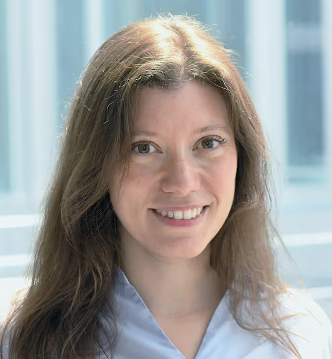
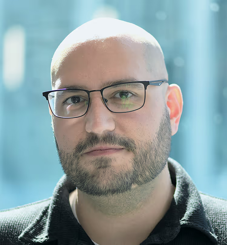
Meet the MxW Team
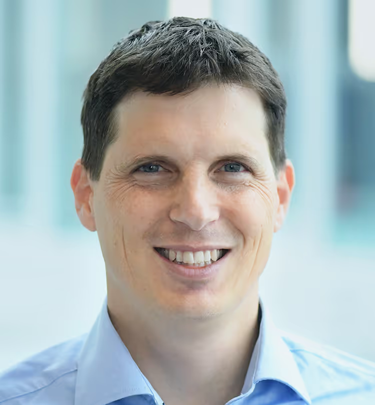
Dr. Urs Frey

CEO | MaxWell Biosystems (Switzerland)

Dr. Marie Obien

CCO | MaxWell Biosystems (Switzerland)

Dr. Laura D'Ignazio

Head of Commercial Excellence and Engagement | MaxWell Biosystems (Switzerland)

Dr. Silvia Oldani

Head of NAM | MaxWell Biosystems (Switzerland)

Elvira Guella

Account Manager | MaxWell Biosystems (Switzerland)

Dr. Tom Dufor

Field Application Scientist | MaxWell Biosystems (Switzerland)

Dr. Carlos Sanchez Priego

Field Application Scientist | MaxWell Biosystems (Switzerland)

Dr. Marian Hruska-Plochan

Scientific Business Developer | MaxWell Biosystems (Switzerland)

MxW Booth

Visit us at booth #636 to explore how MaxWell Biosystems is advancing neuroscience research with next-generation HD-MEA technology, enabling high-resolution, label-free functional insights from network activity down to single-cell and subcellular signals.  

Connect with our team and discover what’s new through live demonstrations of our latest accessories and their applications, a showcase of the latest MaxLab Live software features, and a unique exhibition of art created using brain organoids and our HD-MEA technology, bringing science, technology, and creativity together.

We look forward to welcoming you at booth #636 during Neuroscience 2026!

‍

Poster
Presentations

Dr. Laura D'Ignazio

Head of Commercial Excellence and Engagement | MaxWell Biosystems (Switzerland)
A scalable HD-MEA workflow for multiscale functional phenotyping of human midbrain organoids

Abstract

Human 3D midbrain organoids hold promise as physiologically relevant in vitro models to capture selected features of developing human midbrain tissue, including dopaminergic identity, tissue-like organization, and emerging circuit activity. These properties make them well suited to investigate Parkinson’s disease (PD)- and synucleinopathy-relevant alterations in neuronal excitability, network synchronization, and axonal output, which may precede overt neurodegeneration.

However, resolving these phenotypes in 3D tissues requires non-invasive electrophysiological methods that combine longitudinal access, high spatial sampling, and reproducible acquisition across wells and timepoints to detect low amplitude signals, quantify network bursting, and track axonal propagation at subcellular resolution. Here, in collaboration with STEMCELL Technologies, we used high-density microelectrode arrays (HD-MEAs) to longitudinally profile the functional maturation of commercially available human midbrain organoids. Organoids were plated at 77 days of age onto PEDOT-coated MaxTwo HD-MEA 24-well plates, each containing 26,400 electrodes per well, and spontaneous activity was tracked for approximately one month. Organoids showed progressive strengthening of firing activity, including increased firing rate, spike amplitude, and active area, indicating neuronal maturation and improved tissue-electrode coupling. At the network scale, we tracked the emergence of increasingly organized synchronized bursting, with increased burst cadence and enhanced neuronal recruitment over time. To assess pharmacological responsiveness, organoids were challenged with 4-AP and TTX, which respectively enhanced and suppressed firing activity in a highly reproducible manner. Finally, AxonTracking enabled label-free extraction of soma-to-axon propagation patterns at the organoid-electrode interface, providing a complementary readout of intra-organoid axonal functionality. Together, these results establish a scalable, non-invasive multiscale electrophysiology workflow for human midbrain organoids that captures spontaneous network maturation, pharmacological modulation of excitability, and axonal propagation phenotypes. By supporting consistent acquisition, processing, and interpretation of functional readouts across wells and timepoints, this approach provides a foundation for Parkinson’s disease-relevant phenotyping in 3D human neural models using HD-MEAs.

‍

Biography

Dr. Laura D’Ignazio is Head of Commercial Excellence & Engagement at MaxWell Biosystems AG. She earned her PhD at the University of Dundee and completed her postdoctoral training at the Lieber Institute for Brain Development, where she worked on X-linked Dystonia-Parkinsonism (XDP) and developed the first ventral-forebrain organoid model of the disease. Trained as molecular biologist, she brings hands-on experience in stem cells, neural organoid models, and neuroscience. In her current role, Laura drives user success through commercial excellence, engagement, and community building, helping researchers translate high-density microelectrode array (HD-MEA) data into scientific impact. Her interests include human iPSC-derived neural models, and phenotyping and screening workflows.

Dr. Marian Hruska-Plochan

Scientific Business Developer | MaxWell Biosystems (Switzerland)
Multiscale HD-MEA profiling reveals early hyperexcitability and axonal dysfunction in a human iPSC-derived GRN-FTD model

Abstract

Heterozygous loss-of-function (LOF) mutations in the granulin precursor gene (GRN) are a major genetic cause of frontotemporal dementia (FTD), leading to GRN haploinsufficiency. Reduced neuronal progranulin impairs lysosomal function and proteostasis, promoting synaptic dysfunction and TDP-43 pathology, contributing to altered functional connectivity and selective neuronal vulnerability. Human iPSC-derived models of GRN haploinsufficiency are valuable tools for disease phenotyping and therapeutic discovery; however, translating these models into disease-relevant functional endpoints remains challenging.

Using the MaxTwo High-Density Microelectrode Array (HD-MEA) multiwell platform (MaxWell Biosystems, 26,400 electrodes per well), which enables non-invasive, label-free, real-time electrophysiological recordings, we longitudinally profiled functional phenotypes in a human iPSC-derived FTD model across network, cellular, and axonal scales.  

iCell Induced Excitatory Neurons were co-cultured with normal iCell Astrocytes 2.0 and compared between an Apparently Healthy Normal (AHN) control line and an isogenic line carrying a heterozygous GRN R493X LOF mutation. Spontaneous activity was monitored from DIV 6-50. MaxWell’s ActivityScan and Network assays detected reproducible disease-associated changes across replicates and timepoints, with significantly elevated firing rates and reduced inter-spike intervals emerging by DIV 15. These early abnormalities indicate that HD-MEAs capture disease-related dysfunction before overt structural degeneration. GRN mutant cultures also showed significantly altered network synchrony, including more frequent and robust network bursts, shorter inter-burst intervals, and prolonged bursting, supporting a hyperexcitable network phenotype.  

MaxWell’s AxonTracking Assay reconstructed action potential propagation along individual axonal arbors. GRN mutant neurons displayed reduced axonal branching and shorter neurites, extending the disease-associated phenotype from early network and single-cell alterations to axonal architecture.  

Together, these data identify an early, reproducible, multiscale, and multiparametric electrophysiological signature of GRN haploinsufficiency in a human iPSC-derived FTD neuronal model, longitudinally captured on a single HD-MEA platform with scalable assay workflows. This approach links network-level dysfunction with single-cell and axonal phenotypes, providing robust functional endpoints for therapeutic window definition, progression monitoring, and future therapy-response studies in human neurodegeneration models.

Biography

Social events

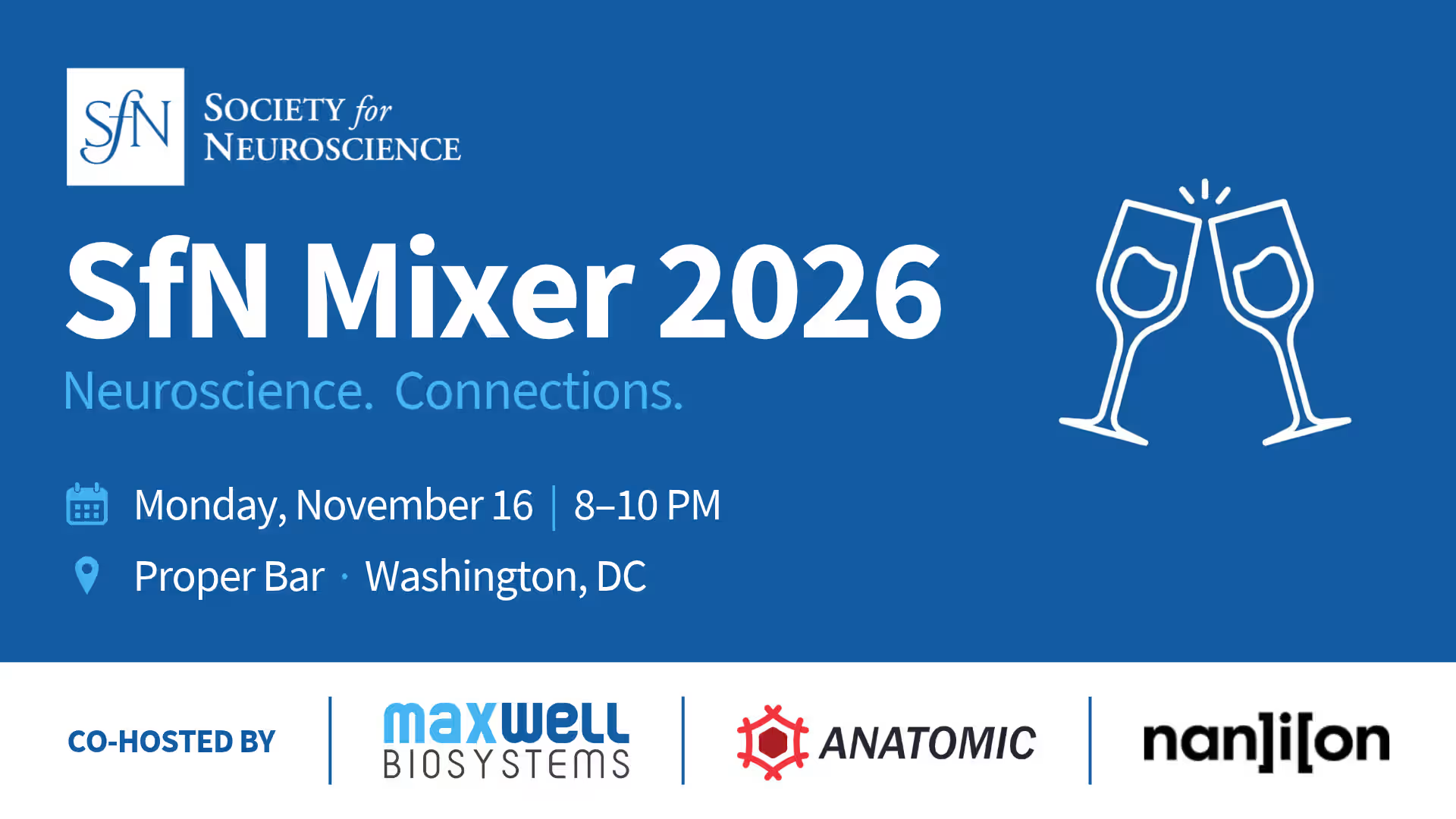
SfN Mixer

Join us for an evening of science and networking at our SfN Mixer, hosted together with Anatomic and Nanion Technologies.

Bringing together researchers from academia and industry, the evening is a chance to connect with the neuroscience and drug discovery community in a relaxed and informal setting. Exchange ideas, discuss experimental approaches, and share experiences around hiPSC-derived neuronal models, ion channel research, automated patch clamp, HD-MEA technology, and drug discovery.  

Whether you are developing your own neuronal models, exploring functional assays, or simply looking to connect with others working across these fields, we would love to meet you and continue the scientific conversations beyond the conference floor. Our signature cocktails will be waiting for you!

Space is limited, so contact laura.dignazio@mxwbio.com to register in advance.
No items found.
MaxWell