
Neuronal Models Symposium (NeuMoS) 2026
Organoids, NAMs, and Neural Circuit Assays for Disease and Drug Discovery
Support ALS research with us
As part of our 10th anniversary celebrations, we are running a fundraising campaign to support ALS research. Join us and make a meaningful contribution to the fight against ALS.
Place
Date
The Neuronal Models Symposium (NeuMoS) brings together the community advancing the future of in-vitro neural systems across neuroscience, cell biology, disease modeling, drug discovery, and biocomputing.
Over three days, the event will feature cutting-edge scientific talks, hands-on workshops, and stimulating discussions designed to spark new ideas and meaningful exchange.
Building on the success of previous MxW Summit editions, this year’s meeting features an expanded scope and updated identity. The new name reflects a deliberate evolution of the symposium, aiming to capture the full ecosystem of in-vitro neuronal network research.
Guided by a dedicated scientific committee, the Symposium offers a unique opportunity to connect with researchers, innovators, and industry leaders driving the next generation of neuroscience research.
Speakers Lineup
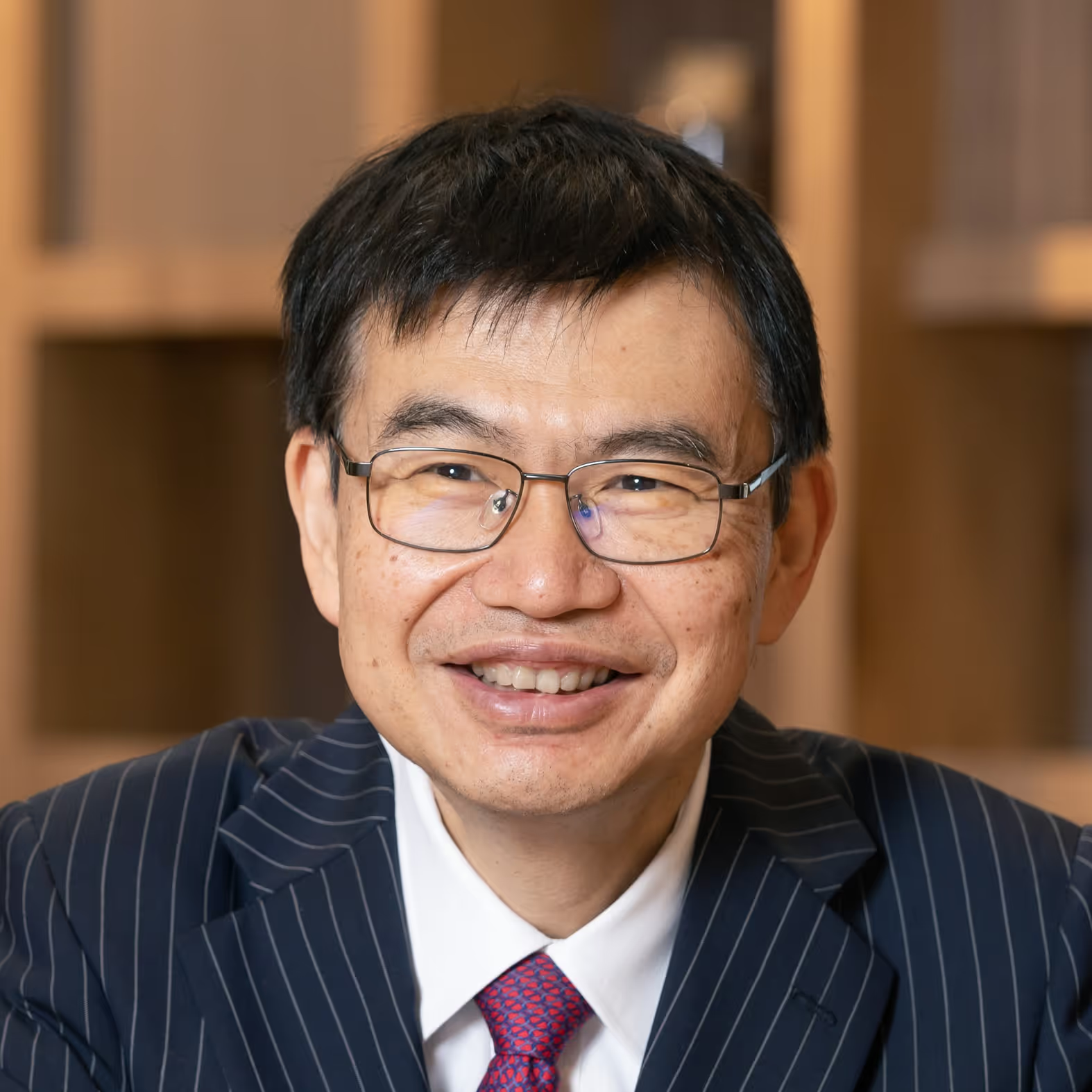
Hideyuki Okano received his M.D. in 1983 and Ph.D. in Medical Science in 1988, both from Keio University. Following a postdoctoral fellowship at the Johns Hopkins University School of Medicine, he became a Professor at the Tsukuba University (1994) and the Osaka University (1997). He returned to Keio University in 2001. He served as a Dean of the Keio University School of Medicine and Graduate School of Medicine from 2007 to 2021 and was appointed Visiting Professor at MIT in 2022. He is currently the Director and Distinguished Professor of Keio University Regenerative Medicine Research Center. He has received numerous honors, including the Medal with Purple Ribbon (2009), the Erwin von Bälz Prize (2014), and the Uehara Prize (2022). In July 2025, he became the president of the International Society for Stem Cell Research (ISSCR). His current research focuses on stem cell therapies for spinal cord injuries, as well as iPSCs-based modeling and drug development for neurodegenerative diseases such as ALS and Alzheimer’s disease.
Human iPSC-Based Neuronal Models for ALS Disease Modeling, Drug Discovery, and Reverse Translation
Human induced pluripotent stem cell (iPSC) technology offers a transformative approach to neurological disease modeling and drug discovery by shifting the starting point of therapeutic development from animal models to human patient-derived cells. This is particularly important for amyotrophic lateral sclerosis (ALS), a clinically and genetically heterogeneous disorder in which many compounds effective in rodent models have failed in clinical trials. Patient-derived iPSCs preserve individual genetic backgrounds and disease risk architectures, enabling “disease in a dish” and, more broadly, “humanity in a dish” as a platform for precision medicine.
In this lecture, I will present our iPSC-based strategy for ALS disease modeling and drug discovery. We established robust protocols to generate spinal motor neurons from ALS patient-derived iPSCs and used these cells to recapitulate disease-relevant phenotypes, including neurite degeneration, mitochondrial dysfunction, oxidative stress, and neuronal hyperexcitability. Screening 1,232 compounds using ALS iPSC-derived motor neurons led to the identification of ropinirole hydrochloride, an approved dopamine D2 receptor agonist for Parkinson’s disease, as a candidate anti-ALS drug. Mechanistic studies showed that ropinirole acts through both D2 receptor-dependent and -independent pathways, including suppression of oxidative stress and modulation of mitochondrial function.
Beyond these mechanisms, our reverse translational studies have revealed a disease pathway linking cholesterol biosynthesis, RNA editing, and motor neuron excitability. Transcriptomic analyses of ropinirole-treated ALS motor neurons indicated suppression of SREBF2-dependent cholesterol biosynthesis. In sporadic ALS iPSC-derived lower motor neurons, cholesterol synthesis-related enzymes were elevated, particularly in ropinirole-responsive lines, suggesting that dysregulated lipid metabolism may define a therapeutically relevant ALS endotype. Ongoing studies further suggest that increased cholesterol biosynthesis can reduce ADAR2 expression and impair A-to-I RNA editing of GRIA2/GluA2, a critical mechanism controlling AMPA receptor calcium permeability. Reduced editing may therefore increase calcium influx, promote motor neuron hyperexcitability, and contribute to degeneration. High-density microelectrode array recordings support this model by demonstrating abnormal firing activity in ALS neurons and its modulation by ropinirole.
Finally, I will discuss how iPSCs derived from participants in the ROPALS phase 1/2a clinical trial enabled reverse translational research linking in vitro drug responsiveness with clinical progression. I will also introduce cortical–spinal assembloids and organoid-based neural circuit assays as next-generation models for integrating dying-forward and dying-back mechanisms in ALS.
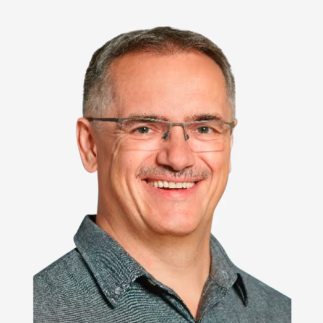
Janos Vörös is a Professor in the Institute for Biomedical Engineering of the University and ETH Zurich (Department for Information Technology and Electrical Engineering) heading the Laboratory for Biosensors and Bioelectronics since 2006. János Vörös has studied Physics at the Eötvös Loránd University in Budapest. After receiving a diploma in Physics in 1995, he was a doctoral student at the Department of Biological Physics of the Eötvös University (in collaboration with Microvacuum Ltd.) where he received his PhD in Biophysics in 2000. Since 1998 he was a member of the BioInterface group in the Laboratory for Surface Science and Technology at the Department of Materials of ETH Zurich as visiting scientist, postdoc, and from 2004 as group leader of the Dynamic BioInterfaces group until 2006. Prof. Vörös is interested in research and teaching in the areas of bioelectronics, biosensors, and neuroscience. His group focuses on the development of novel biosensor techniques for diagnostics and single molecule sequencing; on bottom-up neuroscience; as well as on stretchable biohybrid electronic devices.
Interacting with Neuron Microcircuits 24/7
Networks of neurons fascinate people because we all have them in our heads but we understand them very little. This talk will introduce how PDMS multicompartment microstructures can be used to build neuron microcircuits on HD-MEAs that allow for both stimulating and recording of their activity. A lid with an embedded water compartment assures 100% humidity while a simple custom incubator allow for 24/7 uninterrupted operation over 40 days with a single media exchange (Maurer, et al., 2026). This allows for a closed-loop optimization of stimulation paradigms to achieve specific performace-goals using reinforcement learning (Clément, et al., 2026). The microstructures are compatible with human induced pluripotent stem cells providing an opportunity to address some of the challenges related to investigating the nervous system and its diseases. Three examples of disease models will be introduced:
- The nociceptory system that is often involved in chronic pain can be replicated to study how the dorsal root ganglia modulate signal propagation from the skin towards the dorsal horn (Clément, et al., 2026).
- The system also allows for observing how glioblastoma-axon interaction causes epileptic behavior in networks of healthy neurons.
- A six-layer hierarchical model of the cortex can be used to study how progerin-induced aging influences the response of the network to amyloid-β exposure (Küchler, et al. 2026).
The custom tools (hardware and software) presented in this talk are all publicly available and we are happy to help scientists to implement them in their own labs.
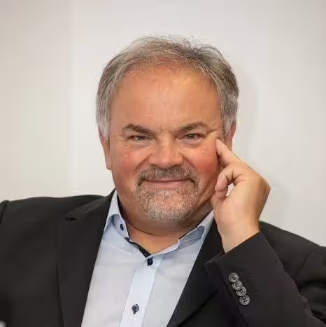
Juergen Knoblich is deputy scientific director at the Institute of Molecular Biotechnology of the Austrian Academy of Sciences (IMBA) and Professor at the Medical University in Vienna. Originally trained as a Drosophila researcher, his research focuses on the development of the human brain and the study of neurodevelopmental disorders. He received his PhD from the University of Tübingen and postdoctoral training in the laboratory of Lily and Yuh-Nung Jan at UCSF, San Francisco. In 2013, the Knoblich group has established cerebral organoids, a groundbreaking new technology that allows reconstitution of human brain development starting from patient iPS cells at unprecedented detail. They have used this system for modelling various neurodevelopmental disorders human brain tissue. They were able to screen through entire sets of disease genes relevant for autism spectrum disorders and could demonstrate that neurodevelopmental disorders can arise from cell types not found in animal models.
Cerebral Organoids: Growing human brain tissue from stem cells to study development and disease
The human brain is unique in both its size and complexity. The development of this remarkable organ involves biological processes that are absent or greatly expanded compared to those of most other species. To study these human-specific aspects of brain development, we developed cerebral organoids—three-dimensional stem cell-derived cultures that recapitulate key features of human brain development in vitro. Using this technology, we have identified developmental processes unique to humans, uncovered mechanisms underlying neurological disorders, and reconstituted functional neural network activity in the laboratory (Lancaster et al., Nature 2013; Esk et al., Science 2020; Eichmüller et al., Science 2022; Li et al., Nature 2023).
My presentation will focus on our most recent findings. We have developed organoid models that capture human-specific stages of brain development and reproduce disease-associated abnormalities at the circuit level. I will present our efforts to combine electrophysiology with barcoded connectomics at single-cell resolution to investigate how neural network activity and network architecture are altered in epilepsy. Finally, I will outline how barcoded prime-editing screens enable the systematic functional interrogation of genetic variants that differentiate Homo sapiens from Neanderthals, providing insights into the genetic basis of human brain evolution.
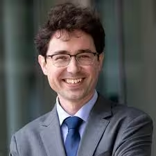
Dr. Tomasz Nowakowski is an Associate Professor of Neurological Surgery, Anatomy, and Psychiatry and Behavioral Sciences at the University of California, San Francisco, and a member of the Eli and Edythe Broad Center for Regeneration Medicine and Stem Cell Research. He earned his Ph.D. in Biomedical Sciences at the University of Edinburgh before joining UCSF for postdoctoral training with Arnold Kriegstein, becoming faculty in 2017.
Dr. Nowakowski's laboratory works to establish the human brain as a primary model system for neuroscience, developing scalable genomic, stem-cell, and viral-vector tools to trace cell lineage, map synaptic connectivity, and study gene regulation directly in human tissue. His research spans human cortical development, brain organoid modeling of neurodevelopmental disorders, and the molecular and circuit-level basis of Autism spectrum disorder.
His work has been published in leading journals and recognized with the Vilcek Prize for Creative Promise in Biomedical Sciences, the Sontag Distinguished Scientist Award, and the Robertson Neuroscience Investigatorship from the New York Stem Cell Foundation, among others. He leads UCSF's role in the CIRM Center for Neuropsychiatric Stem Cell Proteomics and chairs the Developing Brain Working Group of the BRAIN Initiative Cell Atlas Network.
From Genes to Circuits: Convergent Molecular Signatures of Autism.
Autism is one of the most heritable conditions in medicine, and genetic studies have identified dozens of genes that, when mutated, sharply increase a person's chances of being profoundly autistic. But knowing which genes are involved doesn't yet explain how they lead to autism, or whether their effects can be seen in the brains of the much larger group of autistic people whose diagnosis has no identified genetic cause. Our lab is working to close that gap by following the effects of autism risk genes through three stages: from the molecules they interact with, to the postmortem brain tissue of autistic individuals, to the activity of living brain circuits.
We start by asking whether different autism genes affect the brain through shared machinery. Focusing on one gene often mutated in autism, we found that a disease-linked mutation disrupts its normal partnerships with other proteins, and that recreating this mutation in stem-cell-derived brain tissue causes an overproduction of a specific class of brain cells — a defect shared with a second, related autism gene, suggesting the two act through a common mechanism. We then asked whether this kind of shared biology could be found directly in postmortem brain tissue from autistic donors — both those with an identified genetic cause and those without one. Comparing gene activity across thousands of individual brain cells, we found that donors with a known genetic diagnosis and those without one shared a common molecular signature. Finally, we ask whether this molecular signature could be related to how the brain works. Using rare recordings from patients undergoing awake brain surgery, we found that these same inhibitory brain cells respond specifically to the onset of speech, connecting molecular change identified in postmortem tissue to brain circuit involved in language, one of the most commonly affected abilities in autism.
Together, this work traces a path from genetic risk, through shared molecular changes in the brain, to circuit-level function — offering a way to link autism's many genetic causes to its shared biology, and eventually, to its clinical features.

Dr. Ranmal Samarasinghe is an Assistant Professor of Neurology at the David Geffen School of Medicine at UCLA, with clinical interests in epilepsy, autism, and neurophysiologic intraoperative monitoring. He received his medical degree and doctorate from the University of Pittsburgh and completed residency training in adult neurology, postdoctoral research, and a clinical fellowship in neurophysiology, all at UCLA.
His laboratory develops and applies human iPSC-derived brain assembloid models to study neural circuit formation and its disruption in neurological disease. Using a combination of two-photon calcium imaging, local field potential recording, and high-density microelectrode arrays, his group interrogates network dynamics across a spectrum from neurodevelopmental disorders — including Rett syndrome and SCN8A channelopathy — to neurodegeneration. Recent work has extended these approaches to model anesthetic mechanisms and tau pathology in assembloid systems, with findings that recapitulate in vivo electrophysiological signatures of disease. A unifying theme across this research is the use of circuit-level activity as a readout for understanding how molecular and cellular pathology translates into network dysfunction, with direct implications for therapeutic discovery.
Contextualizing molecular pathology through network dynamics in human brain assembloids
A central challenge in translational neuroscience is bridging the gap between molecular pathology and clinically meaningful dysfunction. I will present a network-centric framework for this problem, using electrophysiological and optical recordings from human iPSC-derived brain assembloids across a progression from neurodevelopmental disease to neurodegeneration. Beginning with cortical-subcortical assembloids in Rett syndrome — where two-photon imaging and local field potential recordings first revealed epileptiform network dynamics in MeCP2-deficient circuits — I will trace how this approach has evolved in scope and resolution. Work in hippocampal assembloids extended LFP-based analysis to region-specific circuit identity and its disruption in channelopathy. More recently, high-density MEA recordings have enabled single-unit resolution in assembloid systems, illuminating network-level mechanisms of anesthetic action and, most strikingly, revealing how tau pathology dismantles ensemble coordination in ways that recapitulate in vivo signatures of neurodegeneration. Across these models, a consistent picture emerges: circuit dynamics integrate and amplify molecular and cellular changes, making them a privileged and translationally grounded readout for both disease modeling and therapeutic discovery.
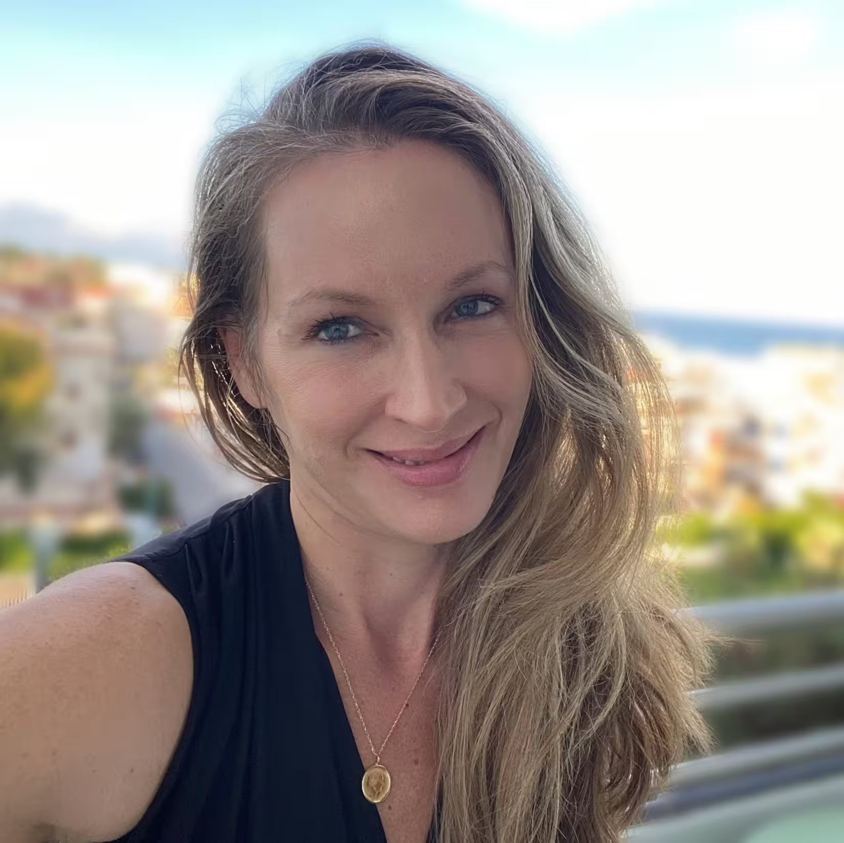
Dr. Keri Martinowich is Chief Scientific Officer and Senior Investigator at the Lieber Institute for Brain Development, where she oversees scientific strategy and research integration for the Institute, and Professor of Psychiatry and Behavioral Sciences at the Johns Hopkins University School of Medicine. She received a B.A. in International Relations from George Washington University and a Ph.D. in Neuroscience from the University of California, Los Angeles, followed by postdoctoral training in translational neuropsychiatry at the National Institute of Mental Health. Dr. Martinowich leads a research program focused on defining the molecular and circuit-level architecture of the human brain and its disruption in neuropsychiatric and neurodegenerative disease. Her laboratory integrates spatial transcriptomics, single-cell genomics, computational biology, and experimental model systems to investigate how gene expression programs are organized across cell types, microenvironments, and interconnected brain circuits. Her work emphasizes the use of postmortem human brain tissue to identify molecular and genetic signatures associated with disease vulnerability, with complementary translation into human iPSC-based models and rodent systems. Through these efforts, her research aims to bridge molecular, cellular, and systems-level mechanisms to advance understanding of complex brain disorders and enable development of biologically informed therapeutic strategies.
Human Spatial Genomics to Stem Cell Models: Circuit Vulnerability in Alzheimer’s Disease
Selective vulnerability of interconnected neural circuits is a defining feature of Alzheimer’s disease (AD), yet the molecular programs linking genetic risk to early circuit dysfunction remain poorly understood. The locus coeruleus (LC) and entorhinal cortex (ERC) are among the earliest brain regions affected in AD, exhibiting early accumulation of phosphorylated tau pathology prior to widespread neurodegeneration and cognitive decline. However, how molecular vulnerability emerges across connected brain regions, and how these processes can be modeled experimentally, remains unclear. To address this, we generated paired spatial transcriptomic and single-cell datasets across the LC–ERC circuit using postmortem human brain tissue from middle-aged donors stratified by APOE genotype, ancestry, and sex. In the LC, we identified APOE- and ancestry-associated alterations in astrocytic and neuromelanin-associated transcriptional programs linked to aging, lipid metabolism, oxidative stress, and neuronal vulnerability. In the ERC, AD risk-associated transcriptional changes localized predominantly to oligodendrocyte populations and white matter-associated spatial domains, suggesting disrupted myelination and glial support programs prior to overt disease onset. Because these datasets were generated from the same donors, ongoing work integrates molecular profiles across regions to define circuit-level programs associated with AD risk and early vulnerability. To translate these findings into experimentally tractable systems, we are generating donor-derived induced pluripotent stem cell (iPSC) models from matched postmortem donors used in the human brain studies. These include LC-like noradrenergic neurons, astrocytes, and emerging multicellular systems designed to model region-specific cellular interactions relevant to AD vulnerability. Using these platforms, we are investigating how APOE-associated molecular programs influence neuromelanin accumulation, cellular activity, and glial interactions. Together, these studies establish a framework integrating human spatial genomics with stem cell-based neural models to investigate mechanisms underlying selective circuit vulnerability in Alzheimer’s disease.
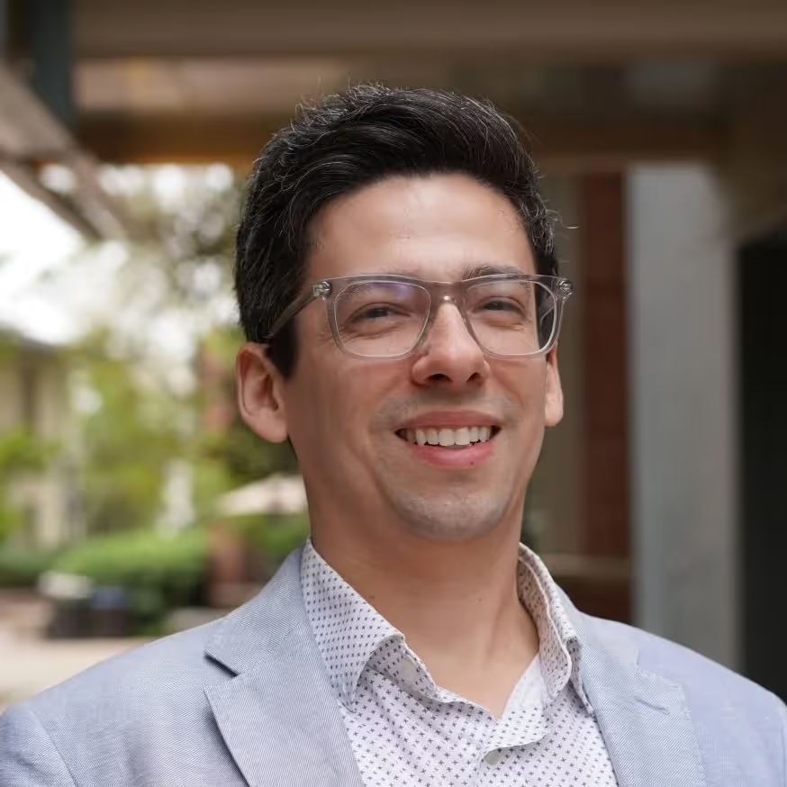
Michael F. Wells, PhD is an Assistant Professor in the UCLA Department of Human Genetics. He earned a PhD in Neurobiology from Duke University in 2015 and completed his postdoctoral training at the Broad Institute in Kevin Eggan’s lab in 2021. During this time, Dr. Wells co-developed the cell village platform that enables high throughput investigations of human genetic, molecular, and cellular phenotypic variation in shared in vitro environments. His research program at UCLA leverages this technology to understand the mechanisms underlying neurodevelopmental disorders of genetic and environmental origin, and is funded by the National Institutes of Mental Health, the Simons Foundation, and the California Institute for Regenerative Medicine.
Village-based proliferation and viability assays
The capacity of cells to proliferate and survive is central to development and disease. Assays that measure cell fitness are therefore a cornerstone of biology, but traditional techniques lack donor diversity and have high technical variability that impedes scale and reproducibility. To overcome these barriers, we designed and validated a “cell village”-based fitness screening approach using pooled cultures of 12–39 genetically distinct human neural progenitor cell (NPC) lines. We also developed Townlet to establish a foundational statistical framework based on Dirichlet regression for analyzing proportional data from cell villages. Applying these systems, we identified hyperproliferation in NPCs harboring the autism risk factor chromosome 16p11.2 deletion, mapped common genetic variants near ZFHX3 associated with NPC proliferation rate, and discovered genetic modifiers of lead (Pb) sensitivity implicating ARNT2. Together, these experimental and analytical tools advance a scalable, genetically diverse in vitro platform for dissecting human variation in cell fitness and gene-environment interactions.
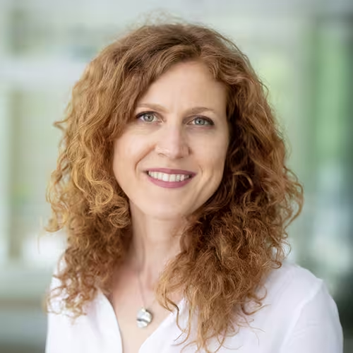
A/Prof. Silvia Velasco leads the Neural Stem Cells Laboratory at the Murdoch Children's Research Institute and is a Principal Investigator at the Novo Nordisk Foundation Center for Stem Cell Medicine (ReNEW) in Melbourne. Her laboratory uses pluripotent stem cell-derived neural organoid models to investigate human brain development and neurological diseases, with the aim of developing safe and effective therapies. A/Prof. Velasco completed postdoctoral training at New York University and the Broad Institute of MIT and Harvard and holds a PhD in Human Biology from the University of Turin.
Brain organoids as platforms for understanding and treating neurological disorders.
Stem cell-derived neural organoids provide a powerful opportunity to study human brain development and disease. We enhanced the utility of these in vitro systems by establishing highly reproducible organoid models that support long-term development and maturation of diverse brain cell types. Using these organoid models in combination with single-cell multi-omic approaches, we investigated cell-type-specific abnormalities associated with neurodevelopmental and neurodegenerative disorders. Finally, we leveraged the reproducibility of these models to develop automated high-throughput organoid screening platforms, enabling the integration of brain organoids into drug discovery pipelines for neurological disorders.
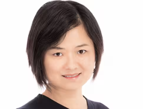
Jen Q. Pan is an Institute Scientist at the Broad Institute of MIT and Harvard and serves as Director of Translational Neurobiology at the Stanley Center for Psychiatric Research. Her research focuses on understanding how dysfunction of genes implicated in psychiatric disorders contributes to disease mechanisms in the central nervous system, with the goal of identifying novel therapeutic approaches.
Dr. Pan leads the Global Research Initiative of Neurophysiology of Schizophrenia (GRINS) Consortium, which investigates sleep and wake EEG recordings from individuals with psychiatric disorders to identify and validate electrophysiological biomarkers. She has also led multiple academic–industry collaborations to advance translational neuroscience and therapeutic development.
Her research has been supported by the Stanley Family Foundation, National Institutes of Health (NIH), the CACNA1A Foundation, the BD2, Laders2Cures, and industry partnerships. Dr. Pan received her B.S. in Chemistry from Nanjing University and her Ph.D. in Neuroscience from Brown University.
Human Neuronal Models Reveal XPO7 as a Regulator of Sodium Channel Function and Network Activity
Schizophrenia is a highly heritable neuropsychiatric disorder, yet how genetic risk variants disrupt human neuronal function remains poorly understood. Here, we use human induced pluripotent stem cell (iPSC)-derived neurons as a disease model to investigate the cellular consequences of loss of function (LoF) of XPO7, a schizophrenia risk gene identified through recent large-scale exome sequencing studies. By integrating high-resolution electrophysiology, transcriptomics, quantitative proteomics, and imaging with neuronal network assays, we identify convergent molecular and functional phenotypes linking XPO7 deficiency to impaired neuronal excitability.
XPO7 LoF alters voltage-gated sodium channel dynamics and availability, resulting in abnormal action potential generation and disrupted synchrony and regularity of neuronal network. These physiological abnormalities are accompanied by widespread molecular changes affecting nucleocytoplasmic transport, ion channel regulation, and synaptic composition. Notably, the voltage-gated sodium channel Nav1.2 (SCN2A) exhibits altered subcellular localization in XPO7-deficient neurons, providing a potential mechanistic link between XPO7 dysfunction and impaired neuronal signaling.
Together, our findings establish XPO7 as a critical regulator of neuronal excitability and network function and demonstrate how human iPSC-derived neuronal models and circuit-level phenotyping can bridge schizophrenia genetics to disease mechanisms. These results highlight sodium channel dysfunction as a potentially actionable pathway for therapeutic development in neuropsychiatric disorders.
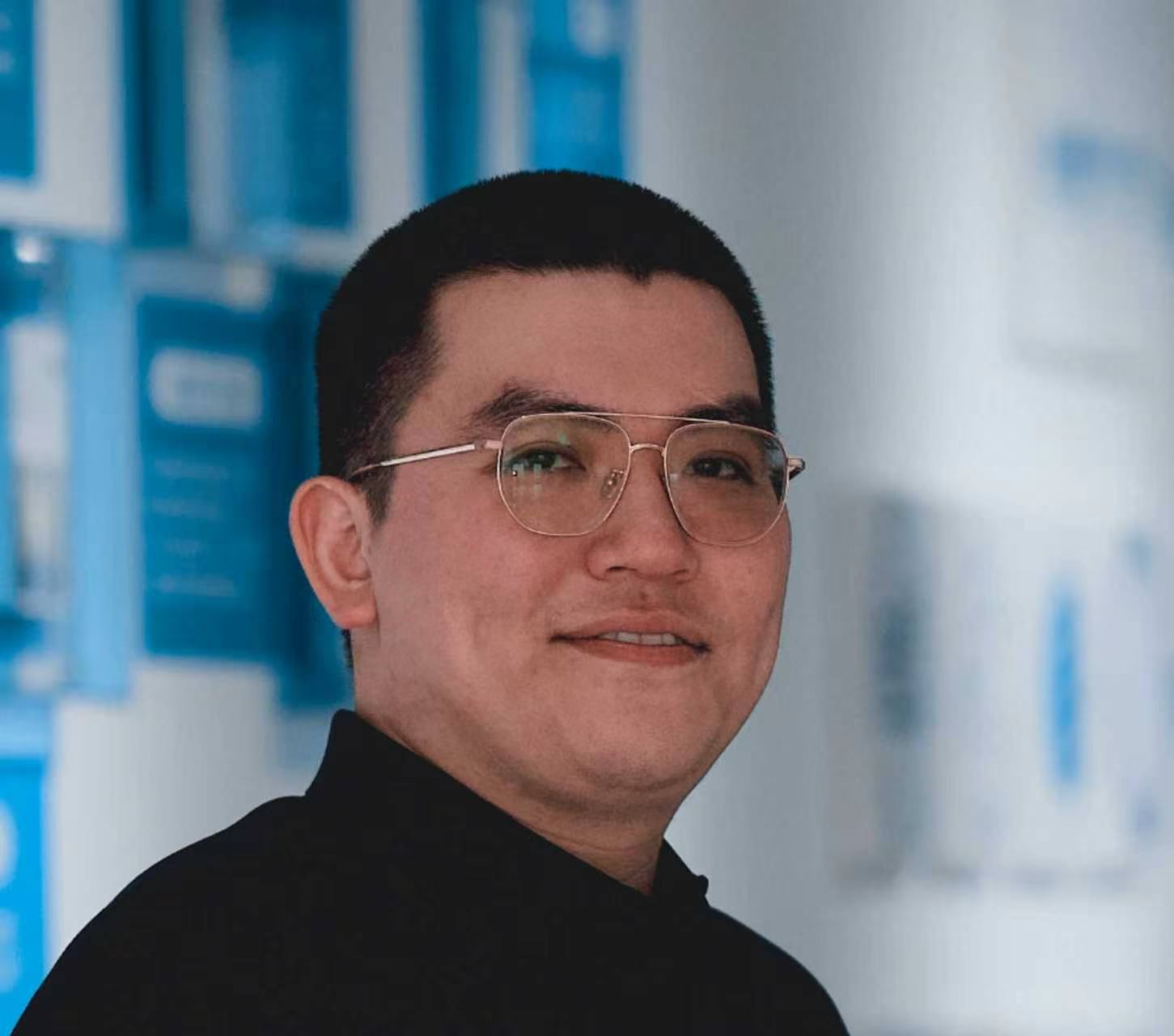
Xinyu Chen is a Ph.D. candidate in the Department of Anatomy, Histology and Embryology at Fudan University School of Basic Medical Sciences. His research focuses on the development of human brain organoid models and their applications in developmental neuroscience and neurological disease research. His work has involved the generation of region-specific brain organoids and assembloid models for studying inter-regional neural connectivity, as well as organoid-based models for investigating neural development, disease-associated phenotypes, and interactions between neural and non-neural cell populations. His current research focuses on developing scalable approaches for quantitative morphological analysis, electrophysiological characterization, neuropharmacological assessment, and neuromodulation in human brain organoids and assembloids.
High-Throughput Phenotyping and Neural Circuit Analysis in Brain Organoids: Morphology, Neuropharmacology, and Neuromodulation
The increasing scale and complexity of brain organoid studies require analytical approaches that support consistent and quantitative comparisons. This presentation will discuss three analytical strategies for morphological characterization, neuropharmacological assessment, and neural circuit analysis in brain organoids.
First, we use CHARM to extend conventional morphological assessment toward high-throughput quantitative analysis. This RGC morphological analysis focuses on image acquisition, feature extraction, and comparison of morphological and histological features across in vitro samples, with particular attention to analytical consistency and sample-to-sample variability. Second, we use high-density microelectrode arrays to examine the neuropharmacological responses of brain organoids. In addition to conventional network-level parameters, we analyze changes in the firing properties of individual neurons following pharmacological perturbation. This strategy allows drug responses to be assessed at both the network and single-neuron levels while retaining information on neuronal response heterogeneity.Finally, we characterize the electrophysiological properties of patient-derived brain organoids carrying DYRK1A mutations and examine neuromodulatory interventions in an assembloid model of interhemispheric cortical connectivity. Local and inter-organoid network activity is recorded before and after intervention. The analysis focuses on identifying electrophysiological features that describe changes in network state and using these features to compare different neuromodulatory conditions.
Together, these approaches highlight the potential of scalable, multimodal analysis to integrate morphological, molecular, and functional readouts in brain organoids and assembloids, enabling a more systematic evaluation of model development, disease-related phenotypes, and responses to intervention.
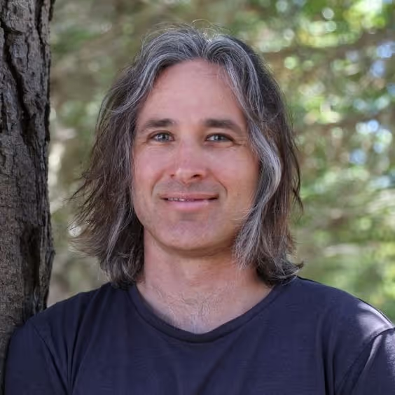
Tal Sharf is an Assistant Professor of Biomolecular Engineering at the University of California, Santa Cruz. He earned his B.S. in Physics from UC Santa Barbara, where he studied turbulence in thermally driven convection in the laboratory of Guenter Ahlers. He then pursued doctoral research with Ethan Minot at Oregon State University, developing biosensors based on carbon nanotube and graphene transistors. Inspired by the BRAIN Initiative, Sharf returned to UC Santa Barbara for postdoctoral training with neurobiologist Kenneth Kosik at the Neuroscience Research Institute, where he was named an Arnold O. Beckman Postdoctoral Fellow. During his postdoctoral work, he generated the first high-resolution map of functional connectivity among neurons in human iPSC-derived brain organoids. In July 2022, Sharf established his laboratory at UC Santa Cruz, where his team integrates physics, biology, and computation to investigate how human brain circuits assemble, wire, and generate function. In 2026, his team reported in Nature Neuroscience the discovery of preconfigured neuronal firing sequences in human brain organoids and the early postnatal rodent brain, suggesting that developing neural circuits contain intrinsic activity patterns that may provide a scaffold for interpreting later experience.
Uncovering the origins of human neural syntax
For centuries, philosophers have sought to understand how human neural tissue self-assembles into a physiologic scaffold that generates thought. Recent evidence suggests that neuronal wiring follows developmentally programmed instructions where neurons born from common progenitors preferentially wire and firing together before being recruited into experience-dependent networks. Despite implications for neurodevelopmental disorders such as autism and schizophrenia, we lack a mechanistic understanding of how developing neuronal circuits operate across spatiotemporal scales relevant to computation (ms precision over large networks). Human organoids provide a unique opportunity to investigate human circuit assembly at these scales because they can be engineered and integrated with technology in ways not possible in vivo. Here, we show that fundamental features of cortical computation (including heavy tailed firing-rate and connectivity distributions, reproducible neuronal firing sequences, and the coexistence of low- and high-dimensional subnetworks) emerge spontaneously in human brain organoids and early postnatal mouse cortex but not 2D cultures. These findings suggest that three-dimensional tissue architecture supports an intrinsic neural syntax that precedes sensory experience. Yet cortical development and function cannot be understood without considering directional communication with subcortical structures, particularly the thalamus, the brain’s primary sensory relay. I will conclude by presenting our bioengineering approach for controlling interactions between region-specific cortical and thalamic organoids. Using microfluidic devices to establish directional organoid-to-organoid axonal connectivity, we aim to determine how anatomically structured communication shapes the emergence and routing of functional activity across developing human neural circuits.
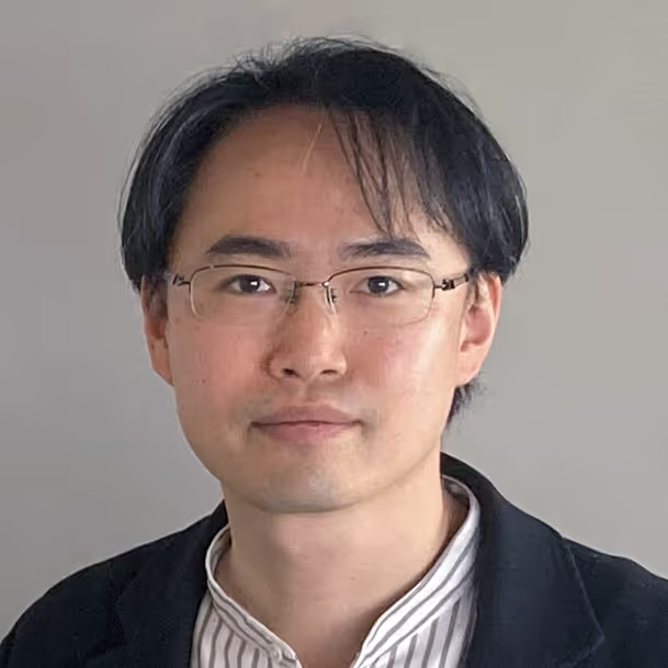
Hideaki Yamamoto is a Professor at the Research Institute of Electrical Communication (RIEC), Tohoku University, Sendai, Japan. He obtained a Ph.D. degree in engineering from Waseda University, Japan, in 2009. He was a JSPS Research Fellow at Tokyo University of Agriculture and Technology, and an Assistant Professor at Waseda University, before joining Tohoku University in 2014. In 2020, he was appointed Associate Professor at the RIEC and was promoted to Professor in 2026. His research interests include the use of engineered neuronal cultures as a model system for understanding brain computation, as well as their applications to brain-inspired computing and biomedicine.
Engineering living neuronal networks: from structure-function relationships to bio-inspired computing
How does the architecture of neuronal networks give rise to robust and energy-efficient computation in the brain? In vitro neuronal networks are effective tools for addressing this question in a physicochemically defined environment. Furthermore, such cell culture technology can be integrated with semiconductor microfabrication to reconstitute networks that more closely resemble the structures in vivo. In this talk, I will demonstrate how protein micropatterns and microfluidic devices can be used as chemical and physical guidance cues to control the growth of cultured neurons. These platforms provide a unique approach for constructively investigating how biological neurons form structured networks and generate high-dimensional dynamics that can be harnessed for bio-inspired computing.
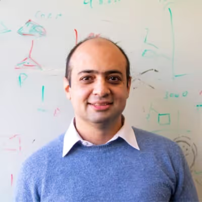
Mohammed Mostajo-Radji is an Associate Research Scientist at the UC Santa Cruz Genomics Institute, where he directs the Live Cell Biotechnology Discovery Lab. His group builds forebrain organoid models and machine-learning tools to define, track, and decode neuronal identity and circuit function from high-density electrophysiology, with applications to neurodevelopmental and neuropsychiatric disease. A parallel thread of his work develops open, cloud-based platforms to make large-scale neuroscience data and analysis broadly accessible, and to bring hands-on experimental science to students in underserved communities worldwide. He earned his PhD in Molecular and Cellular Biology at Harvard University with Paola Arlotta, completed postdoctoral training at UC San Francisco with Alex Pollen, and is a Google Cloud Research Innovator.
Engineering and decoding neuronal circuits.
Engineered forebrain organoids self-organize into functional networks whose architecture reflects their cellular composition, offering a tractable model for how circuits emerge without sensory input. Using longitudinal high-density microelectrode array recordings, I first show that dorsal and ventral forebrain organoids develop distinct network topologies, with refined small-world organization tracking interneuron enrichment. Reading out these dynamics at scale, however, requires extracting what a neuron is, where it sits, and how it participates in a network without features fixed by hand in advance. I then present a representation-learning approach that recovers such structure directly from extracellular waveforms and spiking dynamics, classifying cell types, resolving spatial organization, and exposing network signatures of circuit self-organization and disease.
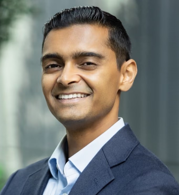
Dr. Arun Sharma, PhD is a stem cell biologist focusing on cardiovascular biology and space biosciences. He is an associate professor at Cedars-Sinai and is the director of the Center for Space Medicine Research. Research in the Sharma laboratory focuses on the applications of human induced pluripotent stem cells (hiPSCs) for modeling cardiovascular disease in-vitro. The lab utilizes hiPSCs, genome editing, cardiac organ-on-chips, and 3D cardiac spheroids/organoids to understand the molecular mechanisms driving cardiovascular disease and heart development. Sharma is also an internationally-recognized leader in the space biosciences field. His laboratory studies the impacts of space on stem cell biology and harnessing microgravity to manufacture unique biomaterials. In 2016, Dr. Sharma led a project that sent human stem cell-derived heart cells to the International Space Station (ISS), the first long-duration cell culture experiment in space. Dr. Sharma and his lab have published articles in leading scientific journals such as Science, Nature Biotechnology, Science Translational Medicine, Circulation Research, Stem Cell Reports, and Cell Stem Cell. He has received numerous awards for his work, including the Sartorius & Science Award in Regenerative Medicine, Forbes 30 Under 30 in Science, the American Heart Association Career Development Award, the Compelling Results Award from NASA, the Igniting Innovation Award from the ISS National Laboratory, and the Donna and Jesse Garber Award for Cancer Research. Dr. Sharma earned his bachelor's degree in biology from Duke University and his PhD in stem cell biology from Stanford University. He completed a postdoctoral research fellowship in cardiovascular genetics at Harvard Medical School.
Stem Cell Research and Biomanufacturing in Space.
Human induced pluripotent stem cells (hiPSCs) provide powerful platforms for modeling cardiovascular disease and advancing regenerative medicine. This talk will highlight how stem cell-derived tissues, organoids, and organ-on-chip systems are being used at Cedars-Sinai to study heart disease, drug-induced cardiotoxicity, and human adaptation to spaceflight. Drawing on pioneering studies performed aboard the International Space Station, Dr. Arun Sharma will discuss how the intersection of stem cell biology and space biosciences is uncovering new insights into human health while creating opportunities for innovation in disease modeling and therapeutic discovery.
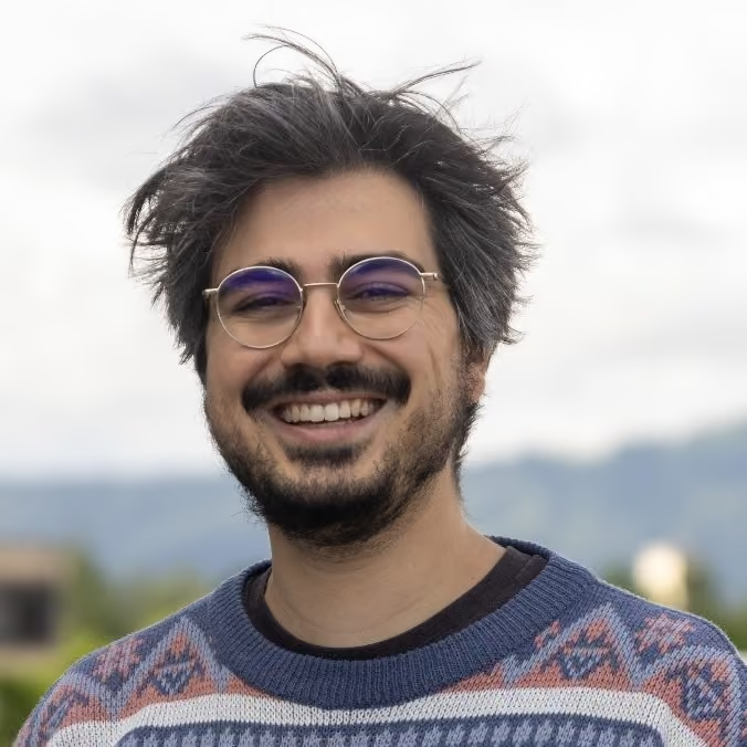
David Colameo is a Senior Scientist at the Electrophysiology Core Facility of the University of Zurich. He specializes in high-throughput electrophysiological characterization of in vitro model systems using automated patch clamp and high-density microelectrode arrays, including both excitable and non-excitable cellular models. He received his Bachelor's and Master's degrees in Biology and Neuroscience from the University of Zurich. In the group of Steven Brown, he used single-molecule RNA-FISH to visualize circadian oscillating transcripts at synapses. During his doctoral research in the group of Gerhard Schratt at ETH Zurich, he described a microRNA-based molecular mechanism that controls excitatory and inhibitory synapse strength during homeostatic plasticity and sleep.
Stem Cell Research and Biomanufacturing in Space.Altered Excitability and Variation of Ion Channel activity in variants of ALS using iPSC-Derived Motor Neurons and High-throughput Automated Patch-Clamp.
ALS is caused by diverse genetic mutations, including C9orf72 repeat expansions, SOD1, and TARDBP, that converge on motor neuron dysfunction. Patch clamp remains the gold standard for characterizing excitable cells, but low throughput has limited its use in disease modeling. We addressed this using high-throughput automated patch clamp (SyncroPatch 384, Nanion) to profile iPSC-derived motor neurons from ALS patients (C9orf72, TDP-43 A382T, SOD1 D109Y) and a healthy donor (Axol Biosystems).
The platform allowed simultaneous, unbiased recording of multiple electrophysiological parameters across genetically defined lines and hundreds of sampled motor neurons. Na⁺ channel gating kinetics were comparable between healthy and ALS neurons, but ALS neurons showed reduced Na⁺ channel current density and decreased excitability, with altered action potential amplitude and firing. Ion channel remodeling extended to reduced K⁺ current density and slightly reduced GABA-induced currents, while glutamate and glycine responses were unaffected.
These results demonstrate that HT-APC can resolve disease-relevant electrophysiological phenotypes in hiPSC-derived neurons at a scale unattainable with conventional patch clamp. This positions HT-APC as a scalable tool for mechanistic ALS research and drug discovery.
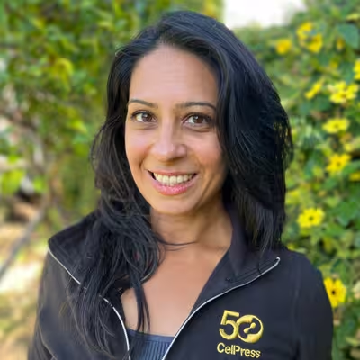
Sheila Chari, Ph.D, is Editor-in-Chief at Cell Stem Cell and Executive Editor at Cell Press. Her primary responsibilities are knowing and publishing the top stem cell discoveries, driving journal publishing strategy, and managing a global editorial staff. She travels to international scientific conferences and research institutions to be on top of the latest developments and meet with authors, reviewers, and readers. She is a proud member of the stem cell community and an ardent supporter of stem cell research. Sheila holds a doctorate from Northwestern University, where she studied transcriptional control of normal and malignant hematopoiesis. She conducted post-doctoral research on epigenetic regulation of cell fate reprogramming at the University of Chicago. Sheila is based in Los Angeles, California, USA.
Publishing with Cell Press: Inside the Editorial Process.
This talk will introduce Cell Stem Cell’s editorial scope, priorities, and role within the broader Cell Press portfolio, with an emphasis on what makes a strong fit for the journal. The session will then focus on practical guidance for navigating the publishing process at Cell Press journals, organized around the questions authors most frequently ask editors. Topics will include selecting the right journal, preparing a cover letter, understanding how editors assess submissions, navigating Cell Press policies on preprints and AI, and responding effectively to reviewer comments. The goal is to demystify the editorial and publication process and provide authors with concrete guidance for preparing and publishing their work in Cell Press journals.
Program & Scientific Topics
The symposium will feature two days of oral sessions, including keynote lectures, invited speakers, and selected short talks from submitted abstracts. The third day will be dedicated to parallel, hands-on workshops focused on practical and emerging methodologies, including microphysiological systems (MPS), spike sorting and data analysis, and organoid culture and plating techniques.
The scientific program is currently being structured around the following tracks:
Brain Development
Disease Modeling
- Drug Discovery
Neurotechnologies & Bio-inspired Computing
Scientific Committee
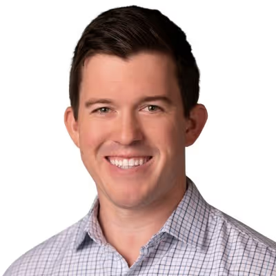
Matt Kelley is a Principal Scientist at Alexion AstraZeneca Rare Disease, where he leads drug discovery programs in rare neurology with the goal of developing innovative and transformational treatments. He also leads Alexion Genomic Medicine's human iPSC-derived neuronal platform, integrating high-throughput molecular readouts with high-density microelectrode array (HD-MEA) electrophysiology to interrogate target engagement and disease-relevant circuit phenotypes on translational human cell models. Prior to Alexion, he held drug discovery program biology lead roles at Pfizer and Voyager Therapeutics, and completed postdoctoral training at Amgen, where he established HD-MEA and in vivo EEG capabilities supporting the Aimovig/erenumab (anti-CGRP) program. He earned his Ph.D. in Neuroscience from Tufts University and his B.S. in Neuroscience from UCLA. He serves on Professional Development Committee for the Society for Neuroscience.
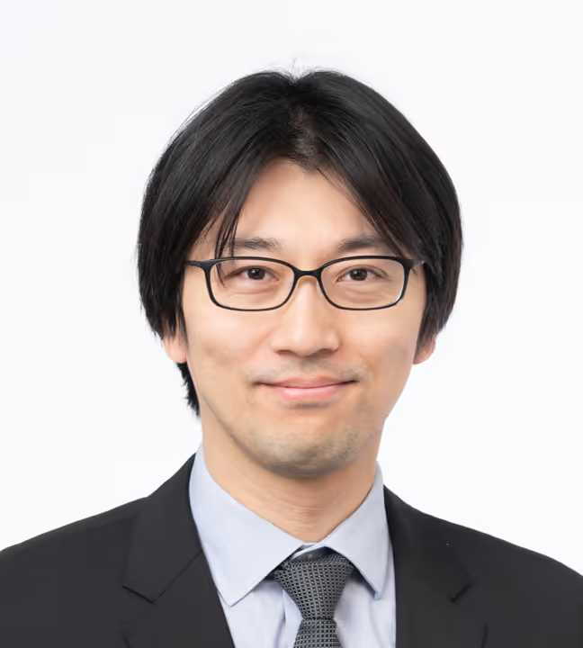
After finishing Ph.D. study on RNA modifications at The University of Tokyo, Yoshiho Ikeuchi conducted post-doctoral research in molecular and cellular neuroscience at the Harvard Medical School and Washington University, St. Louis until 2014. After returning to Japan, he has been conducting research on functional interrogation and engineering of neural organoids as a Principal Scientist at the Institute of Industrial Science, The University of Tokyo.
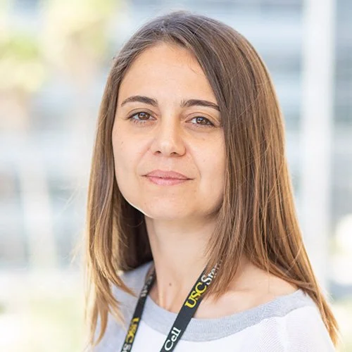
Giorgia Quadrato is an Associate Professor at USC’s Department of Stem Cell Biology and Regenerative Medicine. She is also the Director of the USC CIRM ASCEND Shared Resource Laboratory, dedicated to offering organoids and other stem cell-based models, analyses, consultations, and in-depth education and training. Dr. Quadrato’s lab focuses on understanding the cellular and molecular basis of human brain development and disease. By combining emerging models of the human brain with single-cell omics approaches, her lab aims to identify cell-type-specific disease mechanisms and, above all, new treatments for human neurodevelopmental disorders. Quadrato received the 2019 Donald E. and Delia B. Baxter Foundation Faculty Scholar Award, the 2020 Edward Mallinckrodt, Jr. Foundation Early Career Faculty Award, and the 2022 Broad Innovation Award. Since 2024, she has been a member of the Scientific Advisory Board of Cure Syngap1, and Quiver Bioscience.
Workshops

On September 18, attendees can choose from several parallel workshop sessions. You will be invited to indicate your preferred workshop in advance. As places for some sessions are limited, allocations will be made based on availability, and we will make every effort to accommodate your preference.
Microfluidic Patterning on High-Density Microelectrode Arrays
Led by: Dr. Benedikt Maurer, Joël Küchler
Location: Prof. Janos Voros Lab, ETH Zurich
Combining neuronal networks on microelectrode arrays (MEAs) with microfluidic patterning devices enables powerful experimental paradigms that are not achievable with standard planar cultures. Polydimethylsiloxane (PDMS) microstructures can be bonded directly to MaxWell CMOS high-density MEAs, confining cell bodies while allowing neurites to connect through microchannels.
This enables microcircuit formation with restrained connectivity, co-culture systems for multi-region network models, increases throughput, and prevents cell migration for stable, longitudinal recordings. PDMS membrane placement is recorded through an impedance scan, and cells may be introduced either as single-cell suspensions or pre-formed spheroids. Readouts include axonal conduction assays or analysing the information flow through network architectures.
In this workshop, we will demonstrate the mounting procedure of commercial microstructures, present open-source software based on the MaxWell Python API that facilitates recording workflows, introduce open-source hardware for week-long longitudinal recording, and showcase some applications. Participants will further be able to ask questions regarding their specific experimental requirements.
Plate → Prompt → Publish: Toward Full Lab Automation with Open Culture Science
Led by: Dr. Tjitse van der Molen and Dr. Spencer Seiler
Location: MaxWell Biosystems HQ, Zurich
Open Culture Science's platform provides a team of AI agents you drive entirely through plain-language conversation. A fluidics agent composes and validates microfluidics operations to feed, dose, and maintain your living cultures on your MEAs. An ephys agent writes and runs MaxWell multi-electrode-array experiments for you (activity scans, recordings, stimulation, closed-loop). And SpikeLab builds and runs your whole spike-sorting and analysis pipeline, taking you from raw signals to sorted spikes to manuscript-ready figures. Everything runs through a single chat interface: you make any request in one place, from a one-off analysis to a complete multi-week experiment that keeps cultures healthy, records, stimulates, and analyzes on its own from a plan you approve. Once approved, every subsequent step runs autonomously with no further involvement from you, even overnight while you sleep. In this workshop you'll learn, through guided demonstrations, how to use these agents to automate and accelerate your work end to end: keeping cultures healthy with automated fluidics, designing and running an experiment on the MEA, and turning raw recordings into sorted spikes and publication-quality figures, all without writing a single line of code.
Want to try it on your own data? Bring a laptop with your raw or sorted recording(s), and we'll show you how you can run them through the analysis agents live.
How to Train your Organoid: an Introduction to BrainDance
Led by: Dr. Mircea Teodorescu, Dr. Ash Robbins, and Hunter Schweiger
Location: MaxWell Biosystems HQ, Zurich
BrainDance is a powerful, scalable foundation for neural stimulation experiments with live tissue. Its modular design lets you compose experiments like building blocks: mix, sequence, and reuse experimental phases to match the demands of your research without starting from scratch each time. It offers researchers real-time control over stimulation parameters and native integration with MaxWell CMOS HD-MEAs keeping hardware and software in lockstep.
This workshop will serve as a quick start to setting up neural stimulation experiments with BrainDance.
Unlocking Functional Insights From Neural Organoids
Led by: MaxWell Biosystems' Product & Application Team
Location: MaxWell Biosystems HQ, Zurich
Neural organoids derived from human induced pluripotent stem cells (hiPSCs) are emerging as powerful models for studying brain development, disease mechanisms, and drug response. As the field advances, high-resolution tools to assess the functional properties of these models are becoming essential.
MaxWell Biosystems' High-Density Microelectrode Array (HD-MEA) technology meets this need of capturing label-free neuronal activity at the network, single-cell, and sub-cellular levels with robust, reproducible results. The MaxOne (single-well) and MaxTwo (multi-well) platforms are designed to work across a broad range of scientific applications.
In this workshop, participants will:
- Walk through organoid plating on the HD-MEA chip step by step
- Explore high-resolution functional imaging of iPSC-derived neural models
- Review datasets and analyses from brain organoids representing different regions
- Learn how HD-MEA technology supports compound testing and functional characterization
Registration
Registration to the Symposium includes access to the talks on September 16th and 17th, together with breakfast, lunch and coffee breaks on both event days.
The symposium dinner on Day 2 and the participation to the workshop(s) on September 18th requires a separate ticket, which can be selected during registration.
If registering for the dinner and/or workshop at the same time as registering for the event:
- Click on “Continue” after choosing your ticket type
- Add a ticket labeled “Dinner Registration” and/or "Workshop Day Registration"
- At checkout, fill in your information and press “Continue to Ticket Info”
- Fill in the fields required
- Continue to payment
If registering for the dinner and/or workshop after having registered for the symposium:
- Click on “Skip to More Tickets” when shown the different ticket types
- Add a ticket labeled “Dinner Registration” and/or Workshop Day Registration
- At checkout, fill in your information and press “Continue to Ticket Info”
- Fill in the fields required
- Continue to payment
Discounts
Early bird registration provides a 15% discount and is available until July 31st 2026 using the code “EARLYBIRD” when registering for the event.
Group discounts for labs or companies that wish to register 3 or more people are available upon request by contacting marketing@mxwbio.com.
Cancellation Policy:
Cancellations received 14 days prior to the event will receive a full refund, minus any applicable administrative fees. Cancellations after the deadline will not be eligible for a refund.
Pricing
493 €
580 €
297.5 €
350 €
204 €
240 €
160 €
160 €
110 €
110 €
70 €
70 €
55 €
55 €
Interested in presenting your own work? Make sure the read the next section before you register!
Present Your Research at NeuMoS 2026
Interested in presenting your work? At NeuMoS 2026, all attendees are invited to submit abstracts for poster presentations and short talks. This is a unique opportunity to showcase your latest research, engage with leading experts in the field, and connect with fellow researchers from academia and industry.
To recognize outstanding contributions, attendees and members of the scientific committee will vote for their favorite posters in the Disease Modeling and Neurocomputing categories. The winners will each receive a 500 EUR prize voucher in recognition of their exceptional work and contribution to the NeuMoS community.
We encourage researchers at all career stages to submit their work and join the conversation shaping the future of neuroscience.
How to submit
Abstracts can be submitted after registering via the registration link.
If submitting an abstract at the same time as registering for the event:
- Click on “Continue” after choosing your ticket type
- Add a ticket labeled “Abstract Submission” (free of charge)
- At checkout, fill in your information and press “Continue to Ticket Info”
- Fill in the fields required, including title of the poster/short talk, the list of authors and the abstract
- Continue to payment
If submitting an abstract after having registered for the symposium:
- Click on “Skip to More Tickets” when shown the different ticket types
- Add a ticket labeled “Abstract Submission” (free of charge)
- At checkout, fill in your information and press “Continue to Ticket Info”
- Fill in the fields required, including the title of the poster/short talk, the list of authors and the abstract
Submission Guidelines
- Abstracts must be written in English and may not exceed 2,500 characters, including references
- Figures, images, and attachments are not permitted
- Posters should be printed in A0 portrait format; other portrait formats are also accepted
- Only one abstract per presenting author
- Only registered NeuMoS 2026 participants are eligible to submit
Submission Deadlines
- Short talk consideration: August 1st 2026
- Poster consideration: August 28th 2026
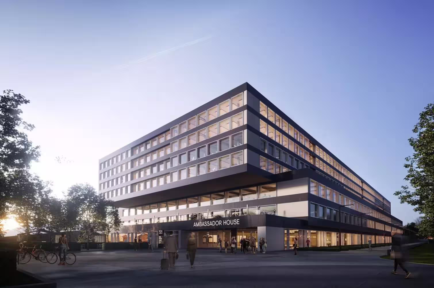
Travel & Accomodation
The conference venue is easily accessible by public transport:
How to get there
From Zurich Airport:
Take the tram number 10 or 12 to the stop Glattpark. The venue is a 5 min walk from there.
From Zurich HB:
Take the tram number 10 to the stop Glattpark. The venue is a 5 min walk from there.
Accommodation
Are you looking for somewhere to stay that is conveniently located close to the venue?
Sponsors




Join us as a sponsor or exhibitor at NeuMoS 2026. This exclusive in-person event is a key meeting point for the global neuroscience community, offering a unique opportunity for sponsors and exhibitors to connect with international experts and showcase their innovations to a diverse, engaged audience. In our previous editions, we have had significant global participation, providing an unparalleled platform for networking and collaboration. For more tailored packages, please contact our Event and Account Manager Ines Blanc Giro (ines.blancgiro@mxwbio.com) or our Head of Commercial Excellence and Engagement Dr. Laura D’Ignazio (laura.dignazio@mxwbio.com)