Publication
Identification of secondary microglial formation centers in the human fetal brain
Abstract
Details
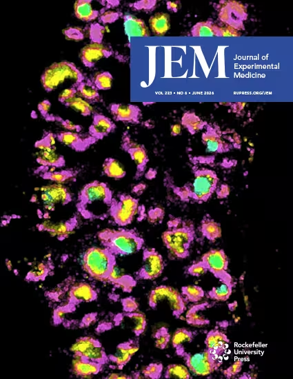
Microglia migrate from the yolk sac and populate the developing brain. How microglia expand rapidly to meet the microglial demand in fast-expanding human fetal brains remains uncharted. Using thick sections in 5−22–gestational week (gw) brains and super-resolution scanning, we identified a large proliferative microglial aggregate (2.129 mm2) near the lateral ganglionic eminence (>12.5 gw), expanding in Down’s syndrome (DS) (4.767 mm2) and Edwards syndrome (ES) (3.437 mm2) fetal brains. Ki67+ microglia within the aggregates accounted for 26.65% (DS: 38.9%; ES: 46.3%) compared with 6.32% (DS: 6.01%; ES: 5.2%) in scattered microglia. This aggregate region contained a distinct microglial population characterized by the absence of phagocytic structures and complex processes, high CSF-1R expression, abundant IL-34+ cells, and some SPP1+ bipolar microglia. We termed this structure the secondary microglial formation center (SMFC). Chimeric microglia–human cortical organoids recapitulated the SMFC in an IL-34– and CSF-1R–dependent manner, indicating that the human SMFC may compensate for the microglial shortage during the fastest expansion period.