Why Flexibility Matters in HD-MEA Recordings
%2520Li.avif)
How Switch Matrix technology combines dense electrode coverage, low-noise recording, and flexible electrode selection for high-content electrophysiology.
Author
Date
Tags
The importance of MaxWell Biosystems’ Switch Matrix technology for high-quality electrophysiology data
Choosing where to look is important.
In medical imaging, for example, an MRI scan is useful because it combines coverage, resolution, and sensitivity. It helps doctors examine the relevant area, detect small structures, and distinguish meaningful signals from background. For scientific data, the same principles matter: you want to observe the right place, with sufficient resolution, and with enough signal quality to trust what you see.
The same is true for electrophysiology data from valuable biological samples. Neuronal cultures, organoids, and other samples often require many days or weeks of preparation. When it is time to record, the system should help you capture the relevant biological activity with high resolution, high sensitivity, and reproducibility.
At MaxWell Biosystems, we are committed to empowering scientists to collect high-quality electrophysiological data from their samples. Our Switch Matrix technology is a key part of this approach.
High spatial resolution: being close to the biology
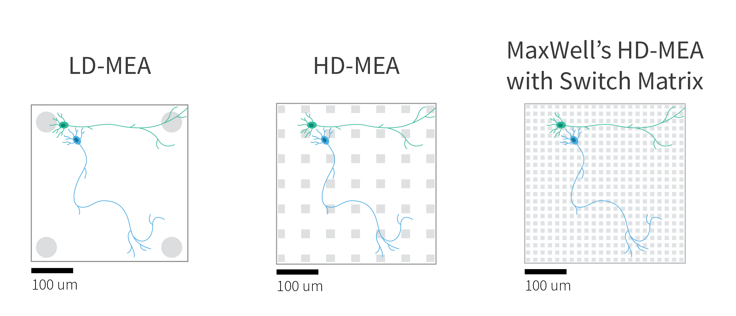
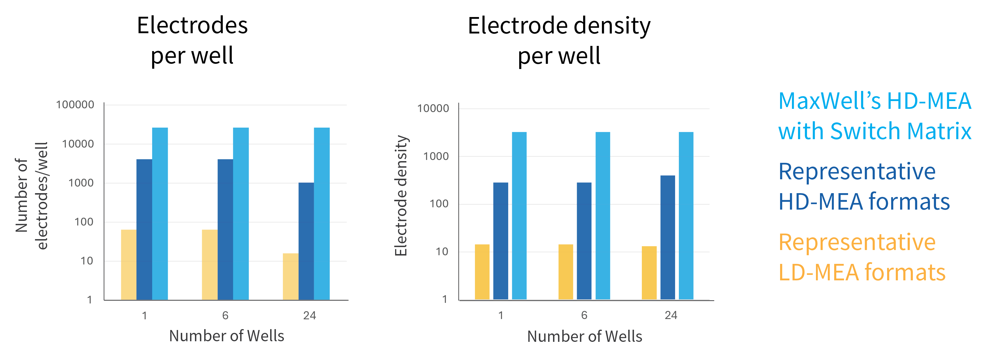
Microelectrode Array technology varies strongly in electrode density. Electrode density describes how closely electrodes are placed to one another. In practical terms, it defines how many possible recording locations are available across the sample.
If electrodes are far apart, relevant signals may fall between them. This is especially important for neuronal samples, where signals may come from individual neurons, thin axons, local network regions, or specific areas of tissue. Extracellular signals are local: the closer an electrode is to the signal source, the more likely it is to capture a strong and informative signal.
A dense array gives the system many possible recording locations. This increases the chance of recording close to the cells, axons, or tissue regions that generate the signals of interest. It also helps reduce spatial averaging of small, localized signals, which is important for resolving features such as axonal activity or single-neuron extracellular action potentials.
In short, electrode density matters because it increases the chance of being at the right spot.
Dense electrode coverage also helps with spike sorting. When the signal from one neuron is captured by several nearby electrodes, the system records not only when the neuron fired, but also its spatial electrical footprint. This additional spatial information helps distinguish activity from nearby neurons and supports spike-sorting approaches, including source-separation methods used in HD-MEA analysis. In other words, dense electrodes do not only help detect signals; they also help assign signals to the right neuronal sources.
High sensitivity: detecting small signals
Being at the right place is only one part of the challenge. The recording system must also be sensitive enough to preserve small biological signals, such as extracellular action potentials, without adding excessive electrode or amplifier noise.
A simple analogy is a camera sensor in low light. If the camera electronics add too much noise, fine details are lost even if the object is in focus. With lower-noise electronics, small details remain visible and can be measured more reliably. Similarly, low-noise electrodes and recording channels help preserve small extracellular signals so they can be detected and analyzed.
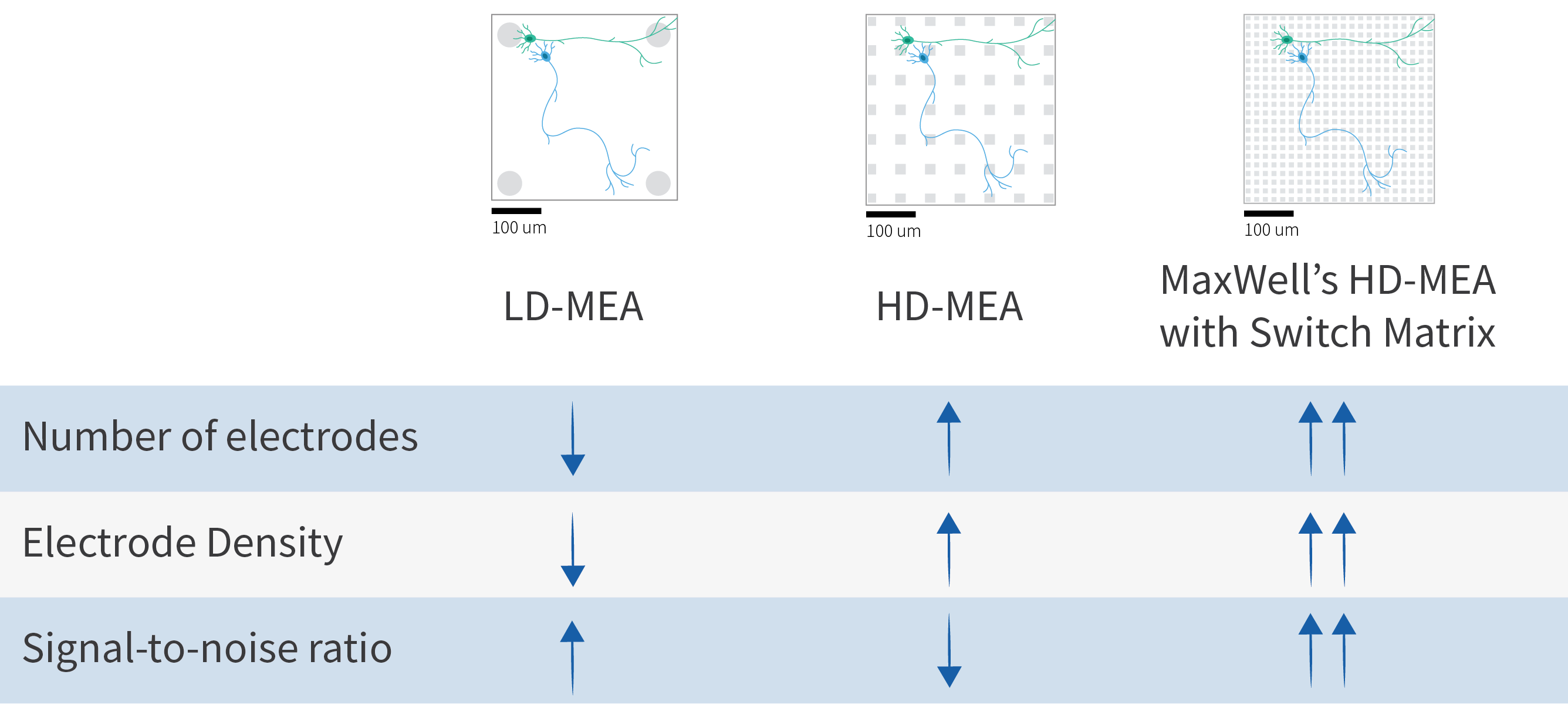
For high-quality MEA data, scientists therefore need both dense spatial coverage and high sensitivity. Dense coverage helps the system record close to the biological signal source. High sensitivity, supported by low-noise electrodes and readout channels, helps preserve small extracellular signals so they remain distinguishable from the noise introduced by the recording chain.

The HD-MEA tradeoff
This creates an important technical tradeoff. Dense electrode arrays need many small electrodes placed close together. At the same time, low-noise readout circuits require area and power, and small electrodes must be matched carefully to the recording electronics to avoid signal loss and excess noise. If too much circuitry needs to be squeezed into every electrode pixel, noise, heat, and bandwidth can become limiting factors.
In other words, more electrodes alone do not automatically mean better data. The architecture of the MEA matters. An HD-MEA should provide many possible recording locations, but also preserve low-noise readout and sufficient flexibility to record from the electrodes that are most relevant for the experiment.
The switch-matrix architecture was developed to address this fundamental challenge. MaxWell Biosystems applies this principle across its HD-MEA platforms.
Instead of permanently wiring every electrode to a fixed channel, the Switch Matrix allows a large number of densely packed electrodes to be flexibly connected to high-quality recording and stimulation channels. This means the system can provide many possible recording locations while maintaining low-noise readout from the electrodes selected for the experiment. The dense electrode layout also provides the spatial information needed for advanced analyses such as spike sorting and axonal tracking.
The switch-matrix architecture has become one of the defining concepts of modern high-density electrophysiology. It underpins state-of-the-art in vitro HD-MEAs as well as in vivo technologies such as Neuropixels. Rather than recording from every electrode simultaneously, a large array of electrodes is dynamically connected to a smaller number of high-performance recording channels. This approach combines extremely dense spatial sampling with low-noise recording, practical power consumption, and efficient use of silicon area.
The underlying technology tradeoffs of MEA and HD-MEA architectures, including fixed-wiring, multiplexed arrays, full-frame readout, and switch-matrix approaches, were reviewed by Obien, Deligkaris, Bullmann, Bakkum, and Frey in the highly cited Frontiers in Neuroscience review “Revealing neuronal function through microelectrode array recordings.”
Scan first, then focus
The practical benefit of Switch Matrix technology is flexibility.
A typical workflow starts by scanning across the electrode array to generate an activity map of the sample. This gives a high-resolution overview of where relevant electrical activity is located. Similar to checking cells under a microscope before starting an experiment, ActivityScan helps users assess whether their sample is active and where that activity is found.
Based on this information, users can then focus on the most relevant electrodes for the next step of the experiment. Depending on the assay, these electrodes may be located around active neurons, along axonal projections, beneath a specific part of an organoid, across a network, or around stimulation sites.
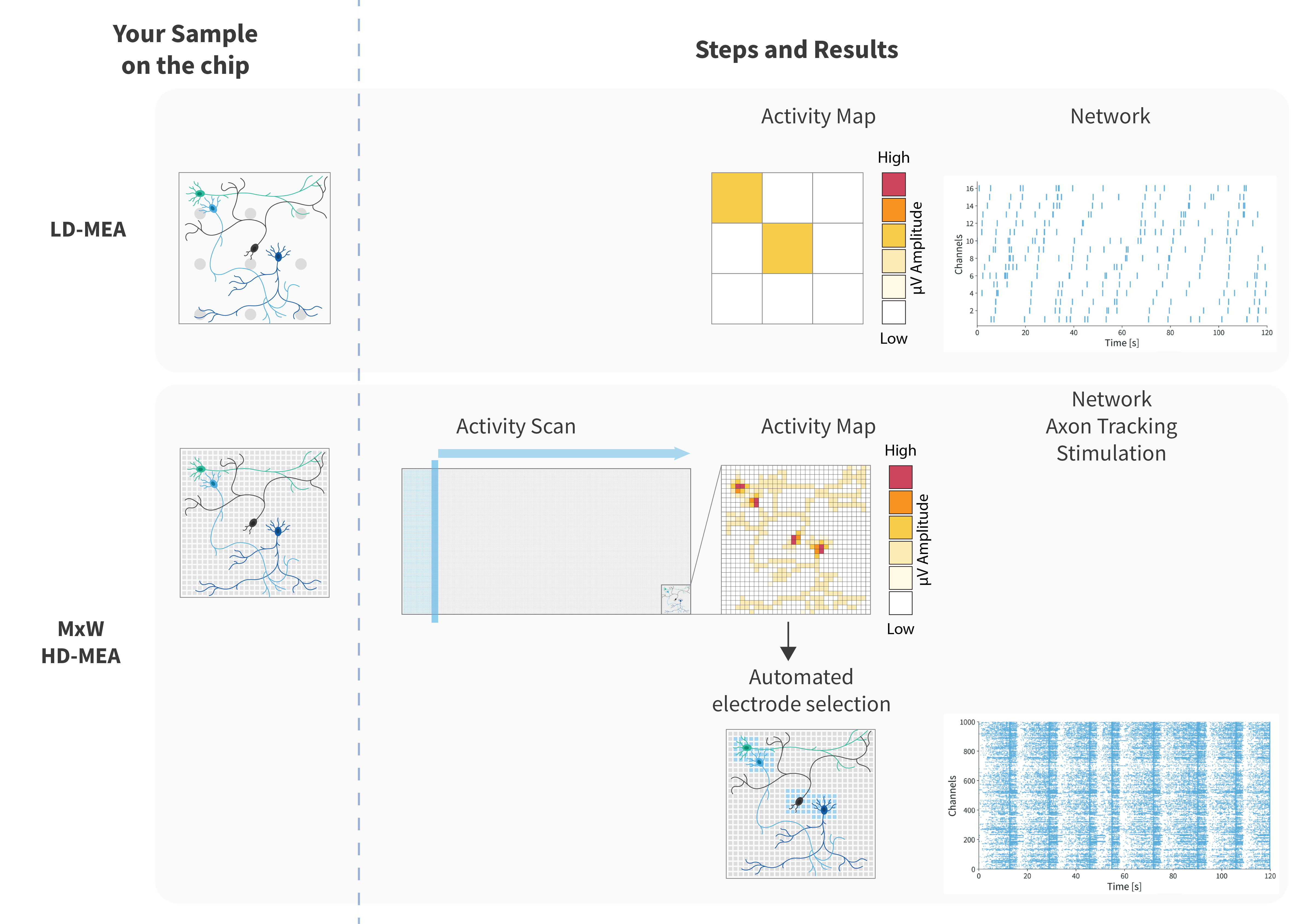
Importantly, this flexibility does not mean that users have to manually configure every experiment. In MaxLab Live, electrode selection is guided by assay workflows and can easily be automated based on activity maps and experimental goals. The same selection logic can be applied consistently across wells, samples, and experiments. This allows users to keep workflows simple and reproducible while still recording from the electrodes that best match the biology.
Fixed grid when you want it, more flexibility when you need it
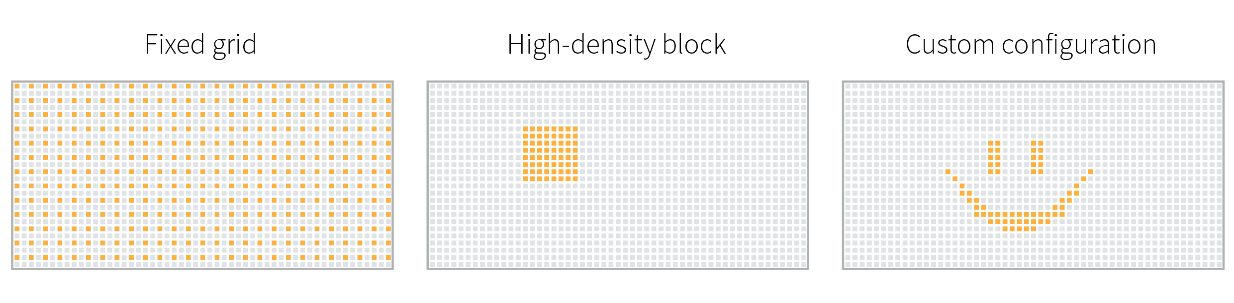
Switch Matrix technology also allows users to record from fixed-grid configurations. This means users can choose familiar electrode layouts when desired.
The difference is that fixed-grid recording is only one option. With the same system, users can also select a high-density block, a sparse network-wide selection, individual electrodes, stimulation sites, or a custom configuration adapted to the sample. In other words, Switch Matrix gives users the option to record in a fixed grid, but does not limit them to one.
This is especially useful because different biological preparations have different requirements. Neuronal cultures may require sparse or dense recording patterns depending on maturation and network structure. Organoids are precious, variable, and three-dimensional, so relevant activity may occur below only part of the tissue. Retina and brain slice experiments often require recording from defined anatomical regions, following axonal signals, or combining recording with stimulation. Microphysiological systems may have engineered layouts such as compartments, channels, or connected modules.
For disease modeling, pharmacology, toxicology, and functional phenotyping, reproducibility is critical. Subtle differences in firing rate, bursting, synchrony, conduction, or network organization should reflect biology, not electrode placement or sampling limitations. Flexible HD-MEA recording helps reduce this risk by allowing each sample to be measured where it is most informative.
High-content electrophysiology across formats
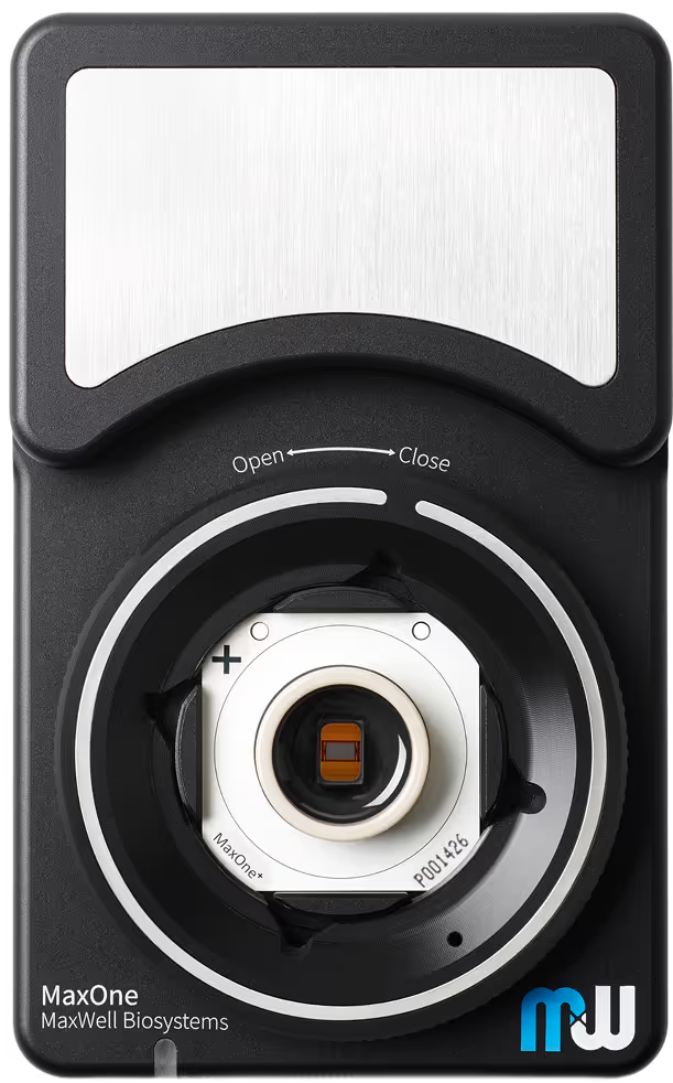
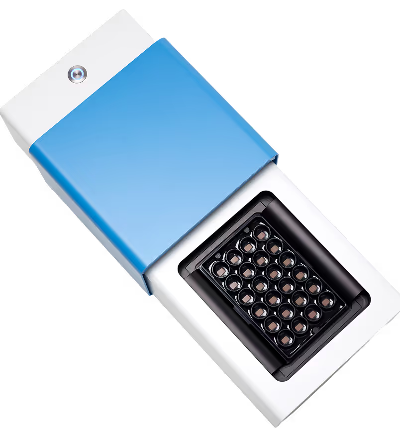
MaxWell Biosystems applies Switch Matrix technology across its HD-MEA platforms. Each MaxOne chip and each well of the MaxTwo 6-Well and 24-Well Plates contains 26,400 electrodes and 1,020 flexible recording channels. This allows researchers to work with the same core recording principle across different experimental formats.
This becomes especially important when HD-MEA technology scales to multi-well plates. More wells mean more possible recording locations, but also more data and higher bandwidth requirements. In any high-density architecture, the system must decide what is recorded continuously, what is selected, and what is reduced before or during acquisition. These choices can affect practical aspects that matter in real experiments, including recording duration, signal quality, and flexibility.
Switch Matrix technology addresses this challenge by selecting the relevant electrodes before detailed recording, instead of forcing the system to stream everything from everywhere all the time. This keeps the data focused on the biologically meaningful signals while preserving long recordings, low-noise readout, stimulation flexibility, and high-content information per well.
For exploratory experiments, users can investigate samples in high detail. For larger studies, they can scale to multi-well formats while maintaining high-content electrophysiological readouts. The goal is not only to record more data, but to record the right data, for long enough, with high signal quality, across the right biological context.

Urs Frey
"When we developed the Switch Matrix concept, the goal was not simply to build an MEA with more electrodes. The goal was to give scientists the freedom to find the biologically relevant signals first, and then record them with the best possible signal quality. This principle has guided the technology from the first switch-matrix HD-MEA prototypes to today’s multi-well platforms: high throughput should not mean giving up signal quality, recording duration, flexibility, or the ability to adapt the experiment to the biology."
Did you know?
Are your samples ready or mature enough to be recorded?
A quick ActivityScan in MaxLab Live can help you check electrical activity in just a few clicks. Just like how you would check under a microscope how well your cells in your cultured sample are adhering or growing, why not check regularly to see how active they currently are?
In which configurations can you record?
With MaxWell Biosystems’ Switch Matrix technology, you can record from a fixed grid when desired, without first running a scan. You can also select individual electrodes, groups of electrodes, high-density blocks, or custom configurations, depending on your experimental question.
How easy is it to scan and select electrodes?
With MaxWell Biosystems’ dedicated MaxLab Live software, users can scan their samples with just a few clicks with our ActivityScan Assay. Automatically choose which electrodes to use in your subsequent recordings based on your experimental needs also with a few clicks with our Network, AxonTracking, and Stimulation Assays.
With our MaxLab Live software, you can also set up your recordings to run in a sequence. Grab a coffee or perform other experiments on the side!
Further readings
For readers who would like to go deeper into the scientific and technical background of Switch Matrix HD-MEA technology, we recommend the following publications:
- Technology overview: A review of MEA signal generation, MEA architectures, and the technology tradeoffs behind high-density electrophysiology.
- Original switch-matrix circuit publication: A detailed circuit description of the first switch-matrix CMOS HD-MEA, combining dense electrodes, flexible routing, recording, and stimulation.
- Application-focused platform paper: An application-focused demonstration of the 26,400-electrode platform for network recordings, single-neuron analysis, axonal tracking, and stimulation.
Related
Resources
Our Technology
Multi-/Micro-electrode Arrays

MaxOne
Versatility and functionality in one compact device
MaxTwo
Maximize your cell functional assays
MaxLab Live
All-in-one Software
Neuronal Cell Cultures
Organoids
Brain Slices
Retina
Cardio and others
Functional Phenotyping
Disease Modeling
Pharmacology & Toxicology
Method Development

Microphysiological Systems
Biocomputing

Revealing neuronal function through microelectrode array recordings

Switch-Matrix-Based High-Density Microelectrode Array in CMOS Technology

High-resolution CMOS MEA platform to study neurons at subcellular, cellular, and network levels