Microphysiological Systems: From Structure to Computation in the Brain

From organoids to engineered microstructures, MPS let researchers build structure into neural circuits rather than just observe it, opening a path to studying brain computation.
Author
Date
Tags
What are Microphysiological Systems (MPS)?
For decades, conventional 2D cell culture has been the workhorse of biological research, enabling countless discoveries. Yet reproducing the complex physical organization that underlies tissue function in vivo remains challenging in a flat, unstructured monolayer.
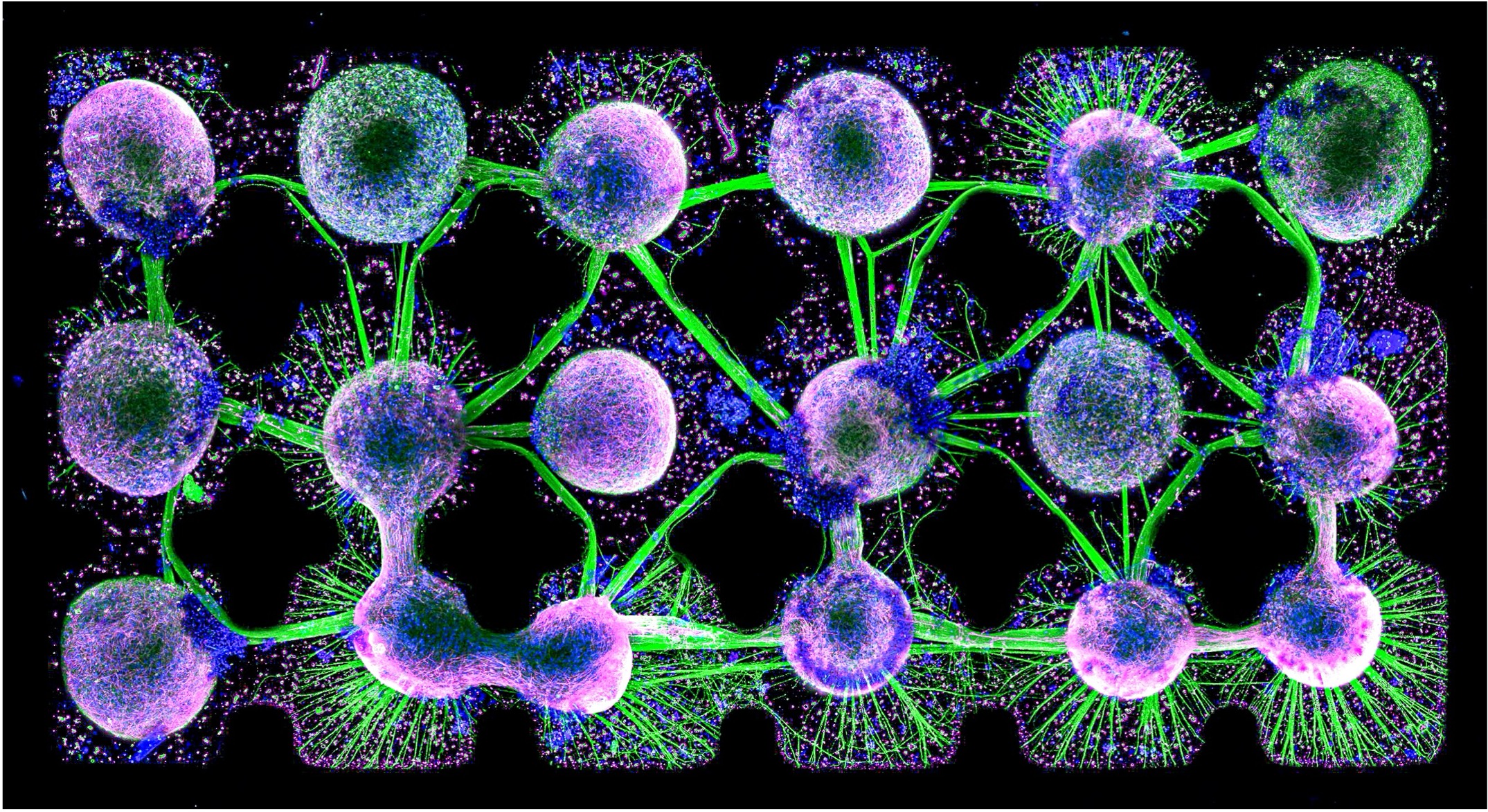
Microphysiological Systems (MPS) have emerged to close this gap by recreating key structural, biochemical, electrical, and mechanical features of the native cellular environment in vitro. Rather than describing a single technology, MPS span a broad and evolving spectrum of in vitro models, from self-organizing organoids to engineered organ-on-a-chip platforms that integrate living tissue with microfluidics. What unites them is architecture: by introducing defined structure and controlled microenvironments, MPS produce models that are more reproducible and more representative of human biology than traditional culture, establishing them as a cornerstone of New Approach Methodologies (NAMs) for basic research, disease modelling, drug discovery, and, increasingly, the study of neural computation.
How structure shapes neural function
Structure and function are closely intertwined throughout biology. The three-dimensional folding of a protein determines its activity; the architecture of a tissue determines how it behaves. The nervous system follows the same principle: how neurons and glia are arranged, connected, and embedded in their microenvironment shapes how information is processed, transmitted, and transformed into function.
This relationship isn't deterministic, though. Network architecture strongly constrains the activity patterns a circuit can generate, but it doesn't uniquely define them. Neural computation stays dynamic and adaptive, emerging from the interplay of topology, cell properties, and experience. Structure conditions function rather than dictating it.
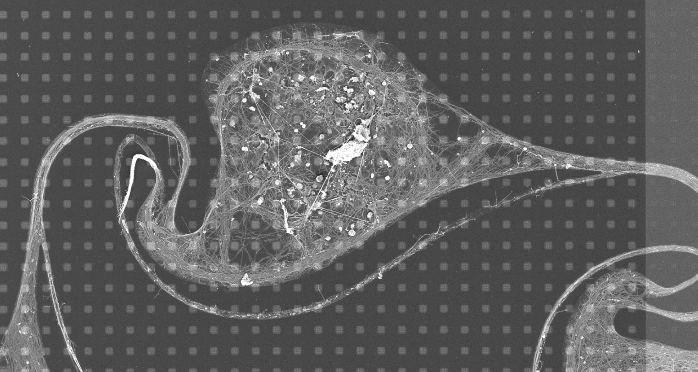
That distinction is what makes MPS such powerful research tools. Function is difficult to engineer directly, but architecture, cell composition, and network topology can be controlled with increasing precision. By defining how a circuit is built, researchers can investigate how structural changes shape connectivity, information processing, learning, and disease progression, turning MPS into a bottom-up framework for studying the link between structure and function.
Why HD-MEAs are ideal for Microphysiological Systems
MPS place new demands on recording technology. As biological models grow more structured and detailed, studying them requires high spatial resolution, low-noise recordings, and the flexibility to accommodate very different samples on a single platform.
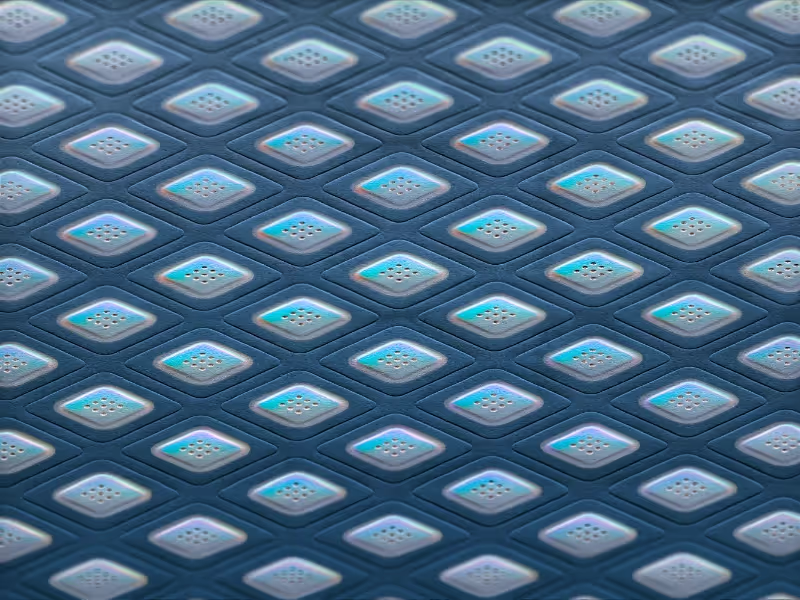
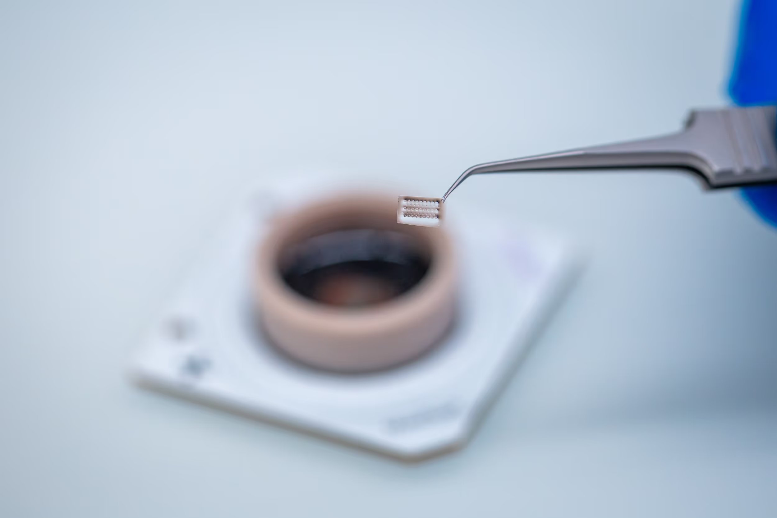
The first challenge is physical integration. Different classes of MPS place different demands on the recording surface: a nearly flat chip topography favors the attachment of PDMS-based microstructures and organ-on-a-chip devices, while a biocompatible PEDOT-coated surface promotes strong cell adhesion and long-term culture stability, well suited to organoids and other 3D neural models. Together, these surface properties support a broad range of MPS on a single platform.
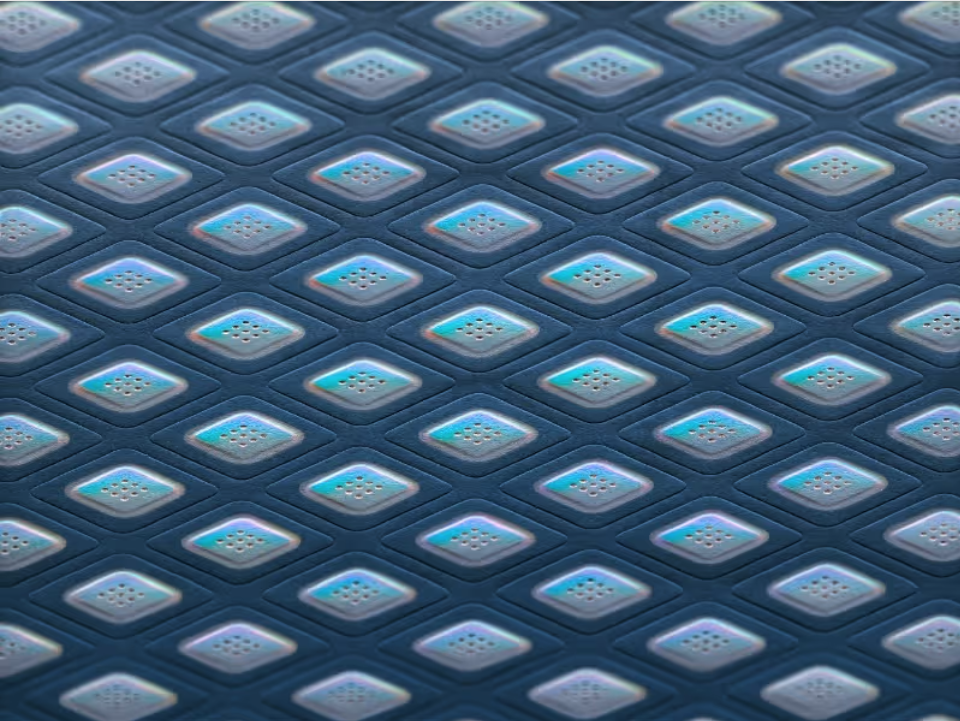
Once a sample is prepared, the next step is locating where to measure. MaxWell Biosystems' Switch Matrix technology, covered in more detail in a previous post, gives researchers this flexibility: an activity scan can rapidly map the array to find active neuronal populations, while the same flexible electrode selection can also identify the open compartments and tunnels within an engineered MPS structure and record there selectively.
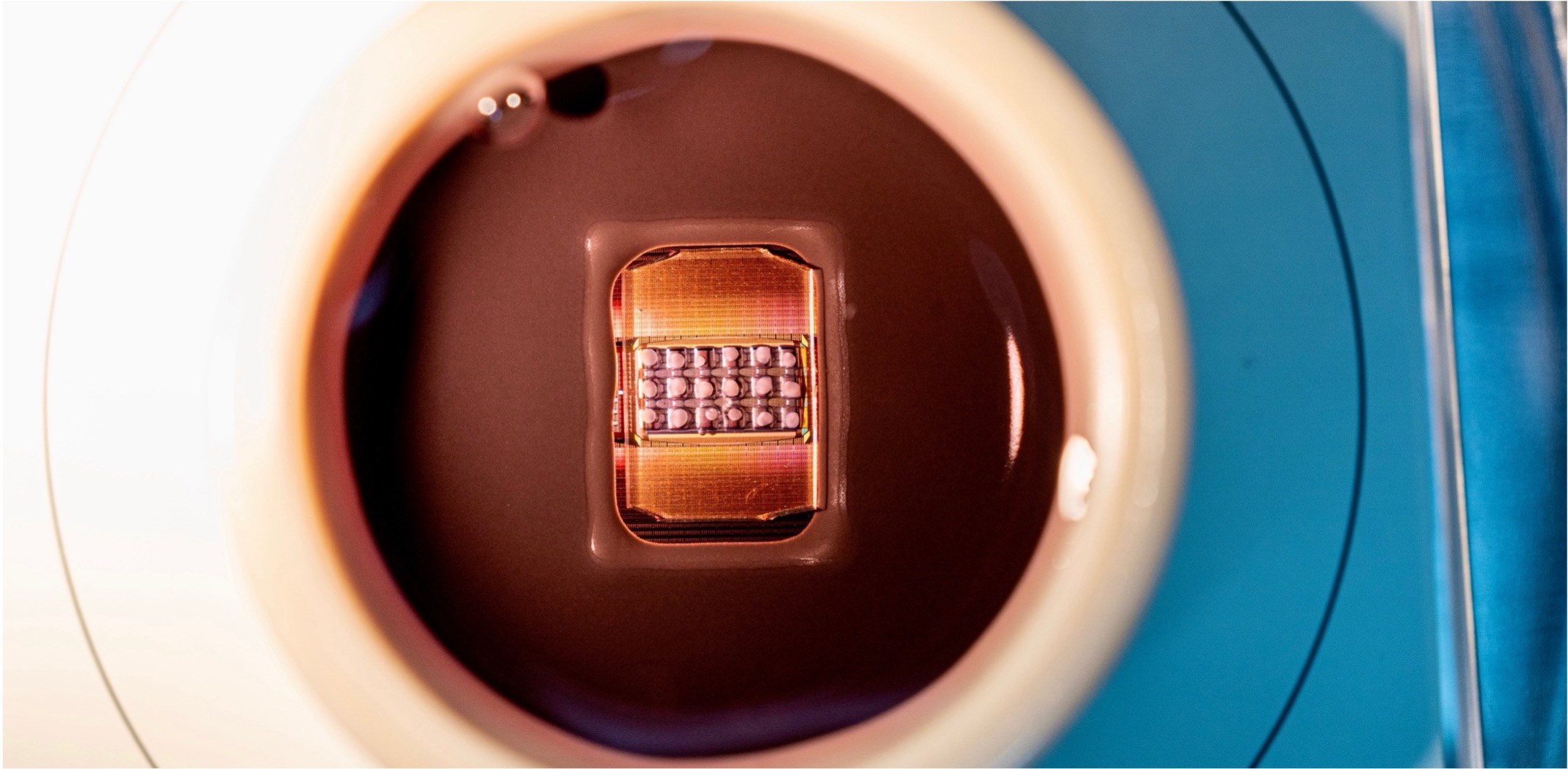
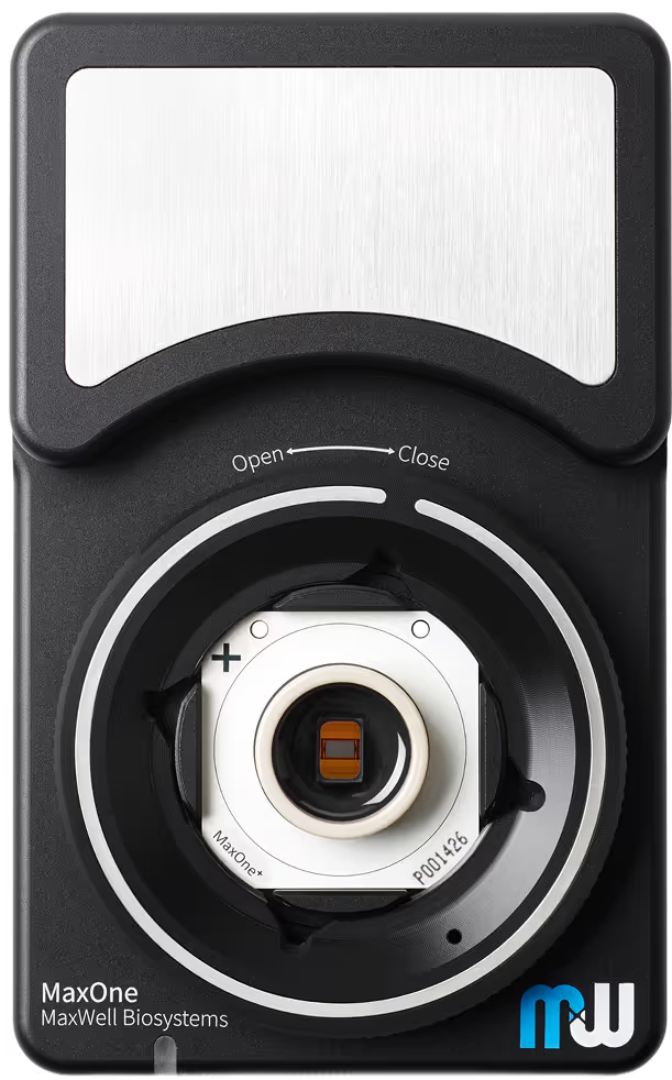
With the sample mapped, the recording configuration can be adapted to it. MaxOne and MaxTwo integrate 26,400 electrodes at a 17.5 μm pitch, dense enough to resolve individual cells and axonal action potential propagation, not just population-level activity. The same Switch Matrix architecture allows any electrodes to be routed for recording or stimulation, so the electrodes used to find cells can then be used to record and stimulate them. Depending on the experiment, researchers can rely on a built-in software suite for standard workflows or a programmable API for custom protocols and closed-loop paradigms.
Rather than constraining the biological model to the recording technology, MaxWell Biosystems' HD-MEAs let the recording technology adapt to the biological model.
Where organoids and engineered circuits meet computation
With biological structure now something researchers can build and read out with precision, a growing body of work is using the full width of the MPS spectrum to study one of the fundamental questions in neuroscience: how neural circuits give rise to computation.
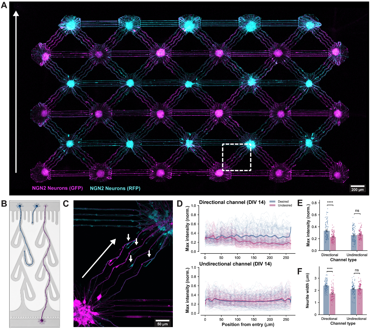
One example of the engineered side of that spectrum comes from a group at ETH Zurich, whose work over the past several years traces a clear progression. Duru et al. (2022) showed that confining neurons within PDMS microstructures could enforce directional signal propagation between compartments, an early demonstration that network topology could be built rather than just observed. Duru et al. (2023) proceeded to use similar structures to quantify input-output relationships between connected populations, and, more recently, Küchler et al. (2025) showed that circuits built this way can perform basic Boolean logic directly in living tissue. Having built up the tools and understanding needed to reliably impose structure on these networks, they have also begun applying that control to a wider range of questions, from biocomputing to disease modelling: in a newer preprint, for instance, Küchler et al. (2026) applied these compartmentalized circuits to study how neurodegeneration spreads through a layered network.
Working with human neural organoids, Alam El Din et al. (2025) recently showed that they spontaneously develop essentially all of the components needed for basic learning and memory: synapse formation, glutamatergic and GABAergic receptor expression, functional connectivity, and synaptic plasticity in response to theta-burst stimulation. Furthermore, van der Molen et al. (2025) reported novel mechanistic insights into how that capacity arises: structured firing sequences, a basic building block of information broadcasting in the brain, appear spontaneously in brain organoids but not in dissociated primary cortical cultures, suggesting they aren't learned through experience but are instead constrained by a preconfigured architecture established during neurodevelopment.
Organoids are also being combined with microfluidic tools, connecting multiple organoids into modular networks known as connectoids. Duenki and Ikeuchi (2026) showed that as more organoids are linked into such networks, the resulting activity becomes progressively richer and more complex, with dynamics shifting closer to the kind of critical state associated with efficient information processing in the brain. In another recent preprint, Chow et al. (2025) found that connectoids consisting of three connected organoids developed the ability to reliably discriminate between two input signals after two weeks of daily stimulation, something neither a single organoid nor a connected pair achieved, pointing to hierarchical, multi-organoid connectivity as a key ingredient for functional learning.
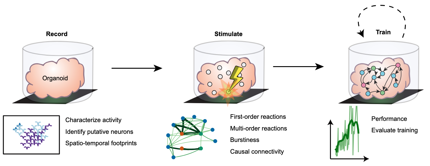
While the study of biological computation has been around for decades, the field has recently gained a lot of traction in both scientific circles and the news. Kagan et al. (2022) interfaced neuronal cultures with MaxWells' HD-MEA to embed them in a simulated game-world ("Pong") and leveraged the free-energy principle to induce goal-directed learning. Cai et al. (2023) showed that a brain organoid connected to a high-density electrode array can function as a biological reservoir computer: by applying spatiotemporal stimulation, they achieved nonlinear and memory-retaining dynamics in the tissue that can be read out for tasks like speech recognition and predicting the behaviour of nonlinear equations, with the organoid learning in an unsupervised manner by reshaping its own functional connectivity. Robbins et al. (2026) demonstrated goal-directed learning in brain organoids, developing a closed-loop framework to embody mouse cortical organoids into a simulated pole-balancing task (“cartpole”). They found that training signals chosen by reinforcement learning improved performance compared to random stimulation, and that the strongest predictor of how well an organoid learned was which neurons were stimulated, based on their connectivity to the rest of the network. Sono et al. (2026) offered a possible reason why controlling connectivity matters so much: using microfluidic devices to impose a defined topology on a cultured network reduced excessive synchrony and increased its dynamic complexity, which in turn allowed the network to be trained to reliably generate specific temporal patterns.
Across the MPS spectrum, progress has depended on the ability to observe and interact with these systems at high spatial and temporal resolution. As MPS models continue to grow in sophistication, HD-MEA technology is helping researchers keep pace, supporting the next stage of in vitro neuroscience as it moves from characterizing biological systems to actively building and testing them. The field is moving quickly, and it's an exciting time to be part of it.
Related
Resources
MaxOne
Versatility and functionality in one compact device
MaxLab Live
All-in-one Software

Our Technology
Multi-/Micro-electrode Arrays
Microphysiological Systems
Biocomputing

Human neural organoid microphysiological systems show the building blocks necessary for basic learning and memory

Preconfigured neuronal firing sequences in human brain organoids

Engineered Biological Neural Networks on High Density CMOS Microelectrode Arrays

Investigation of the input-output relationship of engineered neural networks using high-density microelectrode arrays
Engineered biological neural networks as basic logic operators

A Microfluidic Platform for Spatiotemporal Dissection of Neurodegeneration Across Hierarchical Human Neural Circuits
Brain organoid reservoir computing for artificial intelligence

Goal-directed learning in cortical organoids

Online supervised learning of temporal patterns in biological neural networks under feedback control

In vitro neurons learn and exhibit sentience when embodied in a simulated game-world

Repetitive stimulation modifies network characteristics of neural organoid circuits