 English
 繁體中文
繁體中文 简体中文
简体中文 日本語
日本語
Interview and Featured Publication
Join the Conversation with Prof. Kenneth Kosik and Dr. Tal Sharf
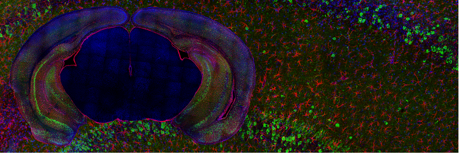
Copyright by Gabriel Luna, Kosik Lab official webpage, https://ken-kosik.mcdb.ucsb.edu
Recently, Dr. Tal Sharf, Prof. Kenneth S. Kosik and colleagues released a pre-print on their novel work entitled, “Human brain organoid networks”, currently under revision. Aiming to explore the physiological behavior of neuronal circuits within organoids, this work demonstrates that human brain organoids have self-organized neuronal assemblies of sufficient size, cellular orientation, and functional connectivity to co-activate and generate field potentials from their collective transmembrane currents that phase lock to spiking activity. These results reveal the potential of brain organoids for the study of neuropsychiatric diseases, drug mechanisms and the effects of external stimuli upon neuronal networks.
Join us in this conversation with Ken and Tal about their fantastic work on human brain organoids and how MaxOne contributed to this exciting story!
In this conversation
 Prof. Kenneth S. Kosik Principal Investigator UC Santa Barbara California, USA |  Dr. Tal Sharf Postdoctoral Researcher UC Santa Barbara California, USA |  Dr. Marie Obien CCO MaxWell Biosystems Zurich, Switzerland |
The interview
Hello Ken and Tal, we are very excited to have this interview with you and thank you for joining us today! To start with, could you tell us about the scientific interests in your group?
 We are very interested in both translational work, that is work that can be applied to medical questions, as well as fundamental basic neuroscience. MaxOne has helped us carry us forward those twin directions.
When MaxOne came out, it was about the same time when we started growing brain organoids in the lab, and so the two technologies converged. While recording the activity of dissociated cultures using MaxOne, we discovered additional directions for further exploration. We emphasized on organoids as this opened so many doors regarding questions of brain wiring, genetics and drug discovery. Organoids gave us access to brain circuitry that cannot be obtained any other way.
We are very much looking forward to hearing about your recent bioRxiv preprint on human brain organoids! What is the key contribution of this work and how did MaxOne play a role in the story?
Organoids are known to be similar in cellular composition and development as the human brain. However, there is little understanding of the functional behavior of neuronal networks within the organoids. One of the things we have been very keen on that MaxOne can do, is to capture broadband signal. This means we were able to look at spikes within organoids with incredible millisecond temporal resolution and high spatial resolution while we can also capture LFPs (local field potentials). The inter-relationship between spikes and LFPs has been very important for people doing animal work for years in which brain encoding is studied. There is a long literature of this kind of phase locking and relevant data in this vein can be acquired using MaxOne.
MaxOne is an amazing platform and is unlocking a lot of possibilities. It can be used to compare what is going on with synchronous neuronal activity, and the signal can be decomposed that into LFPs and spiking. Analyzing these signals allow to understand how human tissue is recapitulating functional phenotypes. In our work, using MaxOne, we provide a roadmap for physiological phenotyping of organoids naturally leading to downstream studies we would like to dive into. This opens a lot of avenues for benchmarking neurodevelopmental disorders in human tissue and screening pharmacological compounds to intervene with pathogenesis with these mapped out phenotypes.
Large-scale acquisition of neuronal network activity enables you to extract a variety of parameters that can be used for phenotyping. Could you elaborate on any novel parameters that you think are important and can only be extracted with high-resolution MEAs (and not possible with other technologies)?
We were using low-resolution arrays before, and this technology can cover a fair amount of real estate, but the problem is that you cannot access functional areas. With MaxOne and its routing capability, we changed the way we can access networks. This opened in our lab the ability to access functional networks, identify regions of interest, select the electrodes with activity, combine that with spike sorting to identify putative single units, and look at statistical relationships between units. Identifying these short latency interactions that are putative synaptic connections allowed us to look at the distribution of probabilities that are occurring between units.
By performing similar experiments with organoids to what was previously reported for 2D cultures by Xinyue Yuan et al (2020, Nature Communications) for short latency interactions, we found something very interesting by looking at the distributions of connection strengths. Those relationships emerged and those distributions of connection strengths are perturbed by the anxiolytic-hypnotic diazepam drug. Also, there is a large amount of evidence that these perturbations should also be manifested in pathological states. This aspect is fundamental to look for and can reveal features that are related to pathological states or genetic mutations.
That’s super cool! For phenotyping and pharmacological studies, reproducibility of results across multiple samples is important. Can you comment on the reproducibility achievable in organoid assays?
With some analytical techniques people claim high reproducibility in developing organoids. However, even then, reproducibility can still be an issue, with regard to cell number distributions, anatomical organization, and other parameters. So, one of the things we wanted to extract were features of organoids that are reproducible over time, regardless of the details of cell layering or cell number distributions.
We have been able to do that, and I do not think we could have done it any way other than with the MaxOne. When we report our data, we do not rely on a single organoid but present recordings from as many as six or seven organoids. When we assemble the circuitry in each of them, we begin to see something that cuts across all the organoids. Those are features like a small number of strong connections and a large number of weak connections. We can look at that distribution and draw a curve and the shape of the distribution was a general feature present in all the organoids which might change in the presence of a drug or a gene mutation.
Another feature is the probability distributions of the inter-spike intervals. We were able to sort out the fitting and show that some of the distributions fit in exponentials, others fit normal curves, and many do not fit any known distributions. The fact that we can establish these kind of probability distributions for inter-spike intervals is another parameter that we can explore when we move to genetics and drugs.
These are unique parameters that you extracted in this work.
How are these translatable?
Yes! Another important question is whether organoids recapitulate functions found in the human brain. We were also able to bridge these parallels: what we see with these distributions and what you see in vivo. Some of the distributions are much more stereotyped and this is giving us the opportunity to dissect the circuit, dissect functional connectivity and phenotype cell types. The MaxOne platform is allowing us to access circuit architecture in a way that is impossible to do in the human brain and the opportunities are really untapped.
We are typically asked if MaxOne can be used to identify different cell types based on activity. Can these parameters you extracted be used to classify cell types?
That is a topic that we are very much looking forward to explore in the future. We will be able to do it, for example, by adding additional technology to the platform. One possibility would be to add cell type specific promoters along with channel rhodopsin and an optogenetic component to the platform. There are other approaches, and this is a direction we certainly will pursue.
It is great to see that your work is opening new avenues in brain research, and we feel very happy to be able to be part of your success and looking forward to your future discoveries! And talking about the future, what is your “dream feature” for MaxWell Biosystems’ HD-MEAs?
I would choose more channels. As reported by Xinyue Yuan et al (2020, Nature Communications) the dual mode chip allows one to record with 19 000 electrodes simultaneously, which is fantastic! Nevertheless, instead of recording a tremendous amount of data, it would be great to be able to do a quick activity scan and then decide to select the top configured channels!
And integrated light stimulation would also be great, which would allow one to combine optogenetic experiments with the high resolution of the system!
Thank you very much! This will for sure be communicated in our next product development meeting. And now, more about you, do you have any hobbies you would like to share with us?
I am a surfer and I enjoy surfing a lot. Since our lab is right at the beach I generally go there quite often, especially when experiments do not work as expected (any scientist understands the feeling).
As Santa Barbara features the best of two worlds: the Pacific Ocean and Santa Ynez Mountains, I do love to go biking in the mountains. The mountains are gorgeous and the view from up there is spectacular!
Thank you so much for sharing! I think all readers, like myself, are now very much looking forward to the publication of this paper and the next groundbreaking results from Kosik Lab!
We at MaxWell Biosystems congratulate Dr. Tal Sharf, Prof. Kenneth S. Kosik, and co-authors for this significant work, which serves as a compelling example of the application of MaxOne/MaxTwo HD-MEA platforms in brain organoids research.
We are very grateful to Ken and Tal for sharing with us their personal insight about this work. We are excited to see how other researchers will build upon this fundamental work towards the future of organoids research.
Join us in our next conversation with another research group. Stay tuned!
Citation
![]()
Tal Sharf, Tjitse van der Molen, Stella M.K. Glasauer, Elmer Guzman, Alessio P. Buccino, Gabriel Luna, Zhouwei Cheng, Morgane Audouard, Kamalini G. Ranasinghe, Kiwamu Kudo, Srikantan S. Nagarajan, Kenneth R. Tovar, Linda R. Petzold, Andreas Hierlemann, Paul K. Hansma, Kenneth S. Kosik, Human brain organoid networks, bioRxiv, September 2021.
bioRxiv preprint doi: https://doi.org/10.1101/2021.01.28.428643
Short Bio
Kenneth S. Kosik is the Harriman Professor of Neuroscience Research and Co-Director of the Neuroscience Research Institute at the University of California, Santa Barbara. Kosik Lab intends to create an intellectual setting conducive to the exploration of fundamental biological processes, particularly those related to the brain and its evolution, and is interested in the underlying molecular basis of plasticity, particularly how protein translation at the synapse affects learning and how impairments of plasticity lead to neurodegenerative diseases.
Tal Sharf is a Postdoctoral Reasearcher in Kosik Lab at the UC Santa Barbara. By utilizing techniques at the intersection of physical science and biology, Tal is developing devices and techniques to investigate neural circuitry with the aim to uncover general rules to explain how they malfunction with disease and mental illness.
Read more about Kosik Lab here.