MaxWell Biosystems HD-MEAs: powered by Switch-Matrix technology
The Switch-Matrix (SM) approach intelligently routes any selected set of electrodes to readout circuits via programmable switches. This enables flexible electrode selection tailored to each sample, enhances signal acquisition at regions of interest, and supports scalable, high-content experimental designs.
In MaxWell Biosystems HD-MEAs (MxW HD-MEAs), any of the 26,400 electrodes can be routed to up to 1,020 readout channels. This flexibility ensures optimal electrode placement to capture neuronal activity where it is most relevant. It also unlocks advanced functional readouts, such as detailed mapping of axonal branches and precise measurement of conduction velocities based on signal propagation.
Up to 32 stimulation buffers on each MxW HD-MEA can be flexibly assigned to any of the 26,400 electrodes, enabling spatially precise electrical stimulation, even at subcellular resolution, while simultaneously recording activity from both nearby and distant neurons. This unmatched flexibility in selecting electrodes for recording and stimulation, combined with exceptional spatiotemporal resolution and electrode coverage, positions MxW HD-MEAs as a transformative platform for neurocomputing. Moreover, the ability to program MaxWell Biosystems’ HD-MEA through a powerful Application Programming Interface (API) empowers users to develop custom systems with the HD-MEA at the core. This level of control and integration supports the creation of next-generation, brain-inspired technologies and advances the pursuit of a true Brain Processing Unit.
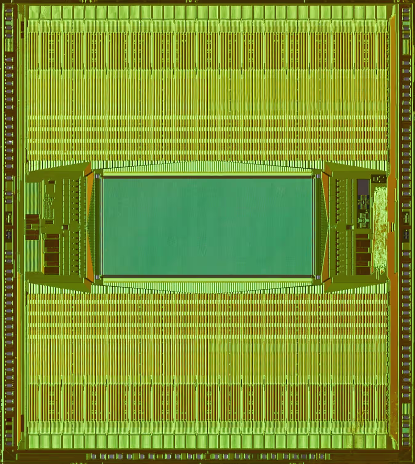
At the core of this capability is MaxWell Biosystems’ Switch-Matrix (SM) technology, a Swiss-engineered architecture that dynamically routes signals across thousands of electrodes. This design achieves an optimal balance of pixel area, power efficiency, and low noise, enabling ultra-high electrode density without compromising signal fidelity.
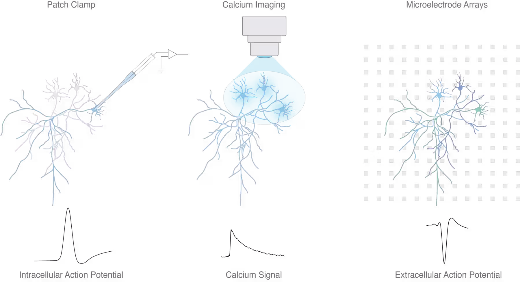
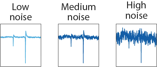
High electrode density ensures single-cell resolution, allowing researchers to precisely capture the most relevant signals at the finest scale. Low noise levels unlock the detection of even the smallest action potentials, which is essential for extracting deep insights and reaching confident conclusions.
For teams in the pharmaceutical and biotechnology sectors, MaxWell Biosystems HD-MEAs empower you to focus on what matters: identifying the most promising signals, capturing a large representative part of the network reducing variability and eliminating noise, driving faster and more confident decision-making in drug discovery and safety testing. For academic researchers, the system offers unprecedented access to the true dynamics of your samples and modulate neuronal activity by stimulation, allowing you to adapt in real time and make new discoveries by uncovering signals that were previously out of reach.
Every cell has a story to tell. With MaxWell Biosystems’ unique HD-MEA, you are empowered to listen to those stories, transforming complex neuronal signals into actionable results, driven by precision and reliability of Swiss innovation.