 English
 繁體中文
繁體中文 简体中文
简体中文 日本語
日本語
MaxOne for Brain Slice Studies 
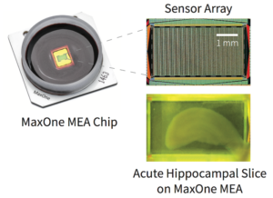
MaxOne enables researchers to perform label-free analysis of intact brain networks in vitro:
- Acute brain slices
- Organotypic brain slice cultures and organoids
- Ex-vivo brain preparation (e.g., eye-brain from turtles)
What You Can Do
Capture Single Neuron and Network-Wide Field Potentials
MaxOne enables recording of neuronal activity across multiple scales at high spatio-temporal resolution.
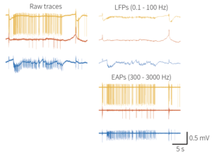
- Both local field potentials and spikes from intact brain networks can be detected simultaneously.
- Low noise signals facilitate the extraction of neuronal activity features from experiments.
- Propagating field potentials across brain areas can be captured and analyzed.
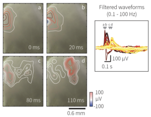
Perform Large-Scale Mapping of Cells and Synaptic Projections
Extract and analyze the action potential spatial fields, axonal projections, and postsynaptic signals of every active neuron in the brain tissue. MaxOne can detect spiking neurons in brain slices and can elicit neuronal activity by electrical stimulation.
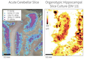
- A neuronal activity map can be extracted to identify areas of the brain slice with spiking neurons.
- Spiking frequency
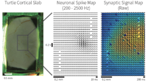
- Postsynaptic events can be revealed by spike-trigerred averaging as a slow +/- signal post-spike. (M. Shein-Idelson, et al., Nat. Methods, 2017)
MaxOne Tissue Holder
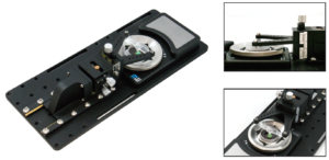
MaxOne Tissue Holder flattens the brain slice on the MEA for stable and reprocible experiments.
The tissue holder keeps the tissue pressed and fixed on the MEA throughout the experiment, in the presence of solution perfusion.
Selected Publications
Kasuba, Krishna Chaitanya; Buccino, Alessio Paolo; Bartram, Julian; Gaub, Benjamin M; Fauser, Felix J; Ronchi, Silvia; Kumar, Sreedhar Saseendran; Geissler, Sydney; Nava, Michele M; Hierlemann, Andreas; Müller, Daniel J Nature Nanotechnology, 2024. Al-Absi, Abdel-Rahman; Thambiappaa, Sakeerthi Kethees; Khanc, Ahmad Raza; Glerup, Simon; Sanchez, Connie; Landau, Anne M; Nyengaard, Jens R Molecular and Cellular Neuroscience, 2022. Kajiwara Motoki; Nomura, Ritsuki; Goetze Felix; Kawabata Masanori; Isomura Yoshikazu; Akutsu Tatsuya; Shimono Masanori; Inhibitory neurons exhibit high controlling ability in the cortical microconnectome Journal Article PLOS Computational Biology, 2021. Obien, Marie Engelene J; Hierlemann, Andreas; Frey, Urs Accurate signal-source localization in brain slices by means of high-density microelectrode arrays Journal Article Scientific Reports, 9 (788), 2019. Shein-Idelson, Mark; Pammer, Lorenz; Hemberger, Mike; Laurent, Gilles Large-scale mapping of cortical synaptic projections with extracellular electrode arrays Journal Article Nature Methods, 14 (9), pp. 882–889, 2017, ISSN: 1548-7091. Viswam, Vijay; Bounik, Raziyeh; Shadmani, Amir; Dragas, Jelena; Obien, Marie Engelene J; Muller, Jan; Chen, Yihui; Hierlemann, Andreas 19th International Conference on Solid-State Sensors, Actuators and Microsystems (TRANSDUCERS) Kaohsiung, Taiwan, 2017, ISSN: 2167-0021. Gong, Wei; Sencar, Jure; Bakkum, Douglas J; Jäckel, David; Obien, Marie Engelene J; Radivojevic, Milos; Hierlemann, Andreas Multiple single-unit long-term tracking on organotypic hippocampal slices using high-density microelectrode arrays Journal Article Frontiers in Neuroscience, 10 , pp. 1-16, 2016, ISSN: 1662453X. Frey, Urs; Egert, Ulrich; Heer, Flavio; Hafizovic, Sadik; Hierlemann, Andreas Microelectronic system for high-resolution mapping of extracellular electric fields applied to brain slices Journal Article Biosensors and Bioelectronics, 24 (7), pp. 2191-2198, 2009, ISSN: 09565663.![]()
title = {Mechanical stimulation and electrophysiological monitoring at subcellular resolution reveals differential mechanosensation of neurons within networks},
author = {Krishna Chaitanya Kasuba and Alessio Paolo Buccino and Julian Bartram and Benjamin M. Gaub and Felix J. Fauser and Silvia Ronchi and Sreedhar Saseendran Kumar and Sydney Geissler and Michele M. Nava and Andreas Hierlemann and Daniel J. Müller },
url = {https://www.nature.com/articles/s41565-024-01609-1},
doi = {10.1038/s41565-024-01609-1},
year = {2024},
date = {2024-02-20},
journal = {Nature Nanotechnology},
abstract = {A growing consensus that the brain is a mechanosensitive organ is driving the need for tools that mechanically stimulate and simultaneously record the electrophysiological response of neurons within neuronal networks. Here we introduce a synchronized combination of atomic force microscopy, high-density microelectrode array and fluorescence microscopy to monitor neuronal networks and to mechanically characterize and stimulate individual neurons at piconewton force sensitivity and nanometre precision while monitoring their electrophysiological activity at subcellular spatial and millisecond temporal resolution. No correlation is found between mechanical stiffness and electrophysiological activity of neuronal compartments. Furthermore, spontaneously active neurons show exceptional functional resilience to static mechanical compression of their soma. However, application of fast transient (∼500 ms) mechanical stimuli to the neuronal soma can evoke action potentials, which depend on the anchoring of neuronal membrane and actin cytoskeleton. Neurons show higher responsivity, including bursts of action potentials, to slower transient mechanical stimuli (∼60 s). Moreover, transient and repetitive application of the same compression modulates the neuronal firing rate. Seemingly, neuronal networks can differentiate and respond to specific characteristics of mechanical stimulation. Ultimately, the developed multiparametric tool opens the door to explore manifold nanomechanobiological responses of neuronal systems and new ways of mechanical control.},
keywords = {},
pubstate = {published},
tppubtype = {article}
}

title = {Df(h22q11)/+ mouse model exhibits reduced binding levels of GABAA receptors and structural and functional dysregulation in the inhibitory and excitatory networks of hippocampus},
author = {Abdel-Rahman Al-Absi and Sakeerthi Kethees Thambiappaa and Ahmad Raza Khanc and Simon Glerup and Connie Sanchez and Anne M. Landau and Jens R. Nyengaard},
url = {https://www.sciencedirect.com/science/article/pii/S1044743122000756?via%3Dihub},
doi = {https://doi.org/10.1016/j.mcn.2022.103769},
year = {2022},
date = {2022-08-18},
journal = {Molecular and Cellular Neuroscience},
abstract = {The 22q11.2 hemizygous deletion confers high risk for multiple neurodevelopmental disorders. Inhibitory signaling, largely regulated through GABAA receptors, is suggested to serve a multitude of brain functions that are disrupted in the 22q11.2 deletion syndrome.
We investigated the putative deficit of GABAA receptors and the potential substrates contributing to the inhibitory and excitatory dysregulations in hippocampal networks of the Df(h22q11)/+ mouse model of the 22q11.2 hemizygous deletion. The Df(h22q11)/+ mice exhibited impairments in several hippocampus-related functional domains, represented by impaired spatial memory and sensory gating functions. Autoradiography using the [3H]muscimol tracer revealed a significant reduction in GABAA receptor binding in the CA1 and CA3 subregions, together with a loss of GAD67+ interneurons in CA1 of Df(h22q11)/+ mice. Furthermore, electro- physiology recordings exhibited significantly higher neuronal activity in CA3, in response to the GABAA receptor antagonist, bicuculline, as compared with wild type mice. Density and volume of dendritic spines in pyramidal neurons were reduced and Sholl analysis also showed a reduction in the complexity of basal dendritic tree in CA1 and CA3 subregions of Df(h22q11)/+ mice.
Overall, our findings demonstrate that hemizygous deletion in the 22q11.2 locus leads to dysregulations in the inhibitory circuits, involving reduced binding levels of GABAA receptors, in addition to functional and structural modulations of the excitatory networks of hippocampus.},
keywords = {},
pubstate = {published},
tppubtype = {article}
}
We investigated the putative deficit of GABAA receptors and the potential substrates contributing to the inhibitory and excitatory dysregulations in hippocampal networks of the Df(h22q11)/+ mouse model of the 22q11.2 hemizygous deletion. The Df(h22q11)/+ mice exhibited impairments in several hippocampus-related functional domains, represented by impaired spatial memory and sensory gating functions. Autoradiography using the [3H]muscimol tracer revealed a significant reduction in GABAA receptor binding in the CA1 and CA3 subregions, together with a loss of GAD67+ interneurons in CA1 of Df(h22q11)/+ mice. Furthermore, electro- physiology recordings exhibited significantly higher neuronal activity in CA3, in response to the GABAA receptor antagonist, bicuculline, as compared with wild type mice. Density and volume of dendritic spines in pyramidal neurons were reduced and Sholl analysis also showed a reduction in the complexity of basal dendritic tree in CA1 and CA3 subregions of Df(h22q11)/+ mice.
Overall, our findings demonstrate that hemizygous deletion in the 22q11.2 locus leads to dysregulations in the inhibitory circuits, involving reduced binding levels of GABAA receptors, in addition to functional and structural modulations of the excitatory networks of hippocampus.![]()
title = {Inhibitory neurons exhibit high controlling ability in the cortical microconnectome},
author = {Kajiwara, Motoki; Nomura, Ritsuki; Goetze, Felix; Kawabata, Masanori; Isomura, Yoshikazu; Akutsu, Tatsuya; Shimono, Masanori; },
url = {https://journals.plos.org/ploscompbiol/article?id=10.1371/journal.pcbi.1008846},
year = {2021},
date = {2021-04-08},
journal = {PLOS Computational Biology},
abstract = {The brain is a network system in which excitatory and inhibitory neurons keep activity bal- anced in the highly non-random connectivity pattern of the microconnectome. It is well known that the relative percentage of inhibitory neurons is much smaller than excitatory neu- rons in the cortex. So, in general, how inhibitory neurons can keep the balance with the sur- rounding excitatory neurons is an important question. There is much accumulated knowledge about this fundamental question. This study quantitatively evaluated the rela- tively higher functional contribution of inhibitory neurons in terms of not only properties of individual neurons, such as firing rate, but also in terms of topological mechanisms and con- trolling ability on other excitatory neurons. We combined simultaneous electrical recording (~2.5 hours) of ~1000 neurons in vitro, and quantitative evaluation of neuronal interactions including excitatory-inhibitory categorization. This study accurately defined recording brain anatomical targets, such as brain regions and cortical layers, by inter-referring MRI and immunostaining recordings. The interaction networks enabled us to quantify topological influence of individual neurons, in terms of controlling ability to other neurons. Especially, the result indicated that highly influential inhibitory neurons show higher controlling ability of other neurons than excitatory neurons, and are relatively often distributed in deeper layers of the cortex. Furthermore, the neurons having high controlling ability are more effectively limited in number than central nodes of k-cores, and these neurons also participate in more clustered motifs. In summary, this study suggested that the high controlling ability of inhibi- tory neurons is a key mechanism to keep balance with a large number of other excitatory neurons beyond simple higher firing rate. Application of the selection method of limited important neurons would be also applicable for the ability to effectively and selectively stimu- late E/I imbalanced disease states.},
keywords = {},
pubstate = {published},
tppubtype = {article}
}

title = {Accurate signal-source localization in brain slices by means of high-density microelectrode arrays},
author = {Marie Engelene J. Obien and Andreas Hierlemann and Urs Frey},
url = {https://www.nature.com/articles/s41598-018-36895-y},
doi = {10.1038/s41598-018-36895-y},
year = {2019},
date = {2019-01-28},
journal = {Scientific Reports},
volume = {9},
number = {788},
abstract = {Extracellular recordings by means of high-density microelectrode arrays (HD-MEAs) have become a powerful tool to resolve subcellular details of single neurons in active networks grown from dissociated cells. To extend the application of this technology to slice preparations, we developed models describing how extracellular signals, produced by neuronal cells in slices, are detected by microelectrode arrays. The models help to analyze and understand the electrical-potential landscape in an in vitro HD-MEA-recording scenario based on point-current sources. We employed two modeling schemes, (i) a simple analytical approach, based on the method of images (MoI), and (ii) an approach, based on finite-element methods (FEM). We compared and validated the models with large-scale, high-spatiotemporal-resolution recordings of slice preparations by means of HD-MEAs. We then developed a model-based localization algorithm and compared the performance of MoI and FEM models. Both models provided accurate localization results and a comparable and negligible systematic error, when the point source was in saline, a condition similar to cell-culture experiments. Moreover, the relative random error in the x-y-z-localization amounted only up to 4.3% for z-distances up to 200 μm from the HD-MEA surface. In tissue, the systematic errors of both, MoI and FEM models were significantly higher, and a pre-calibration was required. Nevertheless, the FEM values proved to be closer to the tissue experimental results, yielding 5.2 μm systematic mean error, compared to 22.0 μm obtained with MoI. These results suggest that the medium volume or “saline height”, the brain slice thickness and anisotropy, and the location of the reference electrode, which were included in the FEM model, considerably affect the extracellular signal and localization performance, when the signal source is at larger distance to the array. After pre-calibration, the relative random error of the z-localization in tissue was only 3% for z-distances up to 200 μm. We then applied the model and related detailed understanding of extracellular recordings to achieve an electrically-guided navigation of a stimulating micropipette, solely based on the measured HD-MEA signals, and managed to target spontaneously active neurons in an acute brain slice for electroporation.},
keywords = {},
pubstate = {published},
tppubtype = {article}
}

title = {Large-scale mapping of cortical synaptic projections with extracellular electrode arrays},
author = {Mark Shein-Idelson and Lorenz Pammer and Mike Hemberger and Gilles Laurent},
url = {http://www.nature.com/doifinder/10.1038/nmeth.4393},
doi = {10.1038/nmeth.4393},
issn = {1548-7091},
year = {2017},
date = {2017-08-14},
journal = {Nature Methods},
volume = {14},
number = {9},
pages = {882--889},
abstract = {Understanding circuit computation in the nervous system requires sampling activity over large neural populations and maximizing the number of features that can be extracted. By combining planar arrays of extracellular electrodes with the three-layered cortex of turtles, we show that synaptic signals induced along individual axons as well as action potentials can be easily captured. Two types of information can be extracted from these signals, the neuronal subtype (inhibitory or excitatory)—whose identification is more reliable than with traditional measures such as action potential width—and a (partial) spatial map of functional axonal projections from individual neurons. Because our approach is algorithmic, it can be carried out in parallel on hundreds of simultaneously recorded neurons. Combining our approach with soma triangulation, we reveal an axonal projection bias among a population of pyramidal neurons in turtle cortex and confirm this bias through anatomical reconstructions.},
keywords = {},
pubstate = {published},
tppubtype = {article}
}

title = {High-density Mapping of Brain Slices Using a Large Multi-functional High-density CMOS Microelectrode Array System},
author = {Vijay Viswam and Raziyeh Bounik and Amir Shadmani and Jelena Dragas and Marie Engelene J. Obien and Jan Muller and Yihui Chen and Andreas Hierlemann },
url = {https://ieeexplore.ieee.org/abstract/document/7994006},
doi = {10.1109/TRANSDUCERS.2017.7994006},
issn = {2167-0021},
year = {2017},
date = {2017-06-18},
pages = {135-138},
address = {Kaohsiung, Taiwan},
organization = {19th International Conference on Solid-State Sensors, Actuators and Microsystems (TRANSDUCERS)},
abstract = {We present a CMOS-based high-density microelectrode array (HD-MEA) system that enables high-density mapping of brain slices in-vitro with multiple readout modalities. The 4.48×2.43 mm 2 array consists of 59,760 micro-electrodes at 13.5 μm pitch (5487 electrodes/mm 2 ). The overall system features 2048 action-potential, 32 local-field-potential and 32 current recording channels, 32 impedance-measurement and 28 neurotransmitter-detection channels and 16 voltage/current stimulation channels. The system enables real-time and label-free monitoring of position, size, morphology and electrical activity of brain slices.},
keywords = {},
pubstate = {published},
tppubtype = {conference}
}

title = {Multiple single-unit long-term tracking on organotypic hippocampal slices using high-density microelectrode arrays},
author = {Wei Gong and Jure Sencar and Douglas J Bakkum and David Jäckel and Marie Engelene J Obien and Milos Radivojevic and Andreas Hierlemann},
url = {https://www.frontiersin.org/articles/10.3389/fnins.2016.00537/full},
doi = {10.3389/fnins.2016.00537},
issn = {1662453X},
year = {2016},
date = {2016-11-22},
journal = {Frontiers in Neuroscience},
volume = {10},
pages = {1-16},
abstract = {A novel system to cultivate and record from organotypic brain slices directly on high-density microelectrode arrays (HD-MEA) was developed. This system allows for continuous recording of electrical activity of specific individual neurons at high spatial resolution while monitoring at the same time, neuronal network activity. For the first time, the electrical activity patterns of single neurons and the corresponding neuronal network in an organotypic hippocampal slice culture were studied during several consecutive weeks at daily intervals. An unsupervised iterative spike-sorting algorithm, based on PCA and k-means clustering, was developed to assign the activities to the single units. Spike-triggered average extracellular waveforms of an action potential recorded across neighboring electrodes, termed ‘footprints' of single-units were generated and tracked over weeks. The developed system offers the potential to study chronic impacts of drugs or genetic modifications on individual neurons in slice preparations over extended times.},
keywords = {},
pubstate = {published},
tppubtype = {article}
}

title = {Microelectronic system for high-resolution mapping of extracellular electric fields applied to brain slices},
author = {Urs Frey and Ulrich Egert and Flavio Heer and Sadik Hafizovic and Andreas Hierlemann},
url = {http://www.sciencedirect.com/science/article/pii/S095656630800643X?via%3Dihub},
doi = {10.1016/j.bios.2008.11.028},
issn = {09565663},
year = {2009},
date = {2009-03-15},
journal = {Biosensors and Bioelectronics},
volume = {24},
number = {7},
pages = {2191-2198},
abstract = {There is an enduring quest for technologies that provide - temporally and spatially - highly resolved information on electric neuronal or cardiac activity in functional tissues or cell cultures. Here, we present a planar high-density, low-noise microelectrode system realized in microelectronics technology that features 11,011 microelectrodes (3,150 electrodes per mm2), 126 of which can be arbitrarily selected and can, via a reconfigurable routing scheme, be connected to on-chip recording and stimulation circuits. This device enables long-term extracellular electrical-activity recordings at subcellular spatial resolution and microsecond temporal resolution to capture the entire dynamics of the cellular electrical signals. To illustrate the device performance, extracellular potentials of Purkinje cells (PCs) in acute slices of the cerebellum have been analyzed. A detailed and comprehensive picture of the distribution and dynamics of action potentials (APs) in the somatic and dendritic regions of a single cell was obtained from the recordings by applying spike sorting and spike-triggered averaging methods to the collected data. An analysis of the measured local current densities revealed a reproducible sink/source pattern within a single cell during an AP. The experimental data substantiated compartmental models and can be used to extend those models to better understand extracellular single-cell potential patterns and their contributions to the population activity. The presented devices can be conveniently applied to a broad variety of biological preparations, i.e., neural or cardiac tissues, slices, or cell cultures can be grown or placed directly atop of the chips for fundamental mechanistic or pharmacological studies.},
keywords = {},
pubstate = {published},
tppubtype = {article}
}
Contact Us
We would love to chat with you! Book a one-to-one call with one of our scientist to discuss how MaxWell Biosystems’ high-content electrophysiology solutions can bring new key insights to your project or request a quote.