.avif)

Achieve ultimate signal quality
Trust your data by capturing even the smallest signals with exceptional clarity, stability, and reproducibility.
High resolution
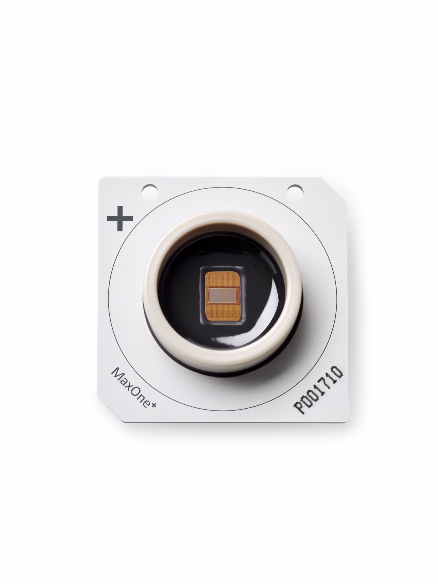
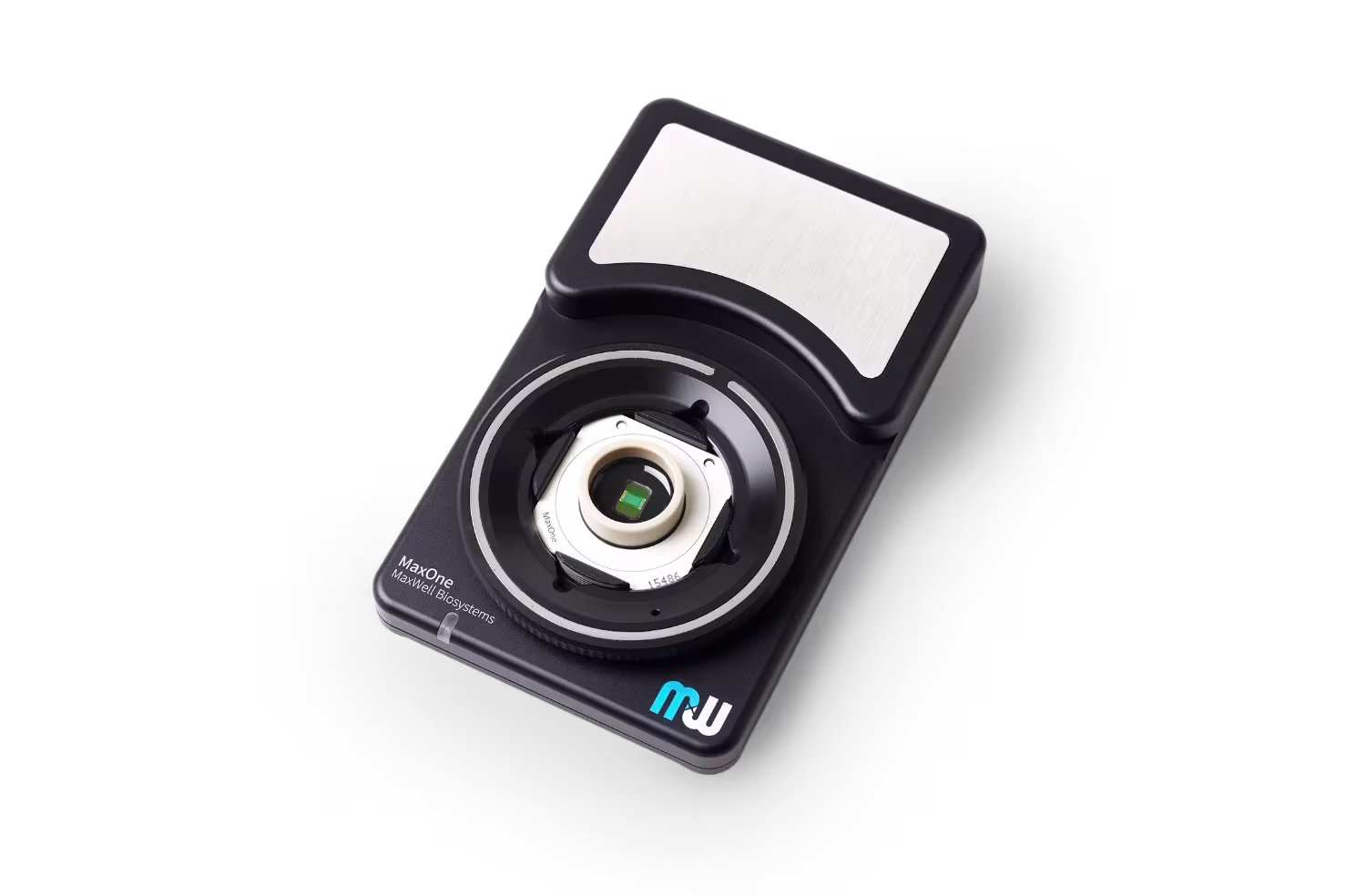
Capture cellular signals with unmatched spatial and temporal resolution, down to single-cell and even sub-cellular detail. MaxOne features 26,400 high-density electrodes at 3,265 electrodes/mm2 with a 20 kHz sampling rate. MaxOne enables detailed insights from individual cells down to axonal signal propagation, ensuring exceptional data quality for your research.
Signal fidelity
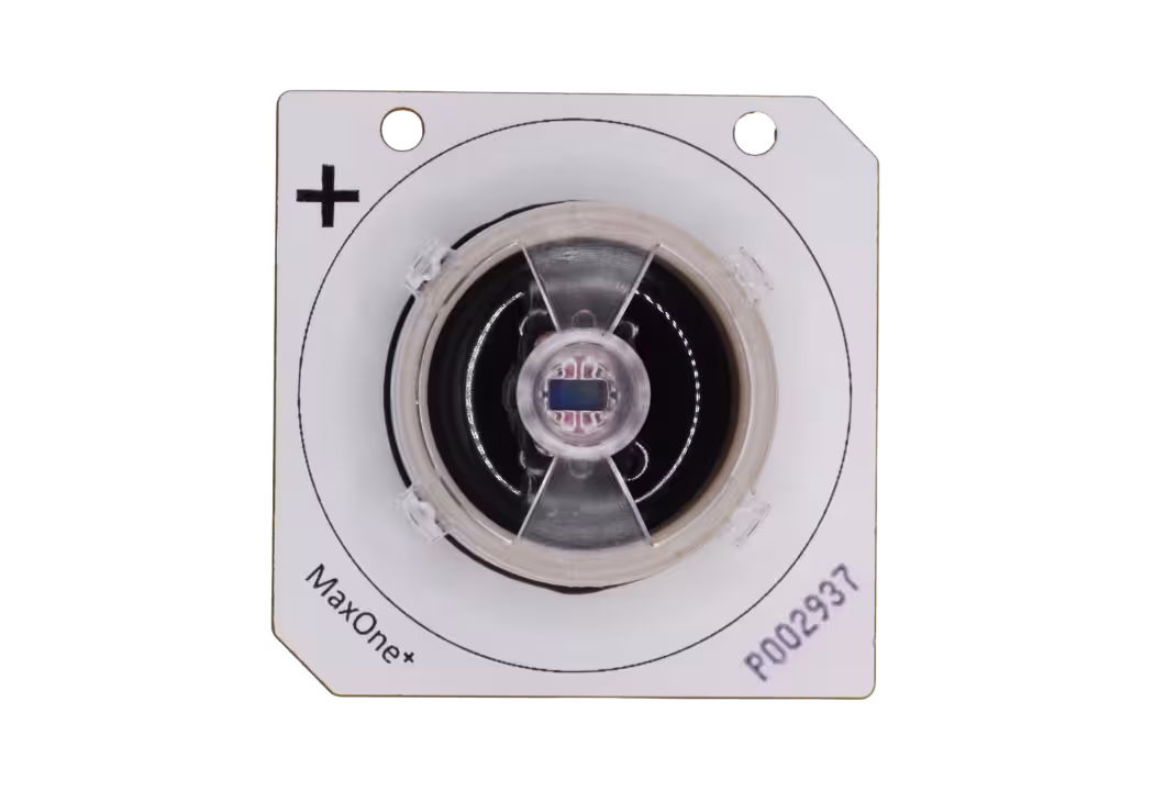
Detect even the smallest signals with exceptional precision and sensitivity. MaxOne delivers industry-leading low-noise performance to ensure accurate data capture, even from weak or rare events.
Signal integrity
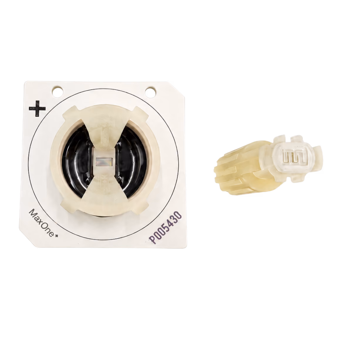
Ensure consistent, high-quality recordings for longitudinal experiments. A soft, biocompatible PEDOT coating covers 44% of the electrode area, creating an optimized surface for reliable signal acquisition over time.
Reproducibility
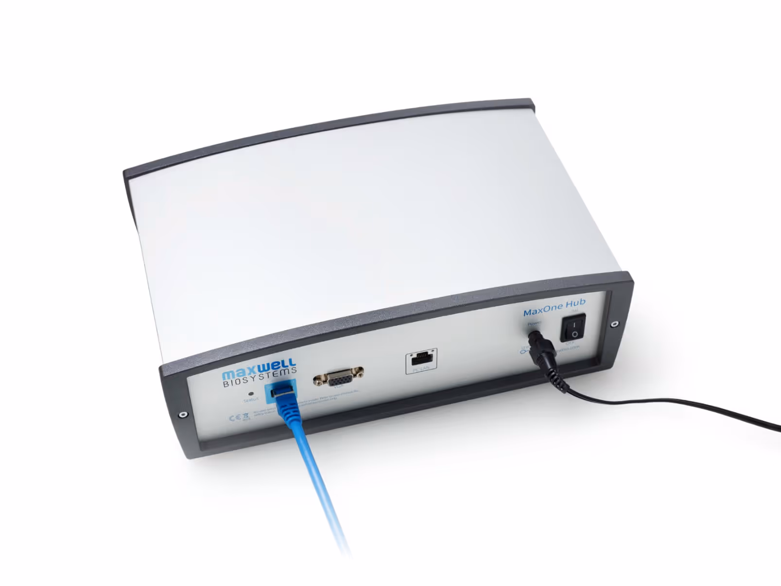
Achieve consistent results across experiments. MaxOne supports in-incubator operation and features automatic chip identification, minimizing variability due to environment and streamlining workflows.
%201.avif)


.avif)