 English
 繁體中文
繁體中文 简体中文
简体中文 日本語
日本語
The Innovation —
CMOS-based high-density microelectrode array technology

The ability to record the electrical activity of neurons has created endless possibilities for advancing neuroscience research.
Extracellular recording is used to measure cellular electrical activity – crucial for studying the function of neurons, such as retinal ganglion cells, cardiomyocytes and other electrogenic cells.
Microelectrodes and recording technologies were developed to capture the electrical signals produced by neurons, without penetrating the cell membrane.
The development of microelectrode arrays made it easier to record electrical signals from larger areas and entire cell populations.
In such a multielectrode array, or MEA, arrays of electrodes are distributed over a large area and can simultaneously record electrical activity from thousands of cells. MEAs can be implanted in vivo or utilized for in vitro cell cultures and tissue preparations.
The need for high-density recording systems
Problem

Typical MEAs measure the average of population activity, while missing signals from the majority of cells in the preparation.
Solution

High-density MEAs (HD-MEAs) allow access to the activity of individual cells at subcellular resolution.
At MaxWell Biosystems, we have engineered an advanced high-resolution MEA platform – for in vitro electrical recording.
MaxWell Biosystems introduces a high-density MEA chip, specifically engineered for in vitro studies.
Using CMOS technology significantly decreases the size of the amplifiers – allowing us to integrate circuitry with thousands of electrodes per square millimeter on the same chip. This makes it possible to conduct experiments requiring cellular (and even subcellular) level resolution.
Using a switch-matrix enables optimal electrode utilization and signal amplification – allowing us to simultaneously achieve exceptional signal-to-noise ratio and high electrode density.
Packaged together with the custom recording unit and proprietary software, MaxWell Biosystems offers an advanced in vitro electrophysiology platform, with unique features.
- Resolution levels needed for high-precision R&D studies
- Highest quality signals
- Flexible electrode configuration, recording and stimulation options
- Versatile culture system for the whole range of in vitro and ex vivo biological preparations
- Compact set-up suitable for long-term measurements
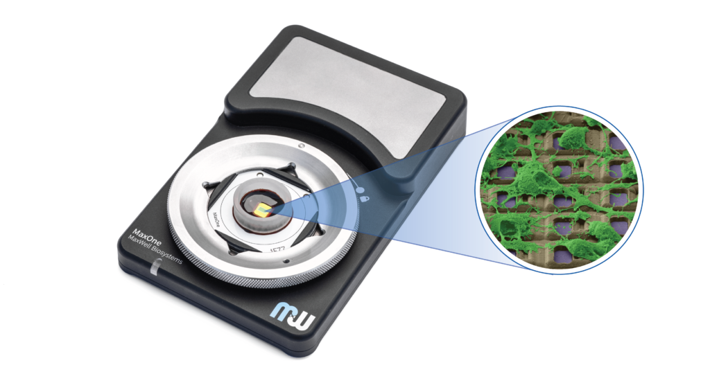
MaxOne System —
High-resolution electrophysiology for in vitro applications

MaxOne MEA Wells
- Culture dish for extracellular electrophysiology
- Multiple ring sizes available to allow for flexibility in experimental design
- Biocompatibility for long-term cultures

MaxOne Recording Unit
- Control system and data acquisition to interface with the recording PC via Ethernet connection
- Compact layout for laboratory space
- Extension modules available to add flexibility for experiments

MaxLab Live Software*
- Custom software suite for setting:
1) electrode configuration and stimulation patterns
2) recording mode
3) data acquisition parameters
4) post-experiment data analysis - Includes 3 custom recording modules and data analysis toolbox
*features listed are standard with MaxLab Live Full
Our HD-MEA platform involves innovative circuitry and software design – built specifically for biological experiments.


Every cell in the network
26’400 electrodes
3.85 mm × 2.10 mm sensor area
17.5 microns electrode pitch

Flexible stimulation
32 stimulation units
Voltage and current mode
Single cell resolution

Data analysis tools
Preprocessing
Visualization
Statistics and reports
How It Works —
From bench experiment to actionable results

-
Harvest cells from primary tissue, such as the brain, retina or heart.
-
When prepared for the experiment, begin to culture cells on MEA chip from MaxWell Biosystems.
-
Begin your experiment: Track results and analyze data on a computer, with the help of MaxLab Live Software.
-
Use the results to guide further experiments, obtain scientific insights, and publish data.
Let high-quality data provide actionable results for your R&D laboratory.
Multiscale recording and analysis —
1. High-resolution, label-free cell activity imaging
Analyze subcellular features, such as full axonal arbors in single neurons.
Electrical signals tracked along neurites enable to investigate novel parameters. High-resolution tracking of axonal action potential propagation allows for investigating changes in axonal conduction velocity.
High-resolution axonal action potential propagation tracking
Tracking axonal branch signals
2. Whole sample activity map
Localize active cells on the MEA during experiments.
The morphological structure of a primary cortical cell culture was imaged using an upright microscope with 10x objective. MAP2 staining was used to visualize the neurons. The optical image closely matches the electrical image obtained using MaxOne. The electrical image provides the location of the cells, as well as information on the activity of the cells, such as spike rate and amplitude.


3. Smart population recording
Identify defined cells for long term recording and further analyses.
Different combinations of parameters can be used to automatically select a defined set of cells to be recorded, such as spike rate, amplitude, etc. The raster plot shows the dynamics of the network activity using 1,024 active electrode sites. Burst features can be investigated in detail (one burst zoomed in).

Applications —
Add depth to your drug discovery, neuroscience, stem cell, and safety pharmacology R&D studies
 Retina

- Identify every retinal ganglion cell (RGC) type
- Record RGC mosaics
- Characterize RGC phenotypes label-free
Brain Slice
 Brain Slice
- Identify defined anatomical / functional regions
- Isolate single-cell activity
- Track oscillations and wave propagations at high-resolution
Neuronal Networks

- Locate all active cells in the whole sample
- Record population activity to extract network connectivity
- Track axonal action potential propagation
Cardiomyocytes

- Visualize beats and wave propagation
- Detect differentiation of embryonic bodies to cardiac cells
- Characterize effect of drugs
iPSCs

- Detect small signals (action potentials) from developing neurons
- Track network development across days and months
- Characterize functional phenotypes in disease models
Scientifically Proven —
Peer-reviewed publications in high-impact journals
- Over 25 peer-reviewed publications in high-impact journals.
- Publications span HD-MEA technology, neuronal network recording, and studies with cardiomyocytes, retinal cells, brain slice, and neurons.

K. Yonehara, M. Fiscella, J. Müller, et al, “Congenital nystagmus gene FRMD7 is necessary for establishing a neuronal circuit asymmetry for direction selectivity,” Neuron, 89(1), pp. 177-193, 2016.

J. Müller, U. Frey, et al., “High-resolution CMOS MEA platform to study neurons at subcellular, cellular, and network levels,” Lab Chip, vol. 15, no. 13, pp. 2767–2780, May 2015.
